| Origin | Body of the pubis Inferior pubic ramus |
| Insertion | Upper third of the linea aspera Pectineal line of femur |
| Action | Adduction of hip joint (primary) Flexion of the hip joint (secondary) Potentially able to rotate the femur (see actions section below) |
| Nerve | Obturator nerve L2-L4 (anterior division) |
| Artery | Deep femoral artery |
Location & Overview
The adductor brevis muscle is a small, somewhat fan-shaped muscle located in the medial compartment of the thigh. The other muscles of the medial compartment of the thigh are: the adductor longus, gracilis, and the adductor magnus [1]. As the smallest muscle in the adductor group, the adductor brevis is positioned between the adductor longus and the adductor magnus muscles and is located deep to the pectineus and adductor longus muscles [2].
The main function of the adductor brevis muscle, as well as the other muscles within the medial compartment of the thigh, is adduction (moving the thigh towards the midline of the body) [3]. The adductor brevis also has secondary actions which will be covered further in the actions section below.
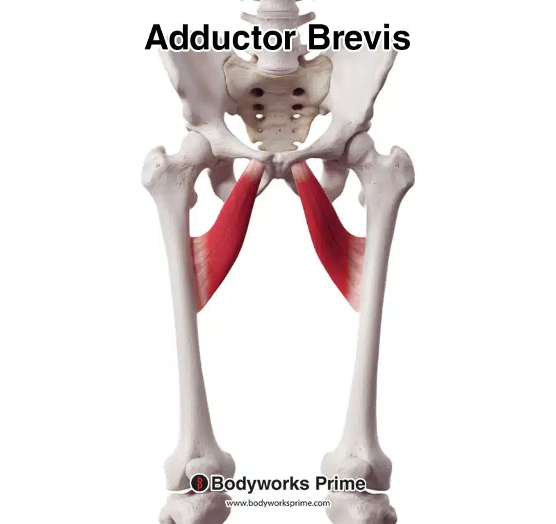
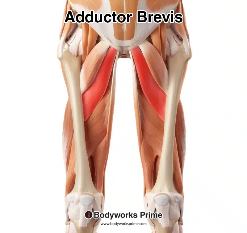
Pictured here we can see the adductor brevis from an anterior view.
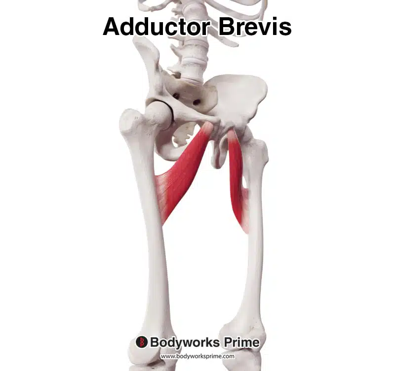
Pictured here we can see the adductor brevis from a anterolateral view.
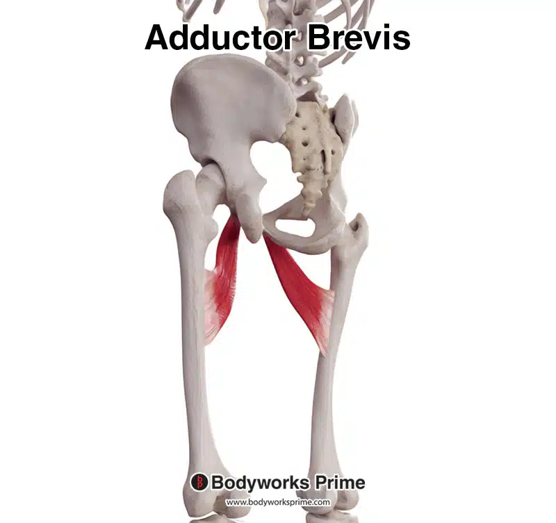
Pictured here we can see the adductor brevis from a postolateral view.
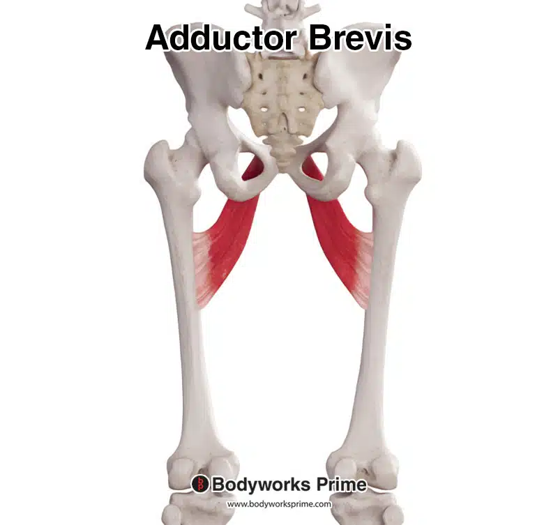
Pictured here we can see the adductor brevis from a posterior view.
Here we can see an image of the adductor brevis muscle highlighted in red. Other muscles of the thigh (such as the quadriceps) have been removed to reveal its location.
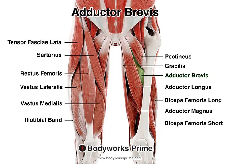
Pictured here, we can see the adductor brevis amongst the other muscles of the thigh. The adductor brevis is displayed on the right side of the image, highlighted in green. Because the adductor brevis is a deep muscle, other muscles have been stripped back so we can see its location. The leg is missing the tensor fasciae latae, sartorius, quadriceps, and iliotibial band. You can see these still in place on the other leg for reference.
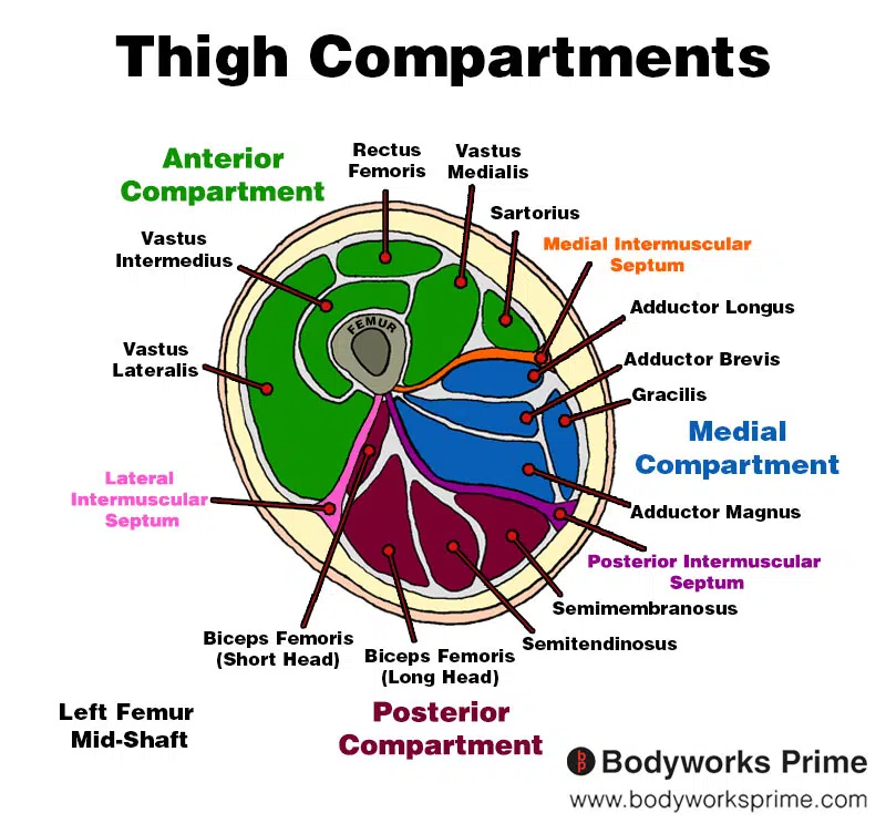
Here we can see an image of the compartments of the thigh. We can see the adductor brevis in the medial compartment, the section coloured in blue.
Origin & Insertion
The adductor brevis muscle originates on the inferior and anterior aspects of the pelvis and inserts onto the medial and posterior side of the femur. The specific origin point on the pelvis is the body of the pubis and the inferior pubic ramus. The body of the pubis is the central, flat portion of the pubic bone that forms the anterior part of the pelvic girdle. The inferior pubic ramus is a thin extension of bone that projects downward and outward from the body of the pubis, connecting it to the ischium [4] [5] [6] [7].
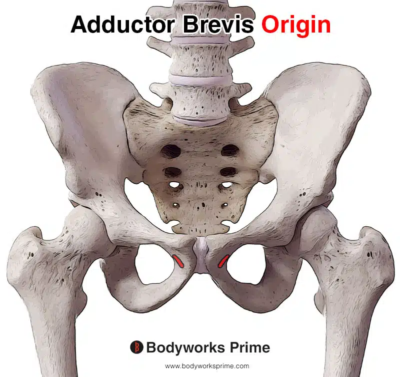
Pictured here we can see the origin of the adductor brevis highlighted in red. The origin is on the body of the pubis and inferior pubic ramus.
The muscle then passes the hip joint and attaches to the femur, specifically on the upper third of the linea aspera, a prominent ridge that runs vertically along the posterior surface of the femur. This insertion extends to the pectineal line, a short, oblique ridge on the proximal posterior surface of the femur that runs between the lesser trochanter and the linea aspera [8] [9] [10] [11]. The pectineal line provides an additional attachment point for the adductor brevis and other muscles, such as: the pectineus and vastus medialis [12].
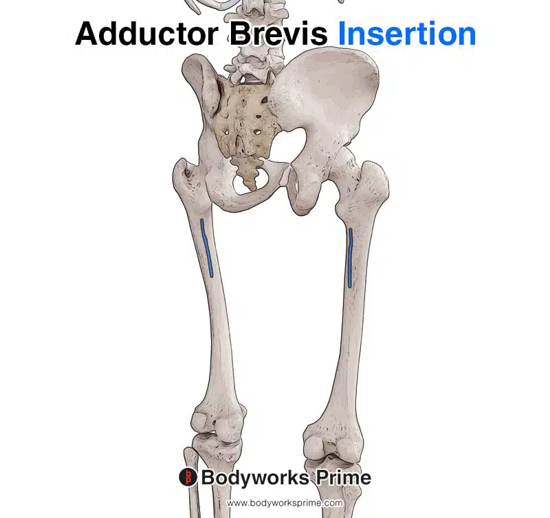
Pictured here we can see the insertion of the adductor brevis highlighted in blue. The insertion is on the upper third of the linea aspera and the pectineal line of the femur.
Actions
The primary action of the adductor brevis muscle is adduction of the hip joint, which involves bringing the thigh towards the midline of the body. A secondary action of the adductor brevis is flexion of the hip joint, helping to bend the hip forward [13] [14].
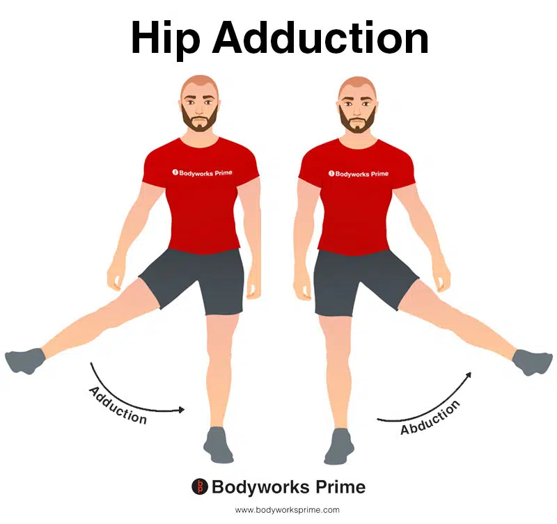
This image shows an example of hip adduction, which involves moving the towards the body (medially). The opposite of hip adduction is hip abduction. The adductor brevis’ primary action is hip adduction.
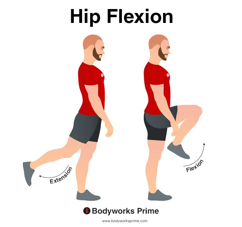
In this image, you can see an example of hip flexion, which is the action of raising your leg up in front of you and causing your hip to bend. If both legs are fixed, the torso will lean forward towards the legs. The opposite movement of hip flexion is hip extension. Hip flexion is a secondary action of the adductor brevis muscle.
Research also suggests that the adductor brevis muscle may play a role in internal and external rotation of the thigh [15] [16]. However, there is limited research on this topic, and most of the available evidence is based on EMG studies, a methodology that has its own set of limitations. The evidence is stronger for external rotation being a direct action of the adductor brevis than internal rotation. Based on the location of its origin and insertion, it does seem likely it could weakly perform external rotation. Some studies indicate that the muscle’s ability to externally or internally rotate the femur may depend on its position. Further research is needed to conclusively determine if the adductor brevis can directly contribute to lateral or medial rotation of the femur. It is important to note that any rotational forces the adductor brevis muscle might provide are likely to be comparatively weak when compared to other hip rotators. Rotation is not considered a primary action of this muscle.
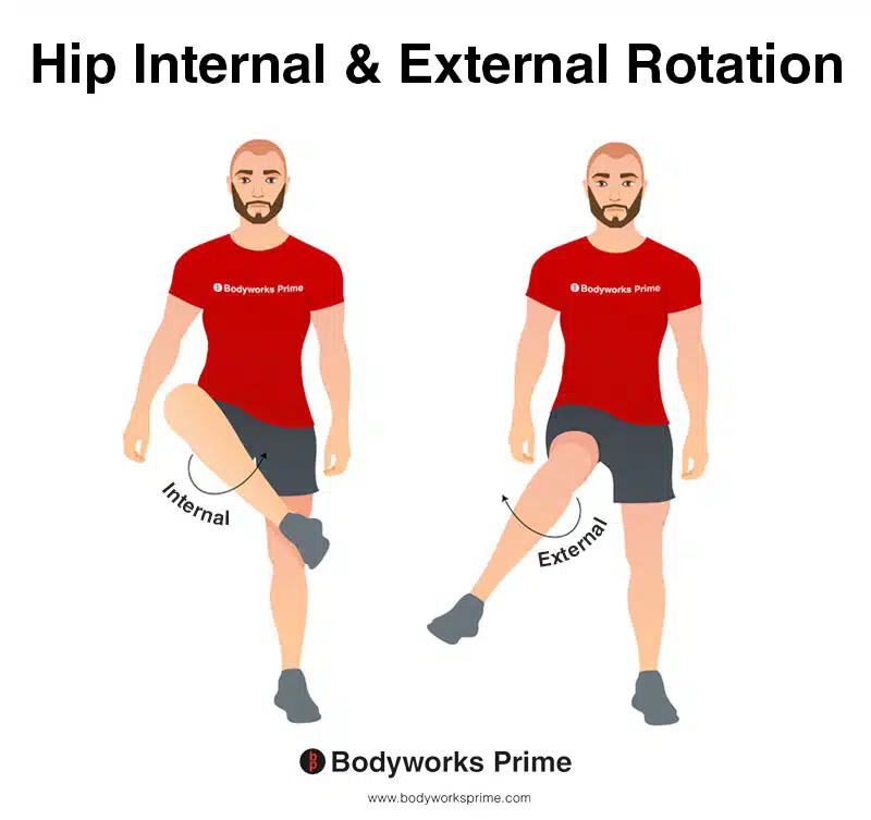
This image shows an example of hip internal (medial) rotation and external (lateral) rotation. Internal rotation involves rotating the leg from the hip joint inwards (medially). The opposite of internal rotation is external rotation, which involves rotating the leg from the hip joint outwards (laterally). A few studies suggest the adductor brevis may be able to assist with internal and external rotation of the hip joint in certain positions [17] [18]. However, more research needs to be done to conclude this action with certainty.
Innervation
The adductor brevis muscle is innervated by the obturator nerve’s anterior division, which arises from the lumbar plexus with nerve roots L2-L4 [19] [20]. The obturator nerve is divided into anterior and posterior divisions as it passes through the obturator canal. The anterior division innervates the adductor longus, adductor brevis, and gracilis muscles, as well as providing innervation to the hip joint. The posterior division innervates the external obturator and adductor magnus muscles, while also supplying innervation to the knee joint. The obturator artery and vein travel with the obturator nerve in the obturator canal [21] [22].
In some cases, the obturator nerve can become entrapped within the thick fascia overlying the adductor brevis muscle, sometimes seen in athletes reporting deep aching pain in the groin and medial thigh, as well as weakness during exercise [23].
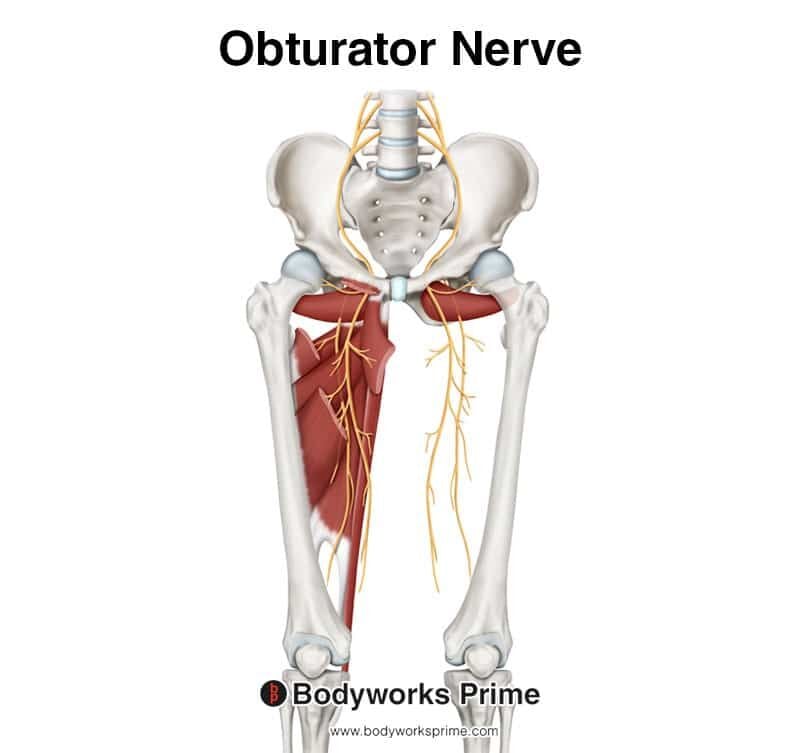
This image shows the obturator nerve arising from the lumbar plexus with nerve roots from L2-L4.
Blood Supply
The blood supply to the adductor brevis muscle primarily comes from the deep femoral artery, which is a branch of the femoral artery. The femoral artery originates from the external iliac artery, becoming the femoral artery inferior to the inguinal ligament. It then divides into superficial and deep (profunda) branches just inferior to the inguinal ligament, with the adductor longus muscle acting as a partition between these two branches. While the superficial branch courses anterior to the adductor longus muscle, the deep branch courses lateral and slightly posterior to it [24] [25].
The deep femoral artery passes distally, first between the adductor longus and adductor brevis muscles, and then between the adductor longus and adductor magnus muscles. During its course posterior to the adductor longus muscle, it gives off the medial femoral circumflex artery and three perforating arteries before terminating at mid-thigh level as the fourth perforating artery. These perforating arteries supply blood to the adductor brevis muscle and the surrounding tissues, and they also have anastomotic branches that lead to other perforators, muscular branches, and cutaneous branches [26] [27].
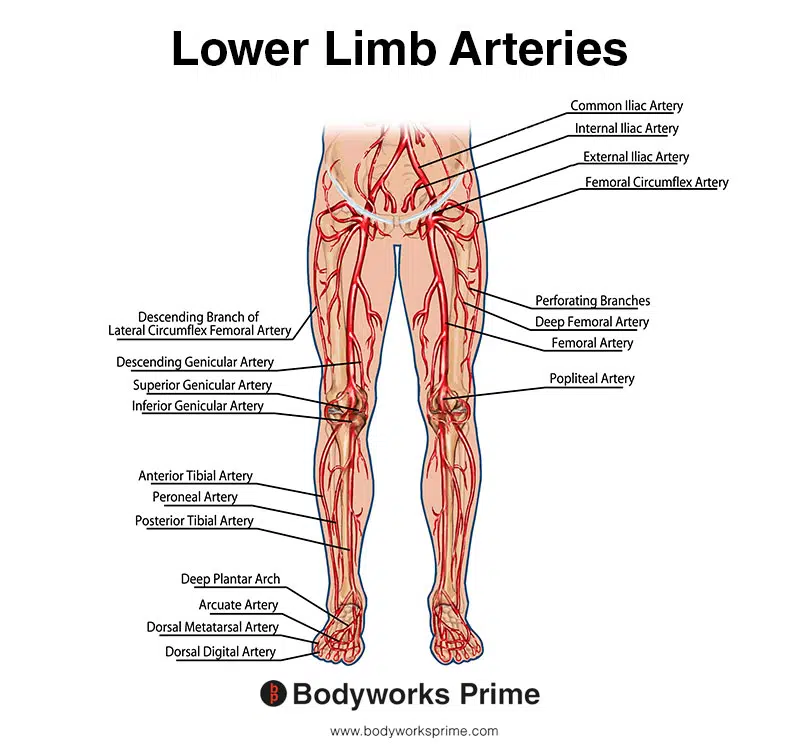
This image shows the arteries of the lower limb. You can see the deep femoral artery located in the thigh region, labelled on the right leg. You can also see how it branches off from the femoral artery. The deep femoral artery provides blood to the adductor brevis muscle.
Want some flashcards to help you remember this information? Then click the link below:
Adductor Brevis Flashcards
Support Bodyworks Prime
Running a website and YouTube channel can be expensive. Your donation helps support the creation of more content for my website and YouTube channel. All donation proceeds go towards covering expenses only. Every contribution, big or small, makes a difference!
References
| ↑1 | Jeno SH, Schindler GS. Anatomy, Bony Pelvis and Lower Limb: Thigh Adductor Magnus Muscle. [Updated 2022 Aug 1]. In: StatPearls [Internet]. Treasure Island (FL): StatPearls Publishing; 2023 Jan-. Available from: https://www.ncbi.nlm.nih.gov/books/NBK534842/ |
|---|---|
| ↑2, ↑4, ↑8, ↑20 | Ransom AL, Sinkler MA, Nallamothu SV. Anatomy, Bony Pelvis and Lower Limb: Femoral Muscles. [Updated 2022 Sep 20]. In: StatPearls [Internet]. Treasure Island (FL): StatPearls Publishing; 2023 Jan-. Available from: https://www.ncbi.nlm.nih.gov/books/NBK500008/ |
| ↑3, ↑12 | Moore KL, Agur AMR, Dalley AF. Clinically Oriented Anatomy. 8th ed. Philadelphia: Lippincot Williams & Wilkins; 2017. |
| ↑5, ↑9 | Kiel J, Kaiser K. Adductor Strain. [Updated 2022 Jun 21]. In: StatPearls [Internet]. Treasure Island (FL): StatPearls Publishing; 2023 Jan-. Available from: https://www.ncbi.nlm.nih.gov/books/NBK493166/ |
| ↑6, ↑10 | Ramage JL, Varacallo M. Anatomy, Bony Pelvis and Lower Limb: Medial Thigh Muscles. [Updated 2022 Aug 29]. In: StatPearls [Internet]. Treasure Island (FL): StatPearls Publishing; 2023 Jan-. Available from: https://www.ncbi.nlm.nih.gov/books/NBK534775/ |
| ↑7, ↑11, ↑14, ↑22, ↑25, ↑27 | Standring S. (2015). Gray’s Anatomy: The Anatomical Basis of Clinical Practice, 41st Edn. Amsterdam: Elsevier. |
| ↑13 | Ransom AL, Sinkler MA, Nallamothu SV. Anatomy, Bony Pelvis and Lower Limb: Femoral Muscles. [Updated 2022 Sep 20]. In: StatPearls [Internet]. Treasure Island (FL): StatPearls Publishing; 2023 Jan-. Available from: https://www.ncbi.nlm.nih.gov/books/NBK500008/ |
| ↑15, ↑17 | Leighton RD. A functional model to describe the action of the adductor muscles at the hip in the transverse plane. Physiother Theory Pract. 2006 Nov;22(5):251-62. doi: 10.1080/09593980600927385. PMID: 17118893. |
| ↑16, ↑18 | Reimann R, Sodia F, Klug F. Die umstrittene Rotationswirkung ausgewählter Muskeln im Hüftgelenk [Controversial rotation function of certain muscles in the hip joint]. Ann Anat. 1996 Aug;178(4):353-9. German. PMID: 8928937. |
| ↑19 | Jeno SH, Schindler GS. Anatomy, Bony Pelvis and Lower Limb: Thigh Adductor Magnus Muscle. [Updated 2022 Aug 1]. In: StatPearls [Internet]. Treasure Island (FL): StatPearls Publishing; 2023 Jan-. Available from: https://www.ncbi.nlm.nih.gov/books/NBK534842/ |
| ↑21 | Weiss, L., Silver, J. K., Lennard, T. A., & Weiss, J. M. (2007). Nerves. In Easy Injections (pp. 105-155). Professor of Clinical Physical Medicine and Rehabilitation, SUNY-Stony Brook College of Medicine; Assistant Professor of Physical Medicine and Rehabilitation, Harvard Medical School, Boston, Massachusetts; Clinical Assistant Professor, Department of Physical Medicine and Rehabilitation, University of Arkansas for Medical Sciences, Little Rock, Arkansas. https://doi.org/10.1016/B978-075067527-7.10006-5 |
| ↑23 | Craig, A. (2015). Nerve Compression/Entrapment Sites of the Lower Limb. In Nerves and Nerve Injuries: Vol 2: Pain, Treatment, Injury, Disease and Future Directions (pp. 755-770). Department of Physical Medicine and Rehabilitation, University of Michigan, Ann Arbor, Michigan, USA. https://doi.org/10.1016/B978-0-12-802653-3.00097-X |
| ↑24, ↑26 | Özkan, Ö., & Coşkunfirat, O. K. (2009). Tensor fascia lata flap. In Flaps and Reconstructive Surgery (SECTION THREE, pp. 561-576). https://doi.org/10.1016/B978-0-7216-0519-7.00038-1 |


