| Origin | Anterior superior iliac spine (ASIS) Anterior aspect of the iliac crest |
| Insertion | Fascia of the iliotibial tract The iliotibial tract inserts onto the iliotibial tubercle (also known as Gerdy’s tubercle) |
| Action | Stabilisation of the hip and knee Hip abduction Hip internal rotation Weak hip flexion Knee flexion past 30 degrees Lateral rotation at the tibia |
| Nerve | Superior gluteal nerve (L4, L5, S1) |
| Artery | Deep branch of the superior gluteal artery |
Location & Overview
The tensor fasciae latae (TFL) is a muscle located in the proximal and anterolateral aspect of the thigh (front and outer part of the thigh). It sits between the deep and superficial fibers of the iliotibial tract, also known as the iliotibial band (ITB). The TFL spans from the anterior portion of the iliac crest to the superior portion of the tibia, it connects to the tibia via the iliotibial tract. The TFL belongs to the muscles of the gluteal region. Other muscles of this region are the gluteus maximus, gluteus medius and gluteus minimus [1] [2].
The tensor fascia latae muscle is important for stabilising the pelvis, especially during activities such as standing, walking, running, jumping, and other lower body movements. It functions in conjunction with the gluteus maximus, gluteus medius, and gluteus minimus muscles. The TFL muscle is connected to the fascia of the iliotibial tract, which in turn provides a link to the gluteus maximus muscle. This is because the gluteus maximus muscle also attaches to the iliotibial tract [3].
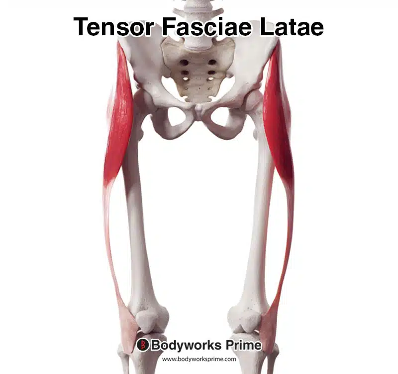
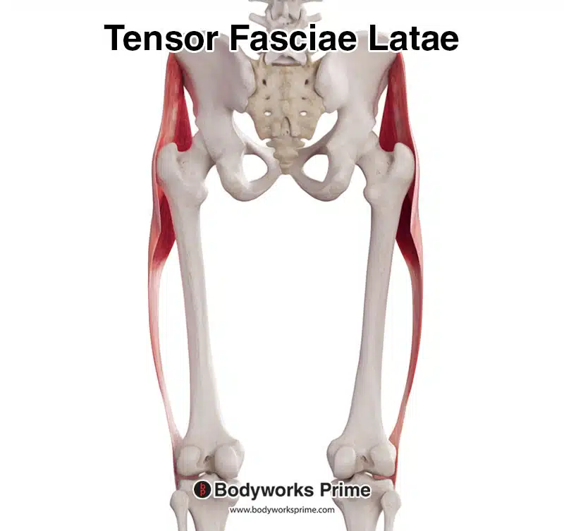
Here we can see the tensor fasciae latae from an anterior view.
Here we can see an image of the tensor fasciae latae and the iliotibial tract together.
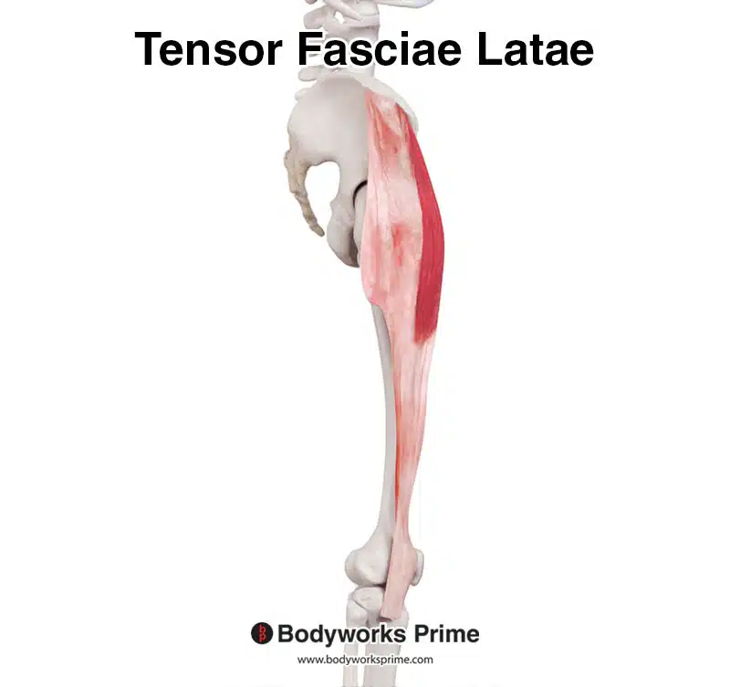
Here we can see the tensor fasciae latae from a lateral view.
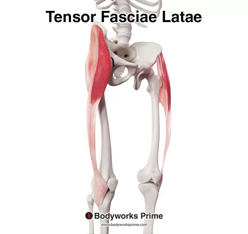
Here we can see the tensor fasciae latae from a posterior view.
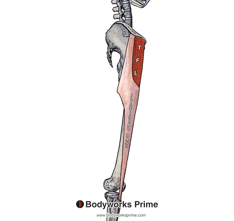
Pictured here we can see the tensor fasciae latae and the iliotibial tract labelled and clearly sectioned.
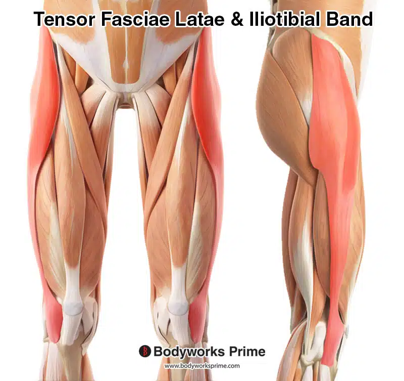
Here we can see both the tensor fasciae latae and iliotibial band highlighted in red amongst the other muscles of the thigh.
Origin & Insertion
The tensor fasciae latae originates from the anterior superior iliac spine (ASIS) and the anterior aspect of the iliac crest. As it passes down the thigh, it attaches to the deep and superficial fascia of the iliotibial band. Due to this, the insertion point for the tensor fasciae latae is the iliotibial band. Once the tensor fasciae latae inserts into the iliotibial band, the iliotibial band continues down the leg, where it eventually crosses the knee joint and inserts onto the iliotibial tubercle on the tibia (also known as Gerdy’s tubercle) [4] [5] [6].
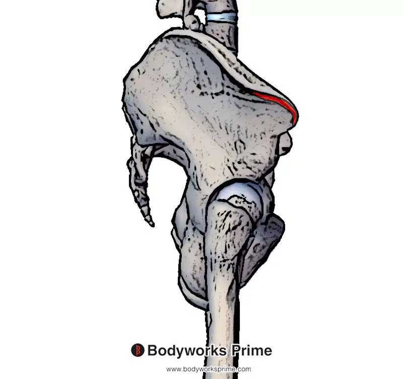
Highlighted in red, we can see the origin point of the tensor fasciae latae on the anterior superior iliac spine (ASIS) and the anterior aspect of the iliac crest.
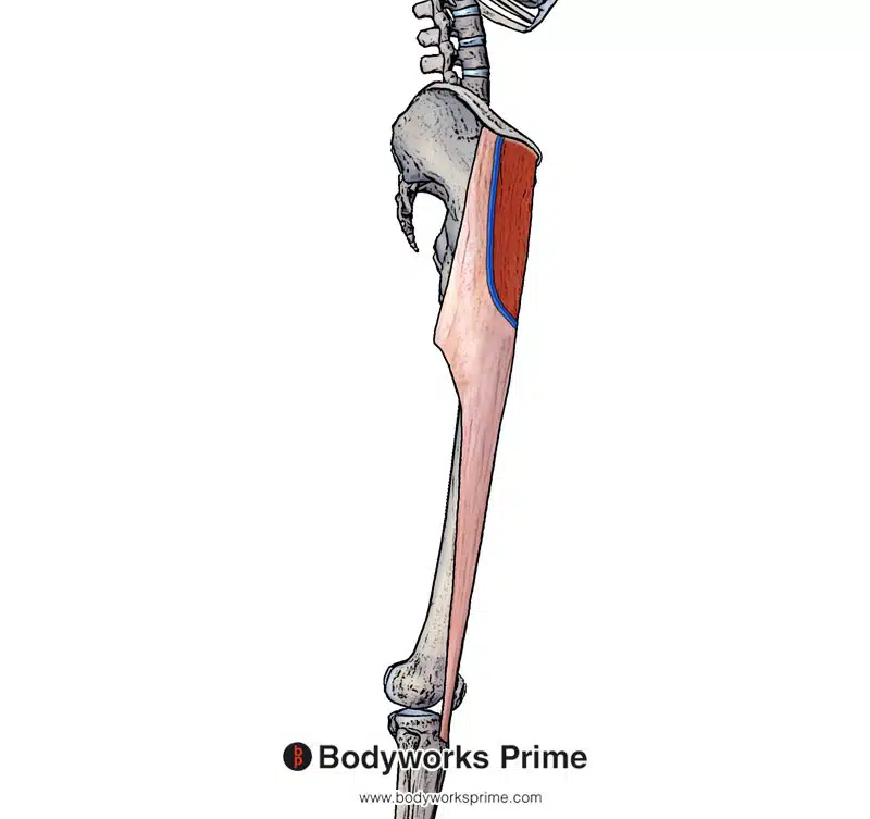
Highlighted in blue we can see the insertion point of the tensor fasciae latae on the fascia of the iliotibial tract.
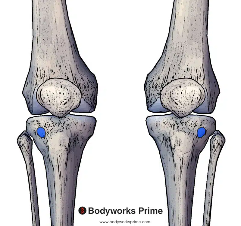
Highlighted in blue we can see the insertion point of the iliotibial tract. The tensor fasciae latae inserts into the iliotibial tract and therefore by proxy has a connection to the same insertion point. The iliotibial tract inserts onto the iliotibial tubercle (also known as Gerdy’s tubercle).
Actions
The TFL muscle assists several muscle groups in stabilising and moving the hip and knee joints. For instance, it works together with the gluteus medius and gluteus minimus during internal rotation and abduction of the hip. Moreover, both the gluteus maximus and TFL are connected to the iliotibial tract via an aponeurosis, enabling the TFL to also help the gluteus maximus in hip abduction. Besides abduction, the TFL can assist with hip flexion as well, although it is relatively weak compared to the primary hip flexors, such as the rectus femoris and iliopsoas muscle group [7] [8].
The TFL connects into the iliotibial tract via an aponeurosis. The iliotibial tract then inserts onto the iliotibial tubercle (also known as Gerdy’s tubercle). Due to this, contraction of the TFL can contribute to flexion of the knee joint (because the iliotibial tract crosses the knee joint). However, the TFL can only contribute to knee flexion beyond approximately 30 degrees. This connection of the TFL to the tibia via the iliotibial tract is how it can assist in stabilisation of the knee joint. The TFL can also act on the iliotibial tract to assist in lateral rotation of the tibia [9] [10].
A function of the TFL is to assist in walking. When the TFL contracts it weakly pulls the ilium inferiorly (hip abduction). This contraction then assists in lifting the hip on the opposite side. This is particularly useful when walking because during the swing phase of gait, it allows the leg to swing through without hitting the ground [11] [12].
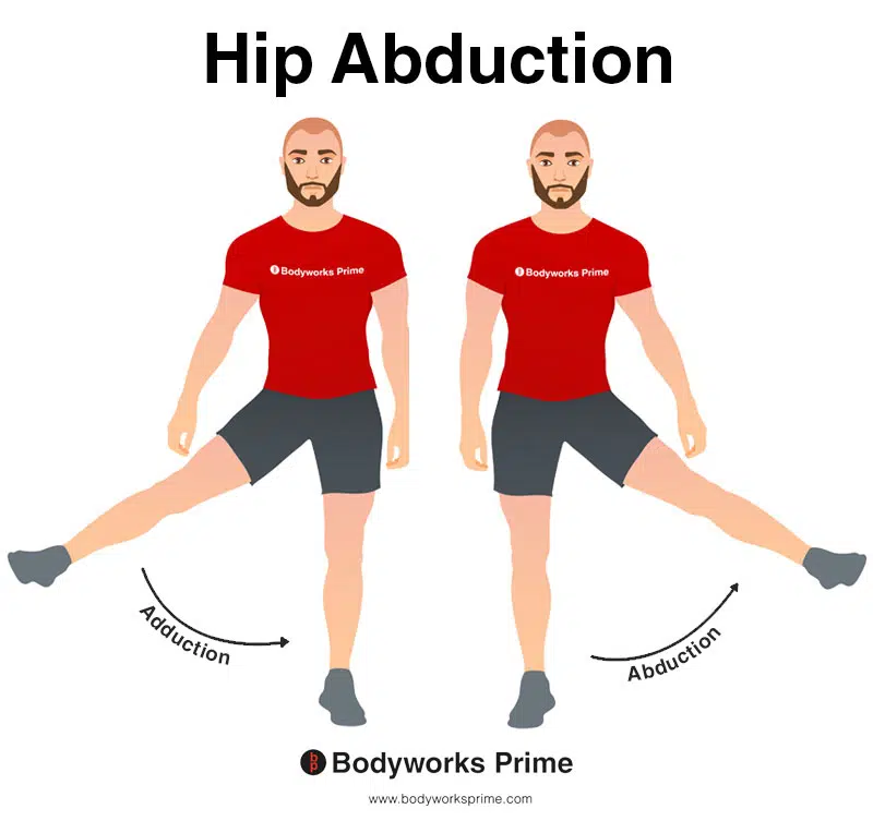
This image shows an example of hip abduction, which involves moving the leg out to the side (laterally). The TFL is a hip abductor. The opposite of hip abduction is hip adduction.
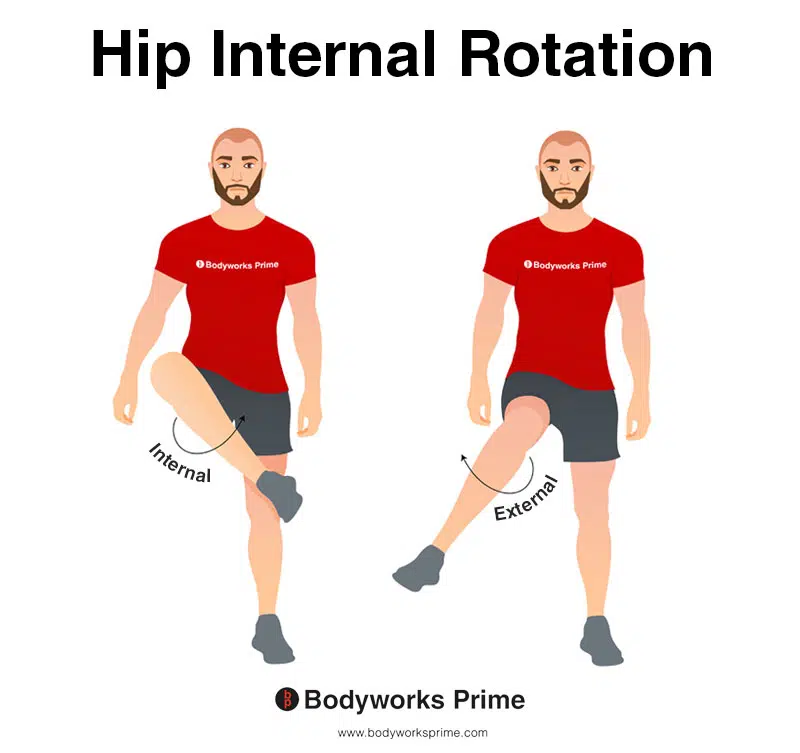
This image depicts an example of hip internal rotation, which involves rotating the leg from the hip joint inwards (medially). Internal rotation is also referred to as medial rotation. The TFL muscle functions as a hip internal rotator. The opposite of internal rotation or medial rotation is external rotation or lateral rotation.
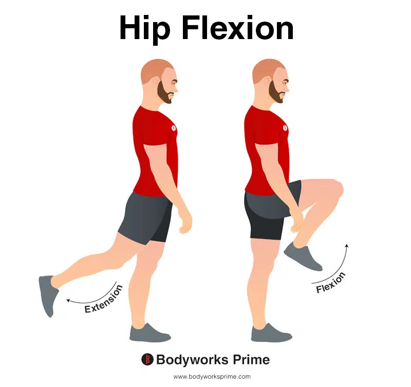
In this image, you can see an example of hip flexion, which is the action of raising your leg up in front of you and causing your hip to bend. The opposite movement of hip flexion is hip extension. Since the TFL is not considered a primary hip flexor, its contribution to hip flexion is relatively weak.
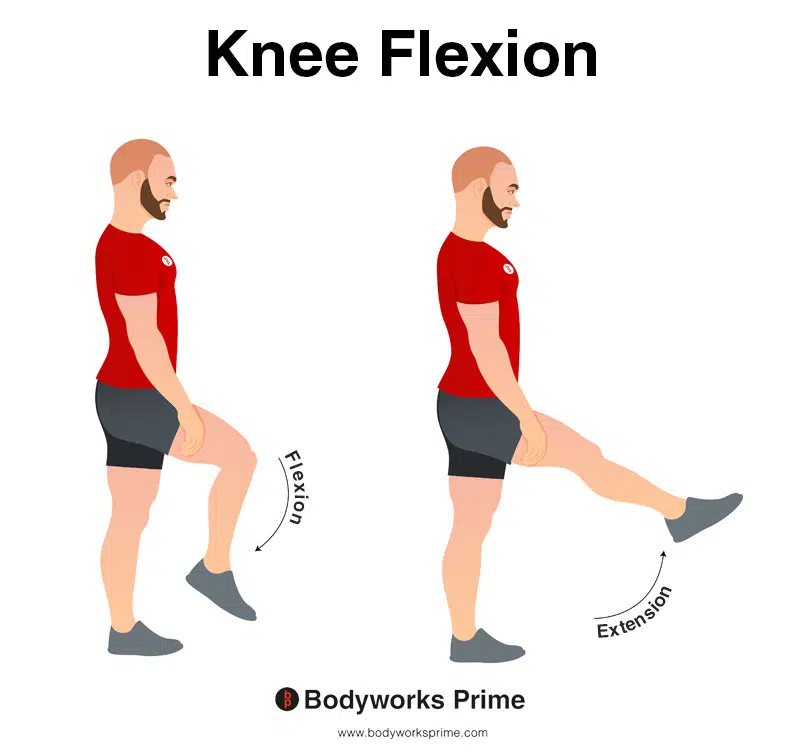
In this image, you can see an example of knee flexion, which is the action of bending your knee. The opposite movement of knee flexion is knee extension. The TFL is involved in knee flexion when the knee is flexed past a 30 degree bend.
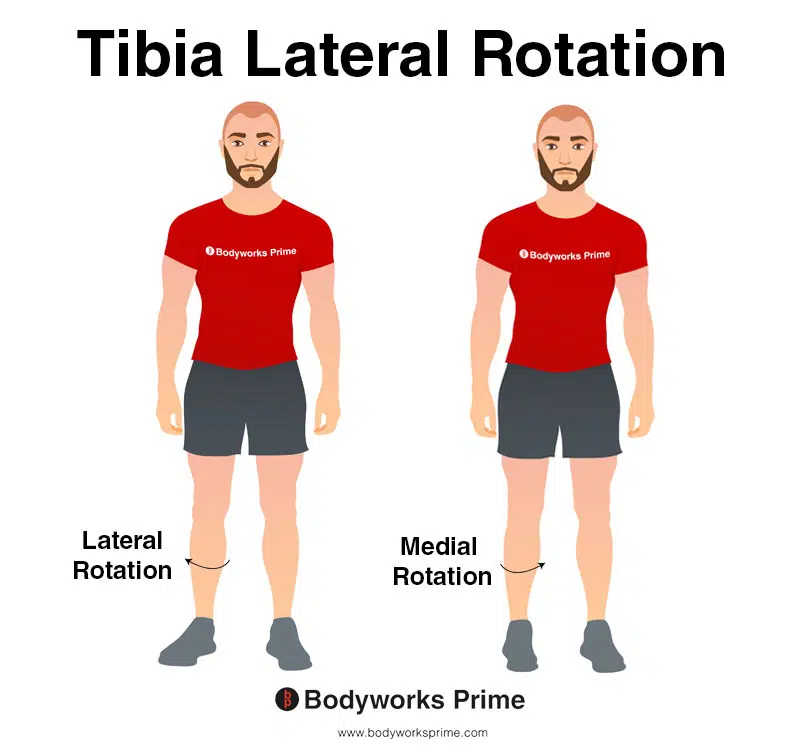
In this image, you can see an example of lateral rotation of the tibia. The TFL is able to laterally rotate the tibia. Lateral rotation of the tibia involves outward rotation of the shinbone away from the midline of the body. The opposite movement of lateral rotation of the tibia is medial rotation of the tibia.
Remember that the TFL muscle plays a role in stabilising the hip and knee joints, but these actions are not depicted in the provided illustrations.
Innervation
The TFL is innervated by the superior gluteal nerve (L4, L5, S1) which is a branch of the sacral plexus [13].
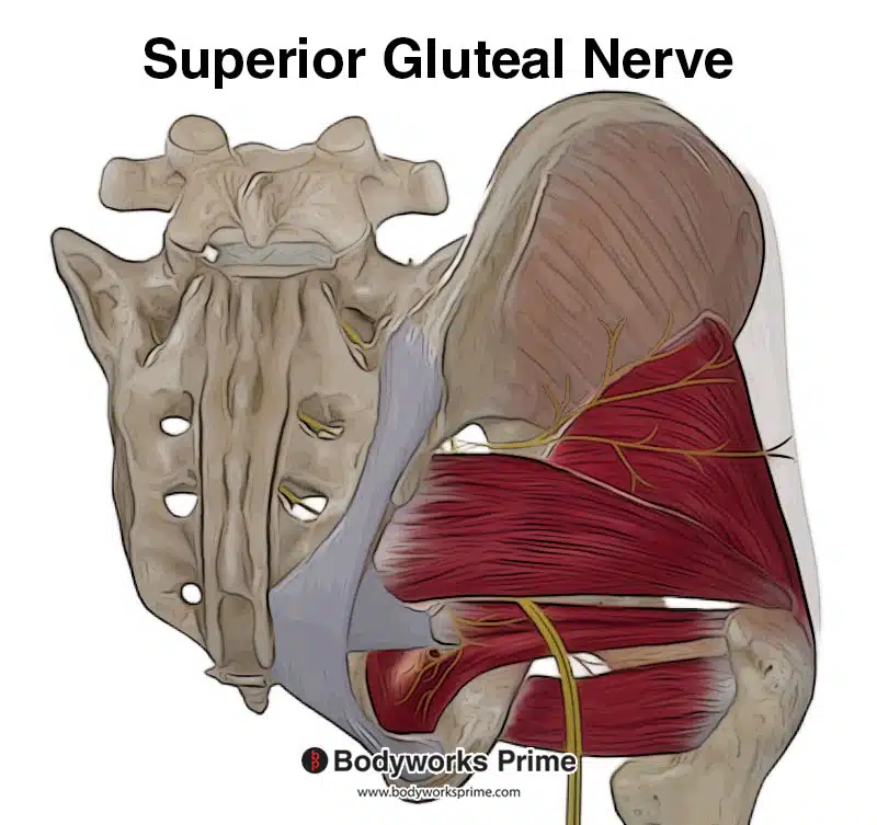
Pictured here you can see the superior gluteal nerve, which innervates the TFL.
Blood Supply
Blood is supplied to the TFL via the deep branch of the superior gluteal artery [14].
Want some flashcards to help you remember this information? Then click the link below:
Tensor Fasciae Latae Flashcards
Support Bodyworks Prime
Running a website and YouTube channel can be expensive. Your donation helps support the creation of more content for my website and YouTube channel. All donation proceeds go towards covering expenses only. Every contribution, big or small, makes a difference!
References
| ↑1, ↑4, ↑7, ↑9, ↑11, ↑13, ↑14 | Trammell AP, Nahian A, Pilson H. Anatomy, Bony Pelvis and Lower Limb, Tensor Fasciae Latae Muscle. [Updated 2022 Aug 8]. In: StatPearls [Internet]. Treasure Island (FL): StatPearls Publishing; 2022 Jan-. Available from: https://www.ncbi.nlm.nih.gov/books/NBK499870/ |
|---|---|
| ↑2, ↑3, ↑6, ↑8, ↑10, ↑12 | Moore KL, Agur AMR, Dalley AF. Clinically Oriented Anatomy. 8th ed. Philadelphia: Lippincot Williams & Wilkins; 2017 |
| ↑5 | Hyland S, Graefe SB, Varacallo M. Anatomy, Bony Pelvis and Lower Limb, Iliotibial Band (Tract) [Updated 2022 Aug 8]. In: StatPearls [Internet]. Treasure Island (FL): StatPearls Publishing; 2022 Jan-. Available from: https://www.ncbi.nlm.nih.gov/books/NBK537097/ |


