| Origin | Body of the pubis (inferior to the pubic crest) |
| Insertion | Middle third of linea aspera of femur (medial lip) |
| Action | Hip adduction Hip flexion |
| Nerve | Obturator nerve (L2, L3, L4) |
| Artery | Obturator artery Medial femoral circumflex artery |
Location & Overview
The adductor longus muscle is situated in the thigh and, as its name suggests, primarily functions in the adduction of the hip joint. It also forms part of the medial wall of the femoral triangle, an inverted triangular space in the anterior part of the thigh that contains important blood vessels, nerves, and muscles [1]. The adductor muscle group consists of three muscles: the adductor longus, adductor magnus, and adductor brevis, [2] with the adductor longus being the most frequently injured muscle, accounting for 62% to 90% of all adductor injuries [3].
The femoral triangle is an essential anatomical landmark for healthcare professionals, as it allows them to access the femoral artery and vein for diagnostic and therapeutic purposes. The triangle is bordered by the inguinal ligament superiorly, the sartorius muscle laterally, and the adductor longus muscle medially [4] [5].
Common pathologies associated with the adductor longus muscle include strains, tendinopathies, and osteitis pubis. Adductor strains are often seen in athletes who participate in sports that require sudden changes in direction or explosive movements, such as football and rugby [6] [7]. Factors that may contribute to an adductor strain include inadequate stretching of the adductor muscle group, muscle exhaustion, previous hip or groin injuries, weak adductors, decreased range of motion, and advancing age [8]. Tendinopathies, on the other hand, are frequently multifactorial, stemming from a combination of intrinsic factors such as anatomy, age, and systemic issues, as well as extrinsic factors like mechanical overload, repetitive overuse of the muscle, and incorrect form or equipment [9]. Osteitis pubis is an inflammatory condition affecting the pubic symphysis and surrounding structures, which can cause chronic groin pain and is often seen in athletes who engage in sports that require repetitive hip adduction [10].
Effective exercises for the adductor longus muscle target hip adduction and stabilisation. One such exercise is the seated hip adduction, which involves sitting with the knees bent and feet placed on the adductor machine’s pads at the gym and squeezing the legs together against the resistance provided by the machine. Another exercise called the ‘Copenhagen adduction exercise’ is recognised as one of the best ways to strengthen the adductors. One of the reasons for its popularity is its adaptability, as it can be executed in almost any location using only your body weight and a suitable item to rest your leg upon [11] [12] [13]. To perform the Copenhagen adduction exercise, first lie on your side, with your lower forearm supporting your weight and the upper leg resting on a bench or similar object. Next, lift your body in a concentric hip adduction movement, while also adducting the lower leg so that your body forms a straight line and your feet touch each other. Then, slowly lower your leg back down and repeat the exercise for the desired number of repetitions.
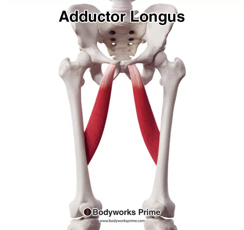
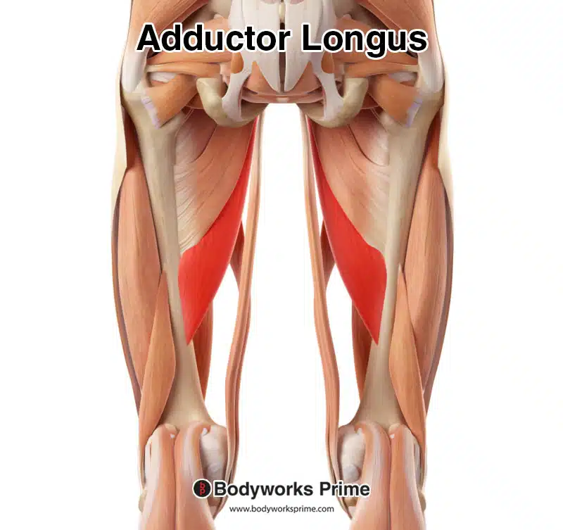
In this image we can see the adductor longus from an anterior view.
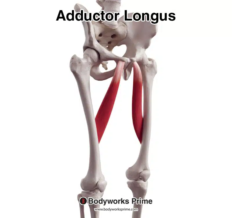
Here we can see the adductor longus from an anterolateral.
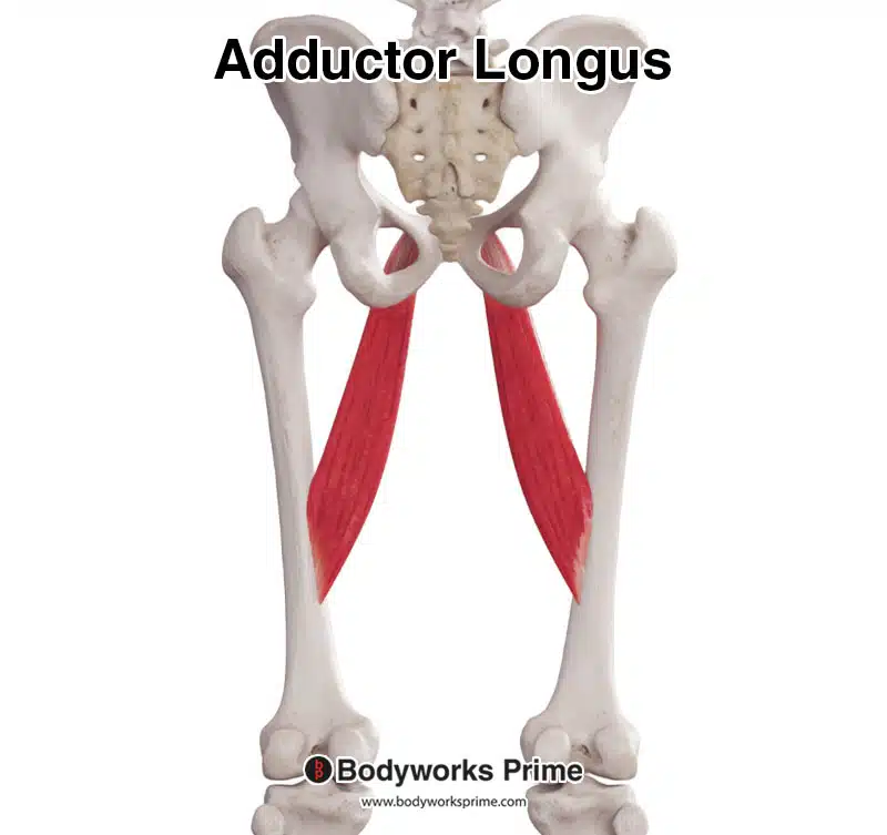
In this picture we can see the adductor longus from a posterior view.
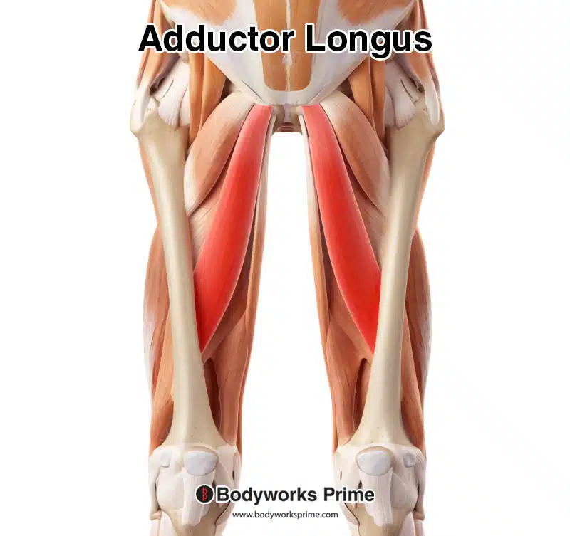
Here we can see the adductor longus highlighted in red, seen from an anterior view. More superficial muscles such as the quadriceps have been removed so we can fully see the adductor longus.
Here we can see the adductor longus highlighted in red, seen from a posterior view. More superficial muscles such as the semimembranosus, semitendinosus, and the adductor magnus have been removed so we can fully see the adductor longus.
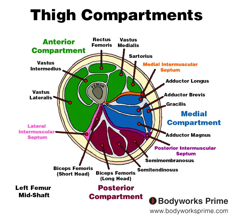
Here we can see an image of the compartments of the thigh. We can see the adductor longus in the medial compartment, the section coloured in blue.
Origin & Insertion
The adductor longus muscle has its origin and insertion points on specific locations within the pelvic and thigh regions. The origin of the muscle is located on the body of the pubis, which is a part of the pelvic bone. The pubis is the front portion of the hip bone, and the body of the pubis is the central area of this bone, slightly below the pubic crest. The pubic crest is a horizontal ridge found on the upper edge of the pubic bone [14] [15] [16].
From its origin, the adductor longus muscle extends diagonally and inserts onto the posterior aspect of the femur, which is the thigh bone. The precise insertion point is on the medial lip of the middle third of the linea aspera. To help visualise this, the femur has a rough, longitudinal ridge on its posterior side called the linea aspera. The linea aspera has an upper, middle, and lower third, and the adductor longus muscle inserts onto the inner edge (medial lip) of the middle third of this ridge [17] [18] [19].
Here we can see pictured the origin of the adductor longus marked in red. The origin is on the medial third of linea aspera of the femur.

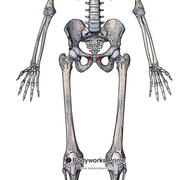
Here we can see pictured the insertion of the adductor longus marked in blue. The insertion is on the body of the pubis (inferior to the pubic crest).
Actions
The adductor longus muscle plays a significant role in the movement and stabilisation of the hip joint. Its primary function is hip adduction, which means it helps bring the thigh toward the midline of the body. This movement can be visualised when you stand with your legs apart and then move one leg inward, closer to the other leg.
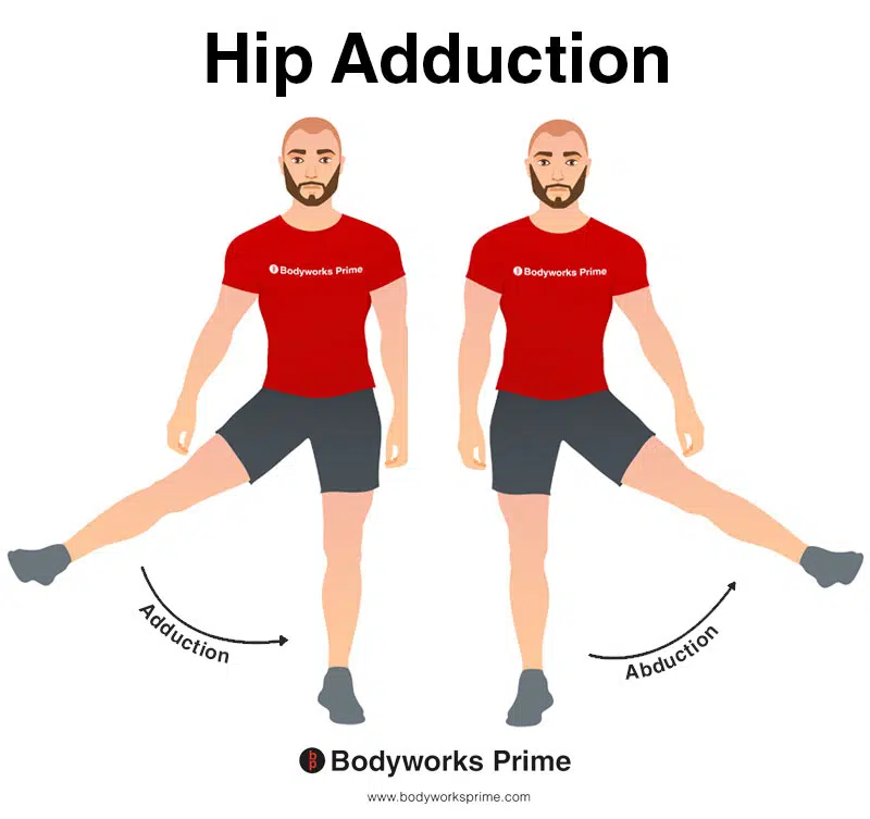
This image shows an example of hip adduction, which involves moving the towards the body (medially). The opposite of hip abduction is hip abduction. The primary role of the adductor longus is hip adduction.
Additionally, the adductor longus also contributes to hip flexion. Hip flexion occurs when the angle between the thigh and the torso is decreased, such as when lifting the knee toward the chest or bending forward at the hip.
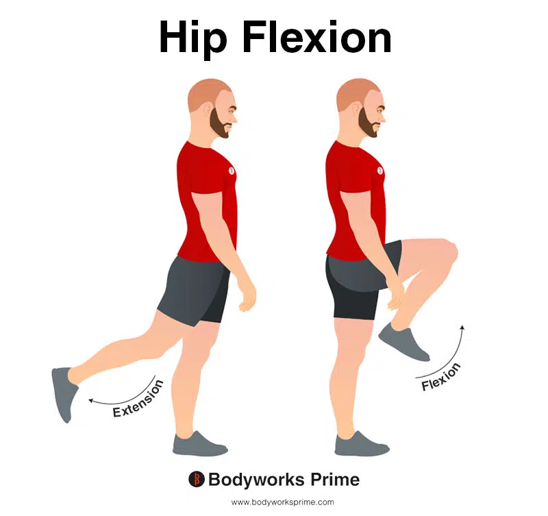
In this image, you can see an example of hip flexion, which is the action of raising your leg up in front of you and causing your hip to bend. If both legs are fixed, the torso will lean forward towards the legs. The opposite movement of hip flexion is hip extension. The adductor longus contributes to hip flexion.
While its primary actions are well-established, there is limited evidence suggesting that the adductor longus might also contribute to internal rotation of the hip joint. However, this role seems to be far less significant and could depend on factors such as hip joint positioning and individual anatomical variations [20] [21] [22]. Limited research also suggests that the adductor longus may weakly contribute to external rotation [23] [24]. However, it is important to consider the limitations of these studies and the scarcity of research specifically examining the adductor longus’s role in both external and internal rotation. At present, it would not be accurate to definitively state that the adductor longus functions as either an external or internal rotator of the hip joint, as more research is needed to clarify its involvement.
Internal rotation (also known as medial rotation) of the hip is the movement in which the thigh turns inward, causing the knee and foot to point towards the body’s midline. This action occurs when the hip joint rotates around its axis, bringing the front of the femur closer to the opposite leg. External rotation (also known as lateral rotation) of the hip is the opposite movement, where the thigh turns outward, causing the knee and foot to point away from the body’s midline. In this action, the hip joint rotates around its axis, moving the front of the femur away from the opposite leg [25].
Innervation
The adductor longus muscle is innervated by the obturator nerve, which arises from the lumbar spinal nerve roots L2, L3, and L4. The obturator nerve is a major nerve in the lower limb that provides both sensory and motor functions. It passes through the obturator foramen (an opening in the pelvis) and enters the medial compartment of the thigh. In addition to the adductor longus, the obturator nerve innervates other adductor muscles, such as the adductor brevis, adductor magnus, and gracilis, as well as the obturator externus muscle. The nerve is responsible for enabling the proper function and movement of these muscles, particularly in actions such as adduction of the hip joint [26] [27].
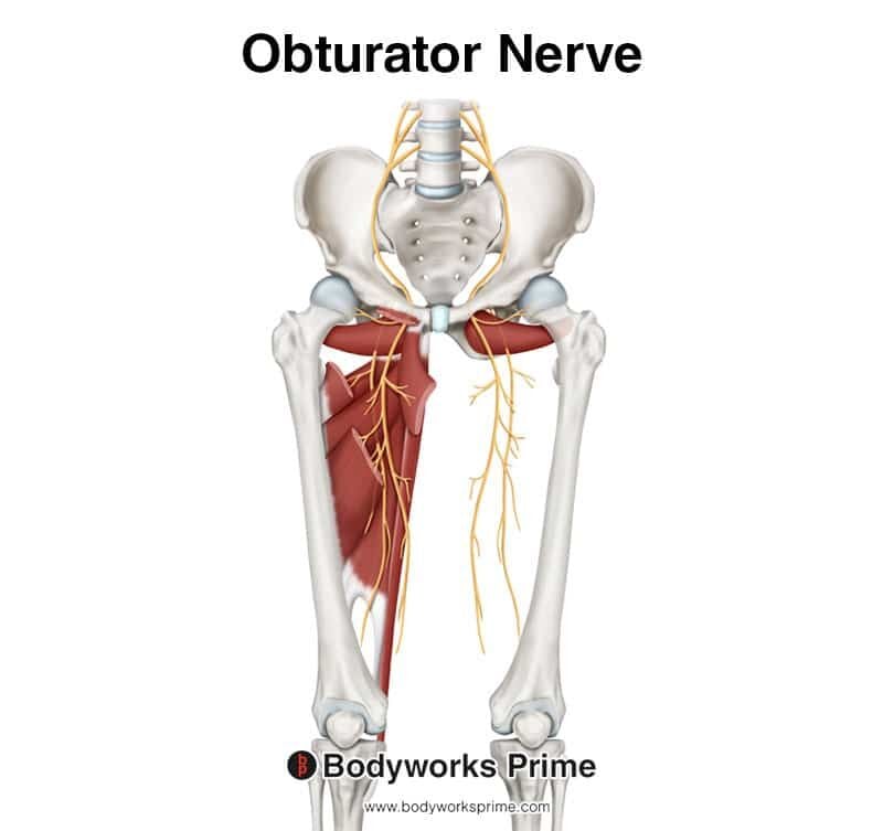
Here we can see an image of the obturator nerve which innervates the adductor longus muscle. The innervation arises from the spinal nerve roots of L2, L3, and L4.
Blood Supply
The adductor longus muscle receives its blood supply from two primary arteries: the obturator artery and the medial femoral circumflex artery. These arteries are branches of the internal iliac artery and the deep femoral artery, respectively [28].
The obturator artery exits the pelvis through the obturator foramen, running along the medial compartment of the thigh. It provides blood to the adductor muscles, including the adductor longus [29].
The medial femoral circumflex artery, another important blood supplier to the adductor longus, branches from the deep femoral artery. This artery wraps around the posterior aspect of the femur and contributes to the blood supply of the hip joint, as well as the surrounding muscles [30].
Want some flashcards to help you remember this information? Then click the link below:
Adductor Longus Flashcards
Support Bodyworks Prime
Running a website and YouTube channel can be expensive. Your donation helps support the creation of more content for my website and YouTube channel. All donation proceeds go towards covering expenses only. Every contribution, big or small, makes a difference!
References
| ↑1, ↑4 | Basinger H, Hogg JP. Anatomy, Abdomen and Pelvis: Femoral Triangle. 2023 Mar 11. In: StatPearls [Internet]. Treasure Island (FL): StatPearls Publishing; 2023 Jan–. PMID: 31082184. |
|---|---|
| ↑2, ↑16, ↑19, ↑25 | Moore KL, Agur AMR, Dalley AF. Clinically Oriented Anatomy. 8th ed. Philadelphia: Lippincot Williams & Wilkins; 2017. |
| ↑3, ↑6 | Eckard TG, Padua DA, Dompier TP, Dalton SL, Thorborg K, Kerr ZY. Epidemiology of Hip Flexor and Hip Adductor Strains in National Collegiate Athletic Association Athletes, 2009/2010-2014/2015. Am J Sports Med. 2017 Oct;45(12):2713-2722. doi: 10.1177/0363546517716179. Epub 2017 Jul 26. PMID: 28745561. |
| ↑5, ↑26, ↑29, ↑30 | Standring S. (2015). Gray’s Anatomy: The Anatomical Basis of Clinical Practice, 41st Edn. Amsterdam: Elsevier. |
| ↑7, ↑8, ↑14, ↑17, ↑22, ↑27 | Kiel J, Kaiser K. Adductor Strain. [Updated 2022 Jun 21]. In: StatPearls [Internet]. Treasure Island (FL): StatPearls Publishing; 2023 Jan-. Available from: https://www.ncbi.nlm.nih.gov/books/NBK493166/ |
| ↑9 | Federer AE, Steele JR, Dekker TJ, Liles JL, Adams SB. Tendonitis and Tendinopathy: What Are They and How Do They Evolve? Foot Ankle Clin. 2017 Dec;22(4):665-676. doi: 10.1016/j.fcl.2017.07.002. Epub 2017 Sep 27. PMID: 29078821. |
| ↑10 | Johnson R. Osteitis pubis. Curr Sports Med Rep. 2003 Apr;2(2):98-102. doi: 10.1249/00149619-200304000-00009. PMID: 12831666. |
| ↑11 | Ishøi L, Sørensen CN, Kaae NM, Jørgensen LB, Hölmich P, Serner A. Large eccentric strength increase using the Copenhagen Adduction exercise in football: A randomized controlled trial. Scand J Med Sci Sports. 2016 Nov;26(11):1334-1342. doi: 10.1111/sms.12585. Epub 2015 Oct 27. PMID: 26508629. |
| ↑12 | Alonso-Fernández D, Fernández-Rodríguez R, Taboada-Iglesias Y, Gutiérrez-Sánchez Á. Effects of Copenhagen Adduction Exercise on Muscle Architecture and Adductor Flexibility. Int J Environ Res Public Health. 2022 May 27;19(11):6563. doi: 10.3390/ijerph19116563. PMID: 35682148; PMCID: PMC9180184. |
| ↑13 | Harøy J, Clarsen B, Wiger EG, Øyen MG, Serner A, Thorborg K, Hölmich P, Andersen TE, Bahr R. The Adductor Strengthening Programme prevents groin problems among male football players: a cluster-randomised controlled trial. Br J Sports Med. 2019 Feb;53(3):150-157. doi: 10.1136/bjsports-2017-098937. Epub 2018 Jun 10. PMID: 29891614. |
| ↑15, ↑18 | Davis JA, Stringer MD, Woodley SJ. New insights into the proximal tendons of adductor longus, adductor brevis and gracilis. Br J Sports Med. 2012 Sep;46(12):871-6. doi: 10.1136/bjsports-2011-090044. Epub 2011 Oct 17. PMID: 22006933. |
| ↑20 | Neumann DA. Kinesiology of the hip: a focus on muscular actions. J Orthop Sports Phys Ther. 2010 Feb;40(2):82-94. doi: 10.2519/jospt.2010.3025. PMID: 20118525. |
| ↑21, ↑24 | Reimann R, Sodia F, Klug F. Die umstrittene Rotationswirkung ausgewählter Muskeln im Hüftgelenk [Controversial rotation function of certain muscles in the hip joint]. Ann Anat. 1996 Aug;178(4):353-9. German. PMID: 8928937. |
| ↑23 | Leighton RD. A functional model to describe the action of the adductor muscles at the hip in the transverse plane. Physiother Theory Pract. 2006 Nov;22(5):251-62. doi: 10.1080/09593980600927385. PMID: 17118893. |
| ↑28 | Jeno SH, Schindler GS. Anatomy, Bony Pelvis and Lower Limb, Thigh Adductor Magnus Muscle. [Updated 2021 Aug 8]. In: StatPearls [Internet]. Treasure Island (FL): StatPearls Publishing; 2021 Jan-. Available from: https://www.ncbi.nlm.nih.gov/books/NBK534842/ |


