| Origin | Adductor part: Inferior pubic ramus & ischial ramus Hamstring part: Ischial tuberosity |
| Insertion | Adductor part: Linea aspera & medial supracondylar line of femur Hamstring part: Adductor tubercle of femur |
| Action | Adductor & Hamstring part: Hip adduction Adductor part: Hip flexion Hamstring part: Hip extension |
| Nerve | Adductor part: Posterior branch of obturator nerve (L2-L4) Hamstring part: Tibial branch of sciatic nerve (L4) |
| Artery | Primary blood supply: Perforating branches of the profunda fermoris artery Superior portion: Medial femoral circumflex artery Inferior portion: Femoral artery, genicular arteries and popliteal artery |
Location & Overview
The adductor magnus muscle is a large, triangular muscle situated on the inner side of the thigh and it is an important member of the adductor muscle group, which helps move the thigh towards the body’s midline [1] [2] [3]. This muscle comprises two unique parts: the ‘pubofemoral’ or ‘adductor part’, originating from the ischiopubic ramus (a bone structure connecting the pubic bone and the ischium in the pelvic region), and the ‘ischiocondylar’ or ‘hamstring part’, arising from the ischium (the lower and back portion of the hip bone). The hamstring part exhibits similarities in structure, proximal attachment, and innervation to the hamstring muscles, although it is not an official member of that muscle group, which includes the biceps femoris, semitendinosus, and semimembranosus muscles [4] [5]. The medial compartment of the thigh houses the pubofemoral or adductor section, while the ischiocondylar or hamstring portion is part of the posterior compartment [6].
The adductor magnus muscle is susceptible to pathologies such as strains, tears, and tightness, which can lead to groin pain or limited hip mobility [7] [8]. Example exercises that target the adductor magnus muscle include sumo squats, wide-stance leg presses, and lateral lunges [9] . However, the ‘Copenhagen adduction exercise’ is recognised as one of the most effective ways to strengthen the adductors. One of the reasons for its popularity is its versatility, as it can be performed nearly anywhere using just your bodyweight and an object to support your leg [10] [11] [12].
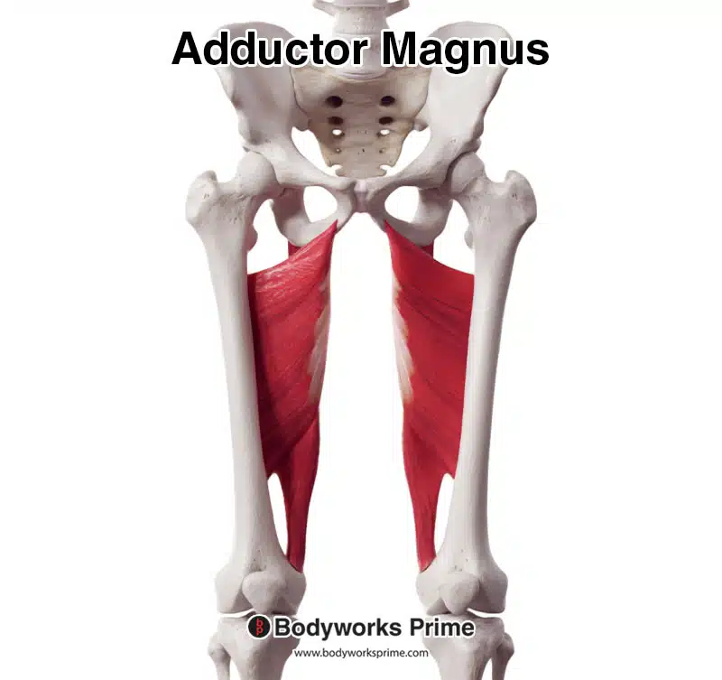
Here we can see the adductor magnus muscle from an anterior view.
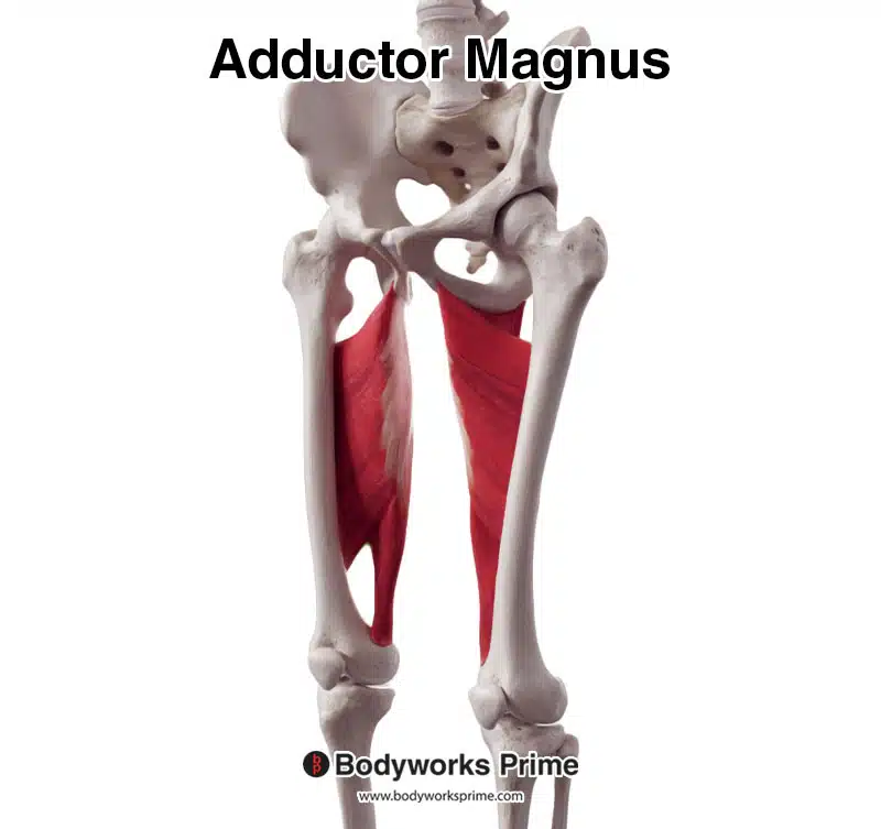
Here we can see the adductor magnus from an anterolateral view.
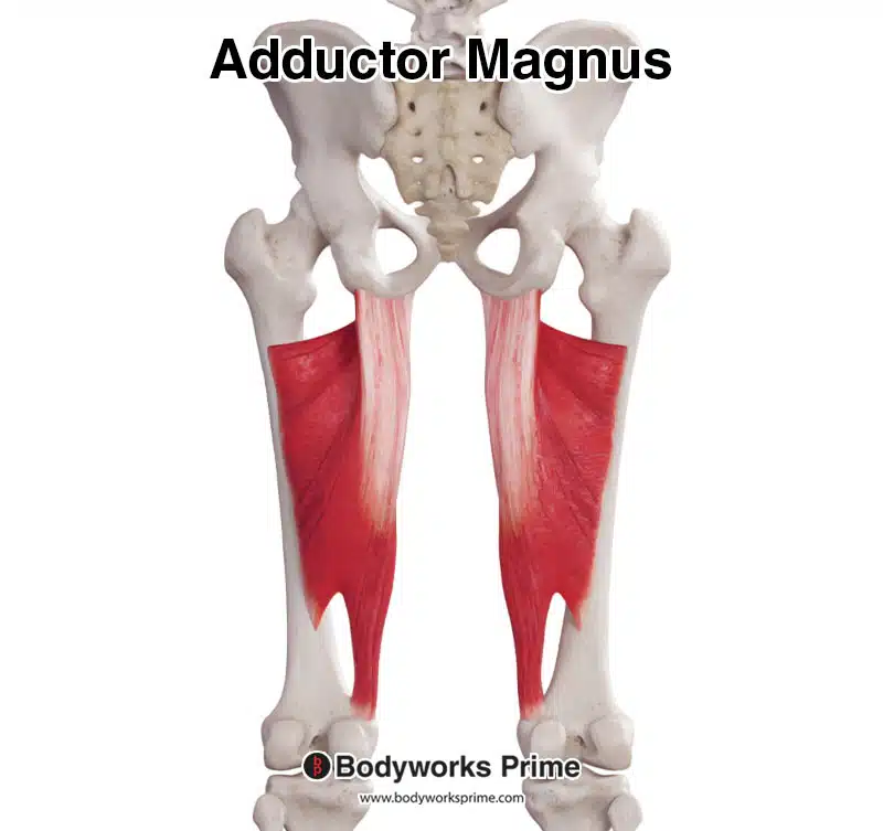
Here we can see the adductor magnus muscle from a posterior view.
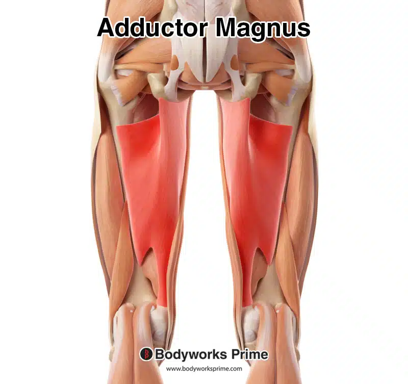
Here we can see the adductor magnus muscle highlighted in red amongst the other muscles of the leg, from a posterior view. Superficial muscles such as the semimembranosus and semitendinosus have been removed to reveal the adductor magnus, due to it being a somewhat deep muscle.
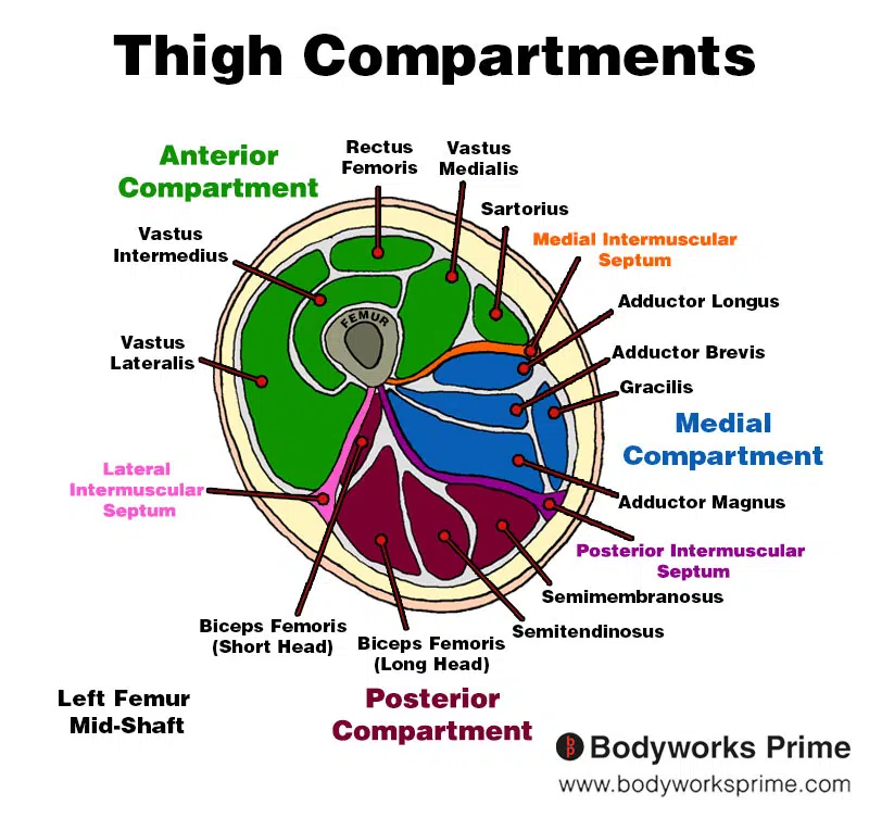
Here we can see an image of the compartments of the thigh. We can see the adductor magnus in the medial compartment, the section coloured in blue.
Origin & Insertion
The adductor magnus muscle has two distinct portions with different origins and insertions. The ‘pubofemoral’ or ‘adductor’ portion and the ‘ischiocondylar’ or ‘hamstring’ portion [13] [14].
The pubofemoral/adductor portion originates from the inferior pubic ramus and the ischial ramus. The inferior pubic ramus is a part of the pubic bone, one of the three bones that make up the pelvis, and the ischial ramus is a thin, flat bone that extends from the ischium, the lower and back part of the hip bone. The pubofemoral/adductor portion of the adductor magnus then inserts onto the linea aspera and medial supracondylar line of the femur. The linea aspera is a long, rough, vertical ridge on the back of the femur, and the medial supracondylar line is a ridge that extends from the linea aspera down to the medial condyle (the rounded prominence at the end of the femur). The portion of the adductor magnus connecting onto the linea aspera also blends with the proximal attachment of the biceps femoris muscle’s short head, allowing the two muscles to work together to stabilise the pelvis and femur[7] [15] [16] [17].
The ischiocondylar/hamstring portion of the adductor magnus originates from the ischial tuberosity, a rough, bony projection on the lower and back part of the hip bone. This portion inserts at the adductor tubercle of the femur, a small and bony prominence located on the medial condyle of the femur. The insertion also connects through fibrous attachments to the supracondylar line of the femur [18] [19] [20].
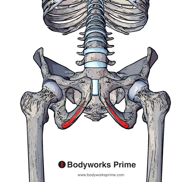
Here we can see the origin of the adductor magnus highlighted in red.
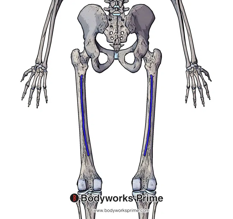
Here we can see the proximal insertion of the adductor magnus on the linea aspera and medial supracondylar line of the femur.
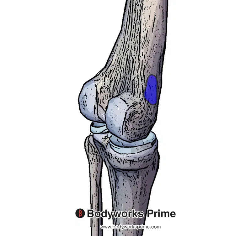
Here we can see the distal insertion of the adductor magnus on the adductor tubercle of the femur.
Actions
The adductor magnus muscle has two distinct portions with different actions, the adductor portion, and the hamstring portion. Both parts contribute to the overall function of the muscle [21] [22].
The adductor and hamstring portions of the muscle are both involved in hip adduction, which is the action of bringing the leg toward the midline of the body. This movement is useful for activities such as walking, running, or maintaining balance when standing on one leg. The adductor portion of the muscle has an additional role in hip flexion, which is the action of lifting the leg upward in front of the body, as in climbing stairs or kicking a football [23] [24]. The hamstring portion of the adductor magnus can extend the hip joint, which involves moving the leg backward, away from the body. A good example of hip extension would be skateboarding, where the leg pushes back the ground below to propel the skateboard forwards [25] [26].
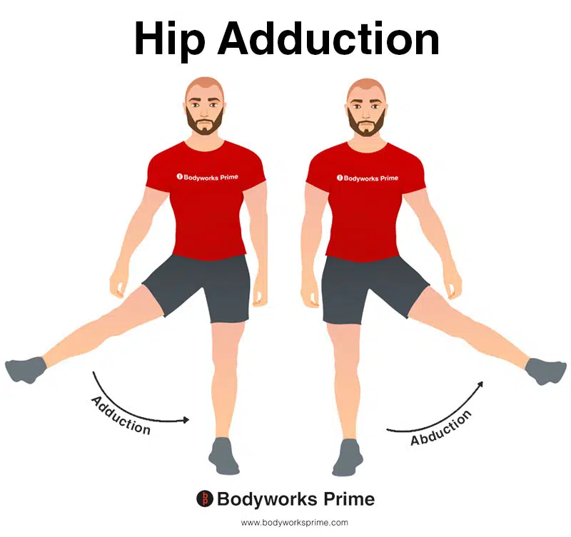
This image shows an example of hip adduction, which involves moving the towards the body (medially). The opposite of hip abduction is hip abduction. Hip adduction is an action of both the the adductor portion and the hamstring portion of the adductor magnus.
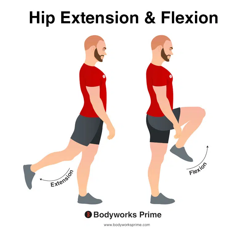
In this image, you can see an example of hip flexion and extension. Hip flexion is the action of raising your leg up in front of you and causing your hip to bend. If both legs are fixed, the torso will lean forward towards the legs. The opposite movement of hip flexion is hip extension. Hip extension is the movement of the thigh or the upper leg backward, away from the body, in the sagittal plane. This action involves increasing the angle between the thigh and the pelvis, causing the hip joint to straighten or extend. Hip flexion is an action of the adductor portion of the adductor magnus, whereas hip extension is an action of the hamstring portion of the adductor magnus.
Innervation
The adductor magnus muscle has a unique innervation pattern because it is composed of two separate portions with distinct functions, each receiving innervation from different nerves [27] [28].
The adductor portion (also known as the pubofemoral part) is innervated by the posterior branch of the obturator nerve. The obturator nerve arises from the lumbar plexus, which is formed by the anterior divisions of spinal nerves L2, L3, and L4. This part of the adductor magnus muscle primarily contributes to hip adduction and flexion [29] [30].
The hamstring portion (also known as the ischiocondylar part) receives innervation from the tibial division of the sciatic nerve. The sciatic nerve is the largest nerve in the body and originates from the lumbosacral plexus, composed of spinal nerves L4 through S3. This portion of the adductor magnus muscle primarily contributes to hip adduction and extension [31] [32].
The dual innervation of the adductor magnus muscle allows it to perform its various actions effectively, as each portion has a specialized function that is controlled by a specific nerve [33] [34] [35].
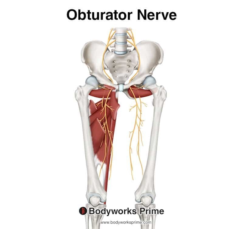
Pictured here we can see the obturator nerve which innervates the adductor magnus muscle.
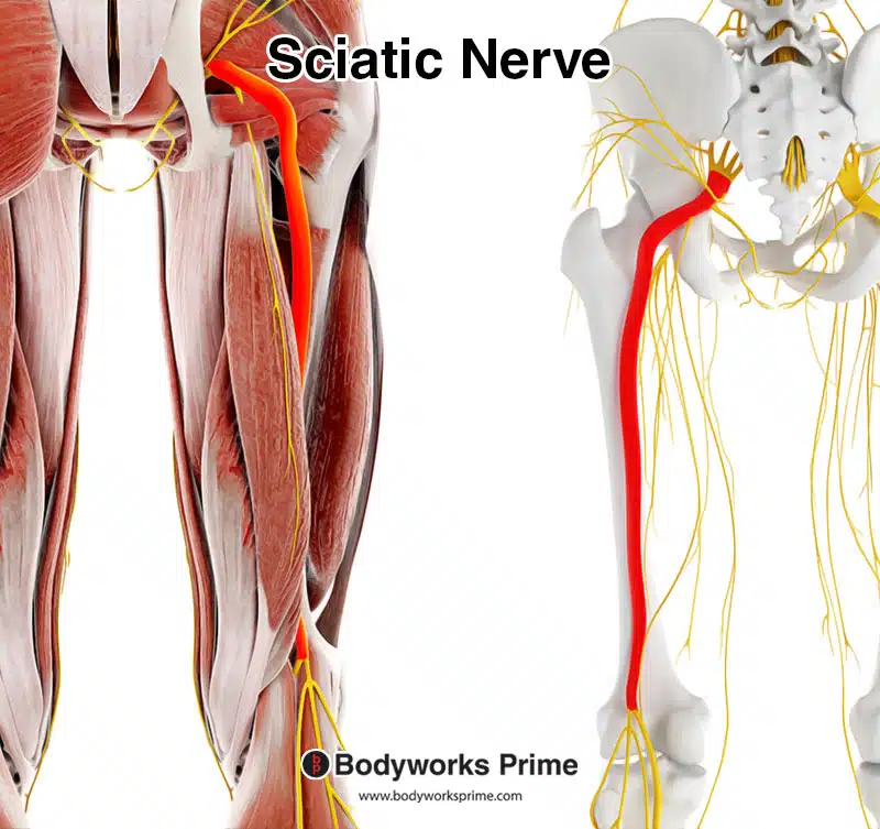
Pictured here we can see the sciatic nerve. The tibial division of the sciatic nerve innervates the adductor magnus muscle.
Blood Supply
The primary blood supply source from the adductor magnus is the perforating branches of the profunda femoris artery. The medial femoral circumflex artery supplies the superior portion of the muscle. The femoral artery, genicular arteries and popliteal artery supply the inferior portion of the muscle. The adductor magnus receives a vascular supply from both is posterior and anterior surfaces[36].
Want some flashcards to help you remember this information? Then click the link below:
Adductor Magnus Flashcards
Support Bodyworks Prime
Running a website and YouTube channel can be expensive. Your donation helps support the creation of more content for my website and YouTube channel. All donation proceeds go towards covering expenses only. Every contribution, big or small, makes a difference!
References
| ↑1, ↑5, ↑24, ↑26 | Jeno SH, Schindler GS. Anatomy, Bony Pelvis and Lower Limb, Thigh Adductor Magnus Muscle. [Updated 2021 Aug 8]. In: StatPearls [Internet]. Treasure Island (FL): StatPearls Publishing; 2021 Jan-. Available from: https://www.ncbi.nlm.nih.gov/books/NBK534842/ |
|---|---|
| ↑2 | Ransom AL, Sinkler MA, Nallamothu SV. Anatomy, Bony Pelvis and Lower Limb, Femoral Muscles. [Updated 2021 Oct 6]. In: StatPearls [Internet]. Treasure Island (FL): StatPearls Publishing; 2021 Jan-. Available from: https://www.ncbi.nlm.nih.gov/books/NBK500008/ |
| ↑3, ↑4, ↑6, ↑13, ↑16, ↑19 | Moore KL, Agur AMR, Dalley AF. Clinically Oriented Anatomy. 8th ed. Philadelphia: Lippincot Williams & Wilkins; 2017. |
| ↑7 | Tyler TF, Silvers HJ, Gerhardt MB, Nicholas SJ. Groin injuries in sports medicine. Sports Health. 2010 May;2(3):231-6. doi: 10.1177/1941738110366820. PMID: 23015943; PMCID: PMC3445110. |
| ↑8 | Kiel J, Kaiser K. Adductor Strain. 2022 Jun 21. In: StatPearls [Internet]. Treasure Island (FL): StatPearls Publishing; 2023 Jan–. PMID: 29630218. |
| ↑9 | Delmore RJ, Laudner KG, Torry MR. Adductor longus activation during common hip exercises. J Sport Rehabil. 2014 May;23(2):79-87. doi: 10.1123/jsr.2012-0046. Epub 2013 Aug 12. PMID: 23945760. |
| ↑10 | Ishøi L, Sørensen CN, Kaae NM, Jørgensen LB, Hölmich P, Serner A. Large eccentric strength increase using the Copenhagen Adduction exercise in football: A randomized controlled trial. Scand J Med Sci Sports. 2016 Nov;26(11):1334-1342. doi: 10.1111/sms.12585. Epub 2015 Oct 27. PMID: 26508629. |
| ↑11 | Alonso-Fernández D, Fernández-Rodríguez R, Taboada-Iglesias Y, Gutiérrez-Sánchez Á. Effects of Copenhagen Adduction Exercise on Muscle Architecture and Adductor Flexibility. Int J Environ Res Public Health. 2022 May 27;19(11):6563. doi: 10.3390/ijerph19116563. PMID: 35682148; PMCID: PMC9180184. |
| ↑12 | Harøy J, Clarsen B, Wiger EG, Øyen MG, Serner A, Thorborg K, Hölmich P, Andersen TE, Bahr R. The Adductor Strengthening Programme prevents groin problems among male football players: a cluster-randomised controlled trial. Br J Sports Med. 2019 Feb;53(3):150-157. doi: 10.1136/bjsports-2017-098937. Epub 2018 Jun 10. PMID: 29891614. |
| ↑14, ↑17, ↑20, ↑22, ↑35 | Standring S. (2015). Gray’s Anatomy: The Anatomical Basis of Clinical Practice, 41st Edn. Amsterdam: Elsevier. |
| ↑15, ↑18, ↑28, ↑30, ↑32, ↑34 | Jeno SH, Schindler GS. Anatomy, Bony Pelvis and Lower Limb, Thigh Adductor Magnus Muscle. In: StatPearls [Internet]. Treasure Island (FL): StatPearls Publishing; 2021 Jan-. Available from: https://www.ncbi.nlm.nih.gov/books/NBK534842/ |
| ↑21, ↑23, ↑25 | Benn ML, Pizzari T, Rath L, Tucker K, Semciw AI. Adductor magnus: An EMG investigation into proximal and distal portions and direction specific action. Clin Anat. 2018 May;31(4):535-543. doi: 10.1002/ca.23068. Epub 2018 Mar 23. PMID: 29520841. |
| ↑27, ↑29, ↑31, ↑33 | Takizawa M, Suzuki D, Ito H, Fujimiya M, Uchiyama E. The adductor part of the adductor magnus is innervated by both obturator and sciatic nerves. Clin Anat. 2014 Jul;27(5):778-82. doi: 10.1002/ca.22274. Epub 2013 Jun 27. PMID: 23813615. |
| ↑36 | Kalinin RE, Suchkov IA, Klimentova ÉA, Shanaev IN. Klinicheskaia anatomiia glubokikh sosudov bedra oblasti bedrennogo treugol’nika [Clinical anatomy of deep femoral vessels in the area of femoral triangle]. Angiol Sosud Khir. 2021;27(1):17-23. Russian. doi: 10.33529/ANGIO2021107. PMID: 33825724. |


