| Rectus Femoris | Origin (Direct head): Anterior inferior iliac spine Origin (Indirect head): Acetabular ridge Insertion: Tibial tuberosity via patellar ligament & quadriceps tendon Action: Extends the knee & flexes the hip Innervation: Femoral nerve (L2, L3, L4) Blood supply: Femoral artery |
| Vastus Lateralis | Origin: Greater trochanter, intertrochanteric line, linea aspera, gluteal tuberosity, lateral intermuscular septum Insertion: Lateral aspect of the patella & tibial tuberosity (via patellar ligament & quadriceps tendon) Action: Knee extension Innervation: Femoral nerve (L2, L3, L4) Blood supply: Femoral artery |
| Vastus Medialis | Origin: Intertrochanteric line, spiral line (pectineal line), linea aspera, medial supracondylar line, adductor longus muscle, adductor magnus muscle, & medial intermuscular septum Insertion: Medial border of patella, & tibial tuberosity (via patellar ligament & quadriceps tendon) Action: Extends the knee Innervation: Femoral nerve (L2, L3, L4) Blood supply: Femoral artery |
| Vastus Intermedius | Origin: Anterior and lateral surface of the femur & lateral intermuscular septum Insertion: Tibial tuberosity via patellar ligament & quadriceps tendon Action: Extends the knee Innervation: Femoral nerve (L2, L3, L4) Blood supply: Femoral artery |
Location & Overview
The quadriceps muscle group is commonly referred to casually as the ‘quads’. This muscle group is comprised of four large muscles located on the front of the thigh, within the thigh’s anterior compartment. A detailed diagram of the compartments of the thigh can be found in the ‘origin and insertions’ section below. The fibers of all four quadriceps muscles converge into a single quadriceps tendon, which attaches to the patella [1] [2]. The quadriceps perform an important role in knee extension and knee stabilisation, allowing for these muscles to play and important role activities such as walking, running, squatting, lunging, and jumping [3] [4]. The quadriceps muscle group is composed of the following four muscles:
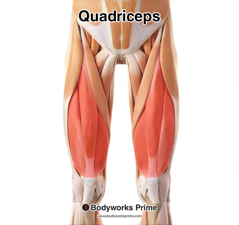
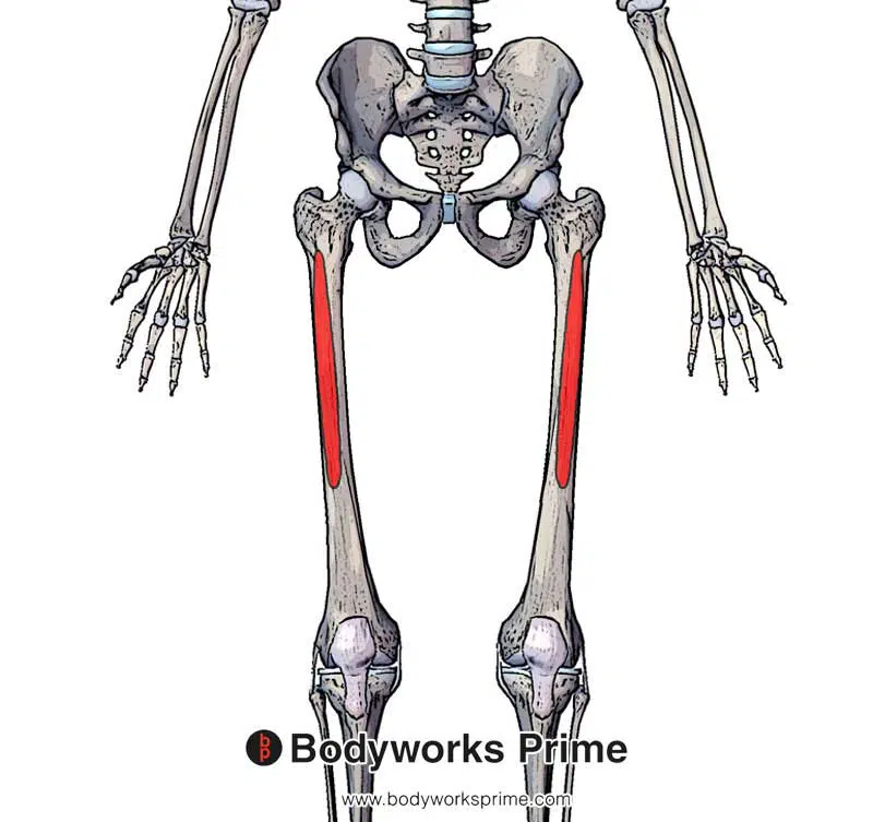
Here we can see all of the quadriceps highlighted in red. The vastus intermedius is deep to the rectus femoris, so it is not visible in this picture.
Rectus Femoris
The rectus femoris muscle is unique within the quadriceps group, as it crosses both the hip and knee joints. It has a central location on the anterior thigh and is responsible for both flexing the hip and extending the knee, due to it being a biarticular muscle [5][6].
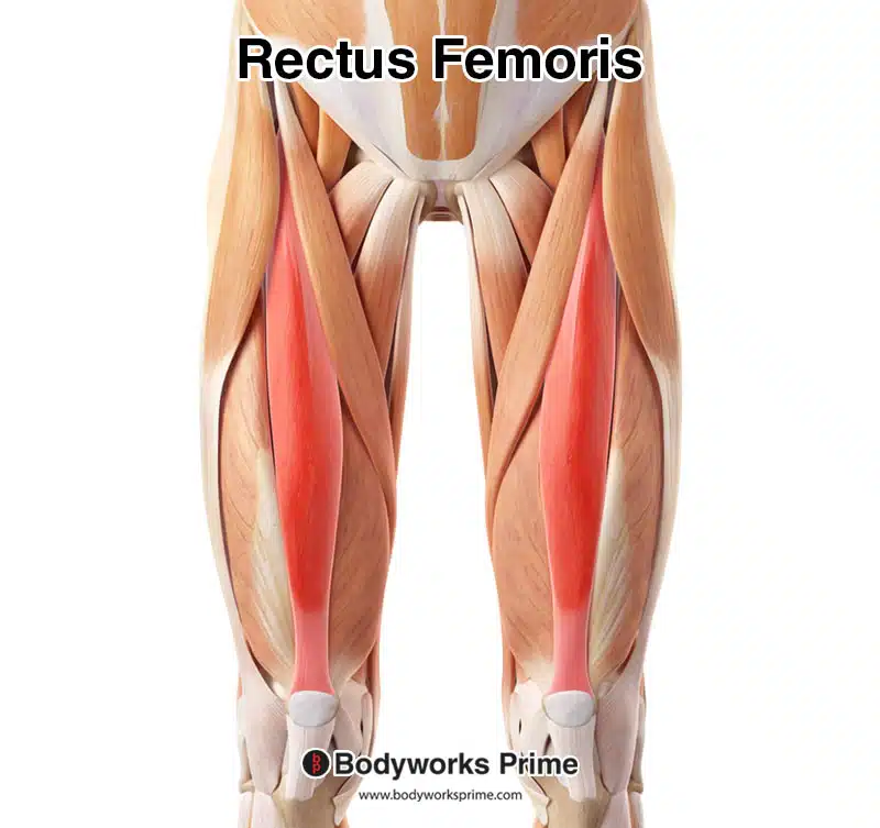
Pictured here is the rectus femoris highlighted in red, seen from an anterior view, amongst the other muscles of the leg.
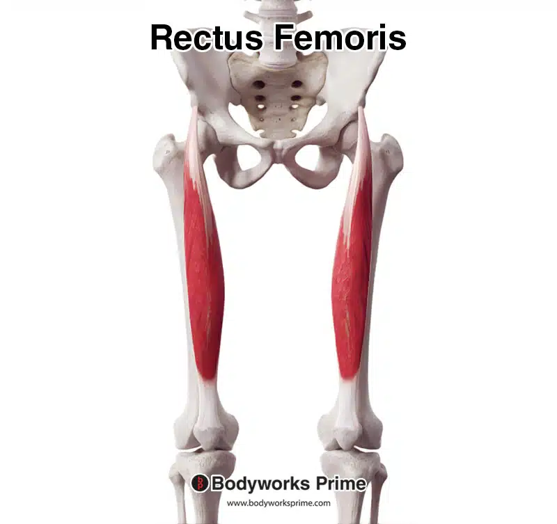
Here we can see the rectus femoris in isolation from an anterior view.
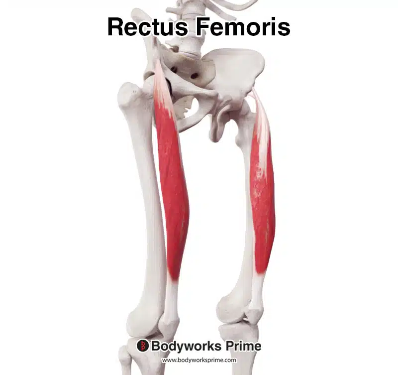
Here we can see the rectus femoris in isolation from an anterolateral view.
Vastus Lateralis
The vastus lateralis is the largest and most powerful muscle of the quadriceps group, it is located on the lateral side of the thigh. It primarily extends the knee [7] [8]. The vastus lateralis is also a common site for intramuscular injections, due to its large size and accessibility [9] [10].
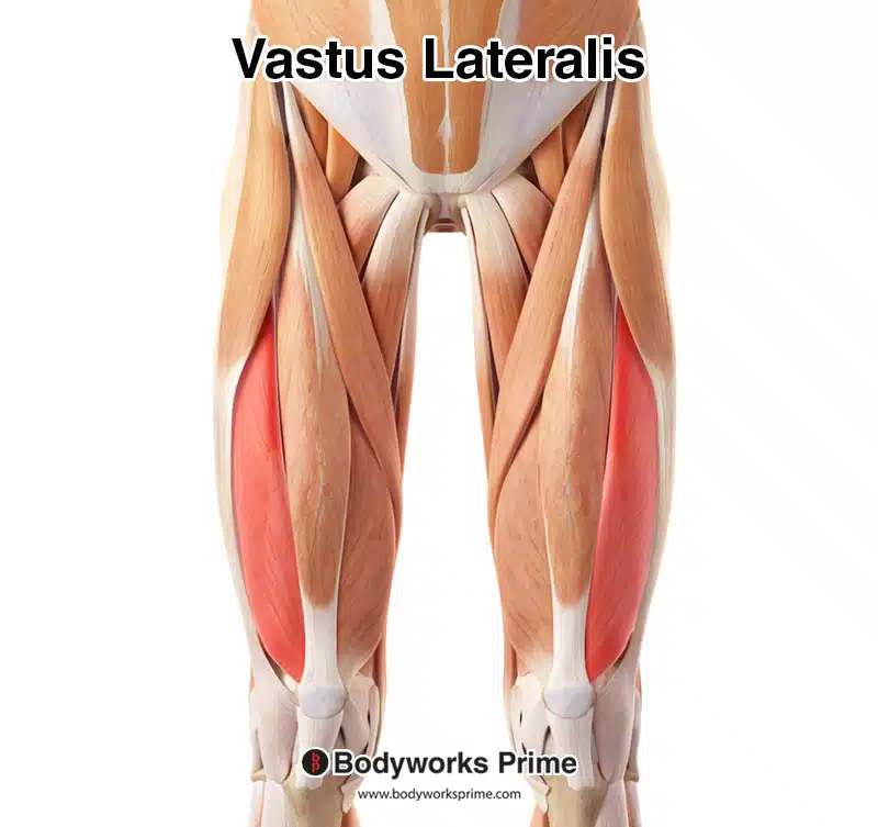
Pictured here is the vastus lateralis highlighted in red, seen from an anterior view amongst the other muscles.
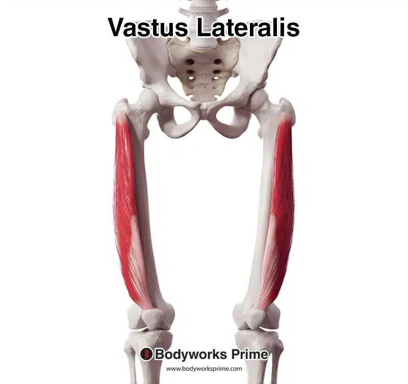
Here we can see the vastus lateralis in isolation from an anterior view.
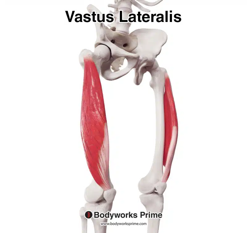
Here we can see the vastus lateralis in isolation from an anterolateral view.
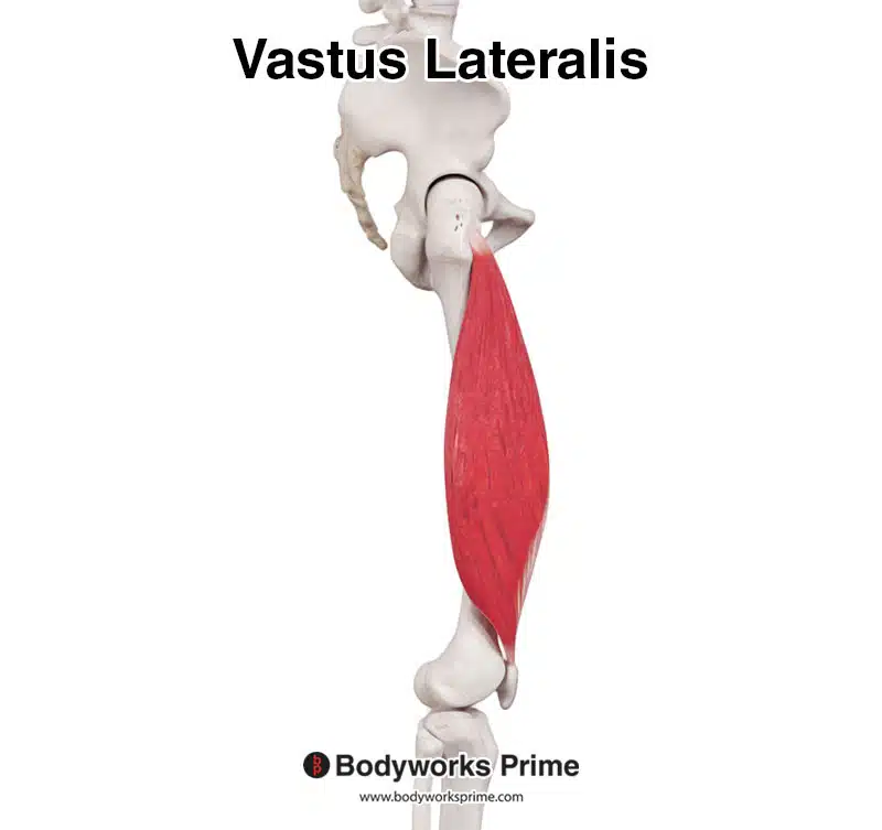
Here we can see the vastus lateralis in isolation from a lateral view.
Vastus Medialis
The vastus medialis is found on the medial side of the thigh, it extends the knee, and its oblique fibers contribute to stabilising the patella [11] [12]. The most distal muscle fibers of the vastus medialis are commonly referred to as the vastus medialis oblique (VMO) [13]. There has been some debate over the years amongst academics as to whether the VMO is a separate muscle, with its own innervation, as some anatomical studies had not found it to be distinct [14] [15] [16] [17]. However, the presence of the VMO is generally accepted by anatomists and surgeons [18] [19]. Research has also suggested that the VMO is innervated by a distinct branch of the femoral nerve compared to the rest of the muscle [20].
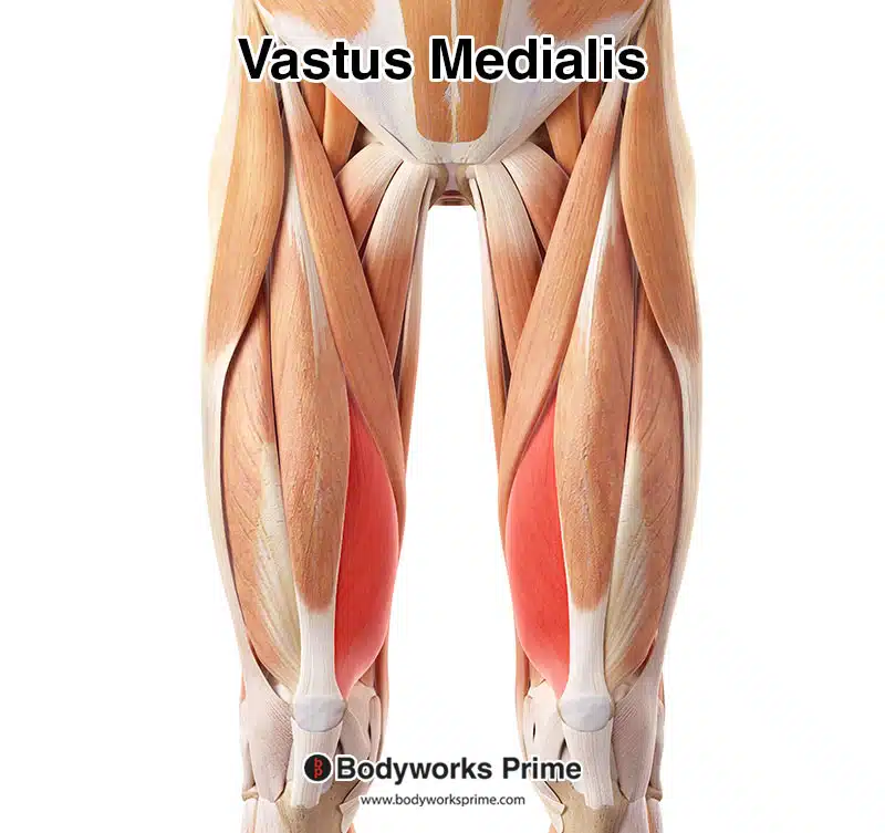
Here we can see the vastus medialis muscle highlighted in red amongst the other muscles of the leg.
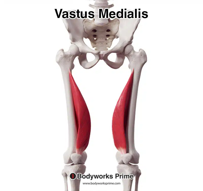
Here we can see the vastus medialis muscle from an anterior view.
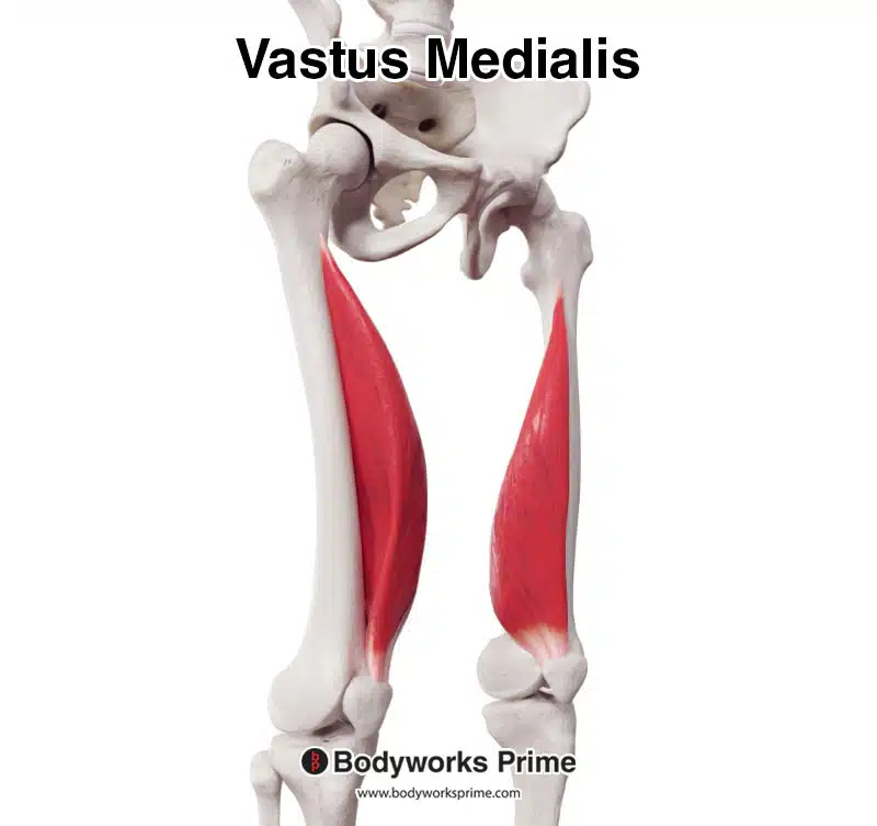
Here we can see the vastus medialis muscle from an anterolateral view.
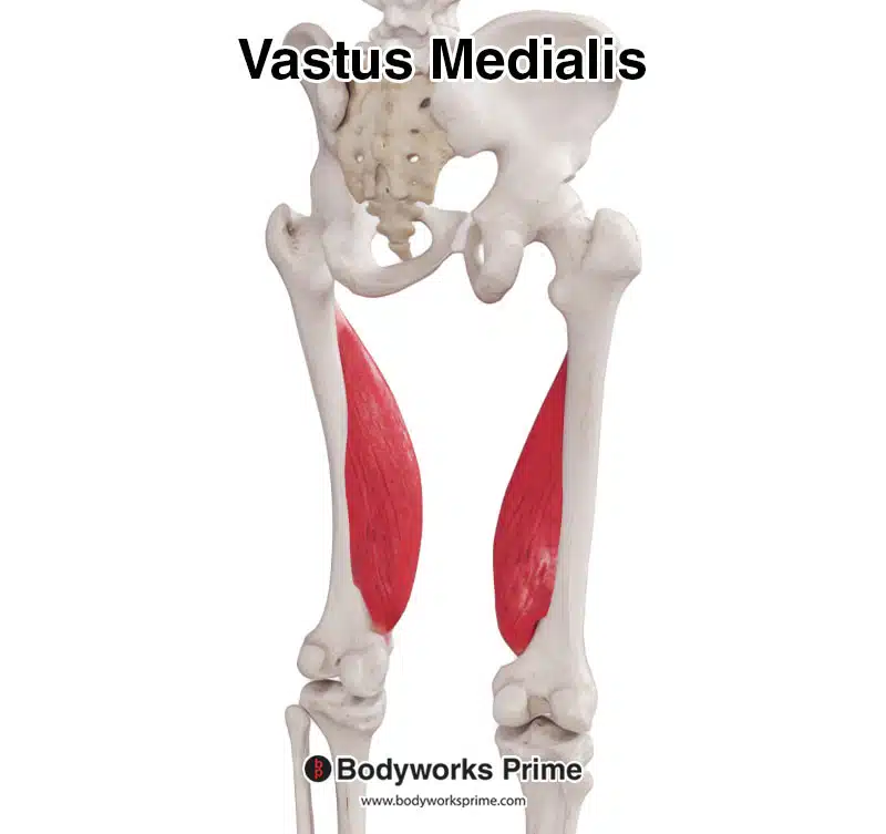
Here we can see the vastus medialis muscle from a posterior view.
Vastus Intermedius
The vastus intermedius is located deep to the rectus femoris and it is another centrally positioned muscle within the quadriceps group. Its primary function is also knee extension [21] [22]. Some studies have reported individual variation where the vastus intermedius and vastus lateralis muscles are fused together, sometimes presenting as an almost complete fusion of the two muscles [23] [24].
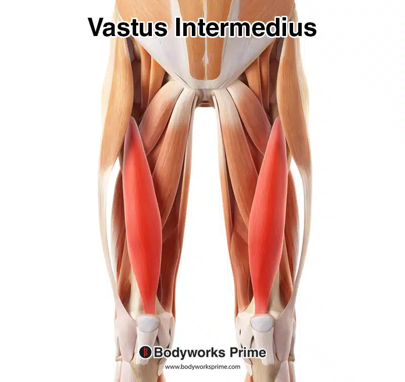
Here we can see the vastus intermedius highlighted in red amongst the other muscles of the thigh. Because the vastus intermedius is a deep muscle, the other quadriceps muscles have been removed from this image to reveal the vastus intermedius.
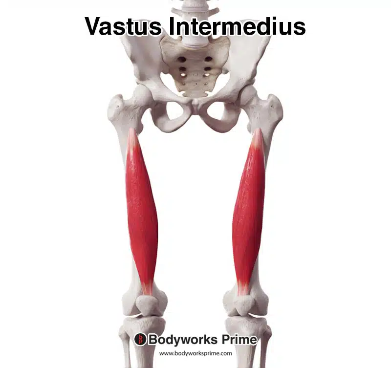
Here we can see the vastus intermedius from an anterior view.
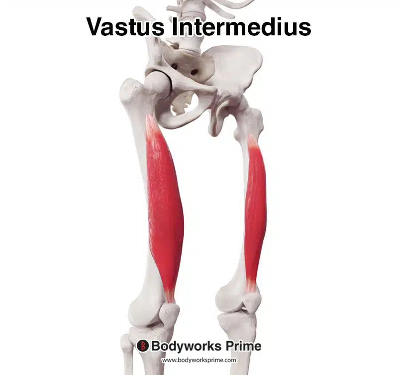
Here we can see the vastus intermedius from an anterolateral view.
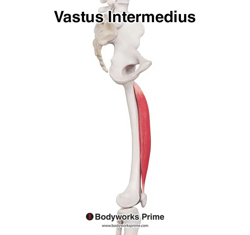
Here we can see the vastus intermedius from a medial view.
Pathologies & Exercises
Some common pathologies which affect the quadriceps muscle group include: strains, tendinopathy and contusions. Quadriceps strains are usually caused by excessive force or overstretching, which can result in damage to the muscle and its tendons [25] [26]. Furthermore, muscle fatigue has also been suggested to increase risk of strains [27]. Contusions often occur from direct trauma to the muscle, which can cause: pain, bruising, swelling, and a reduction in range of motion [28] [29]. Quadriceps tendinopathy is generally an overuse injury involving inflammation or degeneration of the quadriceps tendon, which can result in pain and decreased muscular function [30] [31].
Exercises targeting the quadriceps muscle group can improve strength, hypertrophy, flexibility, and overall function of the lower limb. Some examples of exercises to strengthen and grow the quadriceps muscle group are: squats, lunges, Bulgarian split squats, leg presses, and leg extensions [32] [33] [34] [35] [36].
In the following sections, we will delve deeper into the origin and insertion, actions, innervation, and blood supply of the quadriceps muscle group.
Origins & Insertions
The four quadriceps muscles, namely the rectus femoris, vastus lateralis, vastus medialis, and vastus intermedius, each have distinct origins and insertions which contribute to the overall function of the muscle group. Outlined below are the major landmarks of the femur followed by the origins and insertions for each of quadriceps muscle.
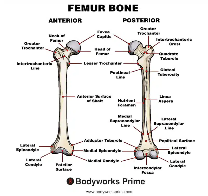
Here we can see the landmarks of the femur. Seeing these landmarks will be useful for understanding the origins and insertions of the quadriceps.
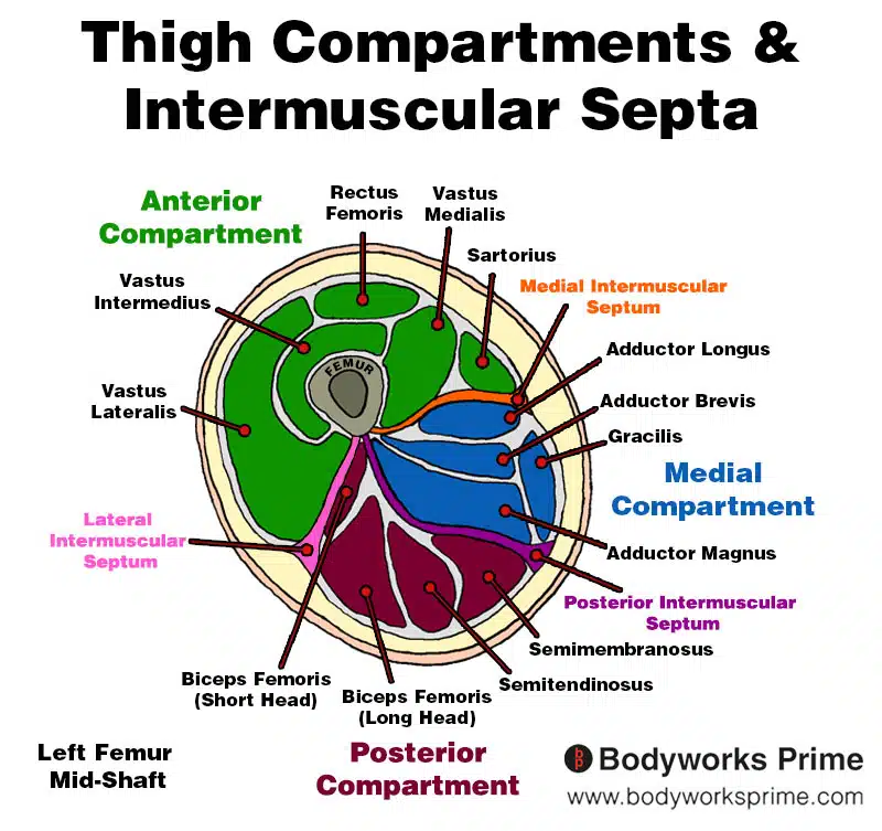
This image shows the thigh muscles, compartments, and intermuscular septa. The image is a cross section view of the thigh at around mid-shaft level of the femur. This diagram will be useful in understanding the origins and insertions of the quadricep muscles listed below.
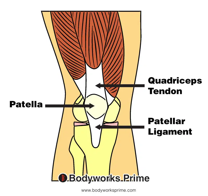
Here we can see the quadriceps tendon, patella, and patellar ligament. The fibers of all four quadriceps muscles converge into a single quadriceps tendon, which attaches to the patella and the patella ligament.
Rectus Femoris Attachments
The rectus femoris muscle has two muscular heads at its proximal end, resulting in two primary points of origin. The first head is the ‘direct head’ which originates from the anterior aspect of the inferior iliac spine. The inferior iliac spine is a bony prominence located on the lower portion of the iliac bone (part of the pelvic girdle) near the hip joint. The second head is the ‘indirect head’ which originates from the acetabular ridge. The acetabular ridge is an elevated margin found on the inner surface of the acetabulum, a socket that articulates with the head of the femur to form the hip joint. The rectus femoris inserts into the patellar tendon, which in turn connects onto the tibial tuberosity at the proximal end of the tibia (top of shinbone) [37] [38] [39].
Anatomical variations in the origins of the rectus femoris have been reported, though they are relatively rare. In some cases, the muscle may have additional heads or slips originating from the ilium or the capsule of the hip joint [40] [41].
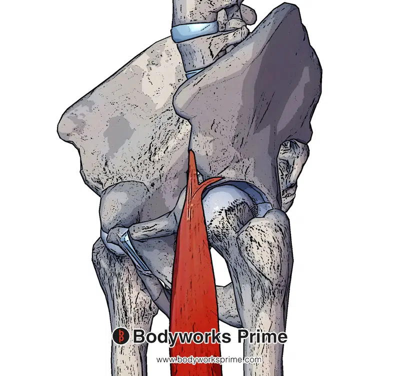
Here we can see the two heads of origin of the rectus femoris. The direct head (originating at the anterior aspect of the inferior iliac spine) and the indirect head (originating at the acetabular ridge).
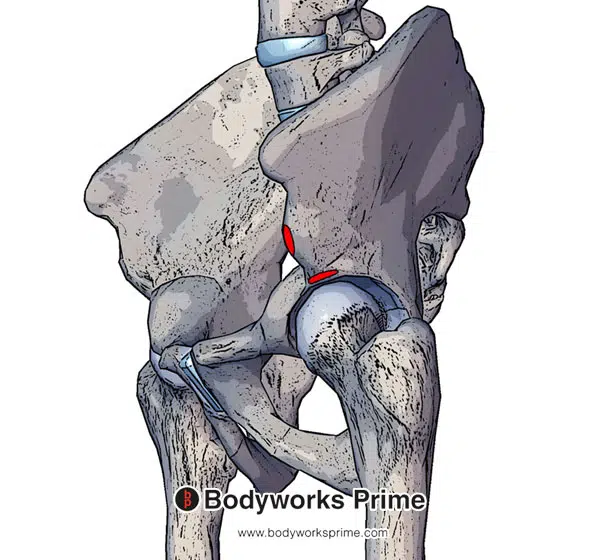
Here we can see the origins of the rectus femoris muscle marked in red. The direct head originates at the anterior aspect of the inferior iliac spine and the indirect head originates at the acetabular ridge.
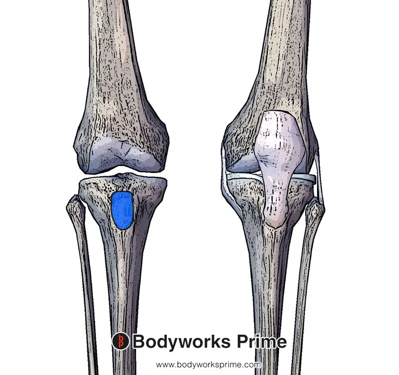
Here we can see the insertion of the patellar ligament on the tibial tuberosity marked in blue. The rectus femoris inserts into the patellar ligament via the quadriceps tendon.
Vastus Lateralis Attachments
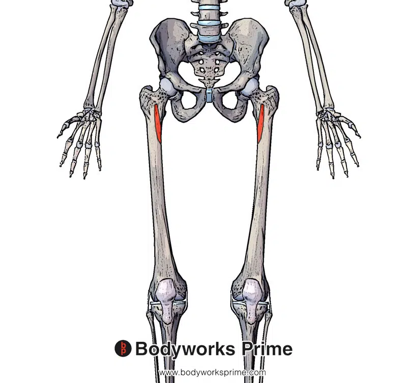
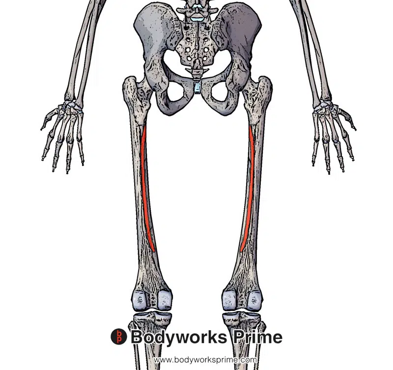
The vastus lateralis muscle has multiple origin points. It starts from the greater trochanter, a large bony prominence at the proximal and lateral end of the femur. The next origin is the intertrochanteric line, a ridge connecting the greater and lesser trochanters—the two prominences on the proximal end of the femur. Another origin is the lateral aspect of the linea aspera, a ridge on the posterior side of the femur. The muscle also originates from the gluteal tuberosity, a roughened area on the femur’s posterior aspect, and the lateral intermuscular septum of the thigh—a deep fascia fold separating the anterior and posterior compartments of the thigh, dividing the vastus lateralis and biceps femoris muscles. The vastus lateralis inserts at the lateral aspect of the patella through the quadriceps tendon, which connects to the patellar tendon and ultimately inserts onto the tibial tuberosity of the tibia [42] [43] [44].
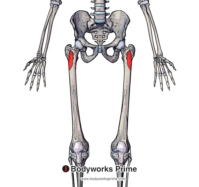
Here we can see the anterior origins of the vastus lateralis, highlighted in red, on the anterior side of the femur (greater trochanter and intertrochanteric line).
Here we can see the posterior origins of the vastus lateralis, highlighted in red, on the posterior side of the femur (lateral aspect of the linea aspera, gluteal tuberosity, and lateral intermuscular septum).
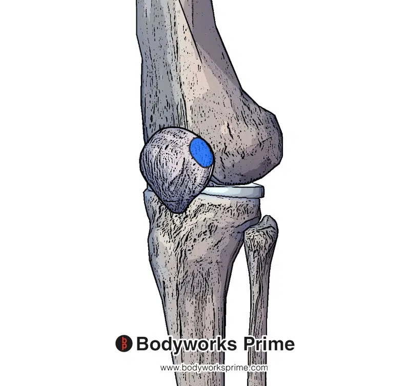
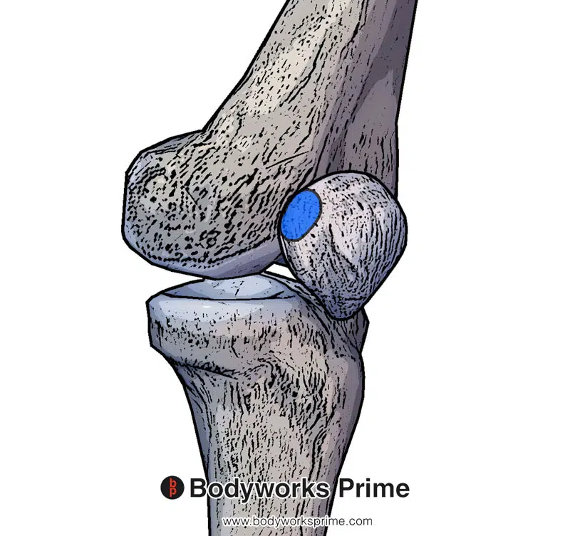
Here we can see an insertion of the vastus lateralis, highlighted in blue, on lateral side of the patellar.

Here we can see the insertion of the patellar tendon at the tibial tuberosity. The vastus lateralis inserts into the quadriceps tendon which then connects to the patella and the patellar tendon.
Vastus Medialis Attachments
The vastus medialis muscle has several origin points. The first origin is the inferior part of the intertrochanteric line, which is the lower portion of a ridge connecting the greater and lesser trochanters on the femur. Another origin is the spiral line (pectineal line), a curved ridge on the medial side of the femur that wraps around from the intertrochanteric line. The next origin is the medial lip of the linea aspera, which is the inner edge of the ridge running along the posterior aspect of the femur. A further origin point is the proximal part of the medial supracondylar line, a ridge on the medial side of the femur located above the medial condyle. Additionally, the medial intermuscular septum serves as another point of origin for this muscle. The medial intermuscular septum is a deep fascia layer in the thigh that separates the medial and posterior muscle compartments, extending from the medial aspect of the linea aspera on the femur down to the medial aspect of the tibia. The final points of origin are on the adductor longus and adductor magnus muscles, both these muscles are part of the adductor group of muscles, located in the medial compartment of the thigh. The vastus medialis then inserts at the medial border of the patella through the quadriceps tendon. The quadriceps tendon also connects to the tibial tuberosity on the proximal tibia via the patellar ligament [45] [46] [47].
Highlighted in red we can see the vastus medialis’ anterior origin points (inferior part of the intertrochanteric line and also the spiral line, which is known as the pectineal line).
Highlighted in red we can see the vastus medialis’ posterior origin points (medial lip of the linea aspera and the proximal part of the medial supracondylar line).
Highlighted in blue here we can see an insertion point of the vastus medialis, on the medial side of the patella.
Highlighted in blue, we can see the insertion of the patellar ligament. The vastus medialis inserts into the quadriceps tendon which then connects to the patella and the patellar ligament.
Vastus Intermedius Attachments
The vastus intermedius muscle originates from the anterior and lateral surfaces of the femur (the front and outer sides of the thigh bone). It it also originates from the lateral intermuscular septum, a deep fascia fold separating the anterior and posterior compartments of the thigh [48]. The vastus intermedius then inserts into the quadriceps tendon, alongside the other quadriceps muscles. The quadriceps tendon connects to the patellar ligament, which inserts onto the tibial tuberosity located on the proximal tibia [49] [50] [51].
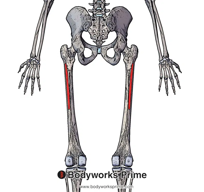
Here we can see the origin of the vastus intermedius on the anterior and lateral surface of the femur.
Here we can see the insertion of the patellar tendon on the tibial tuberosity. The vastus intermedius fibres connect into the quadriceps tendon which then connects to the patella and the patellar ligament.
Actions
Collectively the quadriceps muscle group is responsible for actions at the hip and knee joints. Together, these muscles primarily function in knee extension and hip flexion. Each muscle within the quadriceps group contributes to these actions in a unique manner [52]. The quadriceps’ collective actions are essential for activities such as: walking, running, climbing stairs, and standing up from a seated position.
Collectively, the quadriceps muscle group function as an antagonist to the hamstring muscles at both the hip and knee joints. The hamstrings are primarily responsible for hip extension and knee flexion, so their actions oppose those of the quadriceps [53] [54].
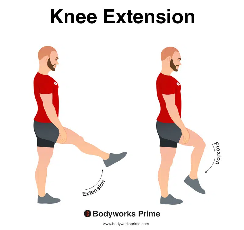
In this image, you can see an example of knee extension, which is the action of straightening out your leg at the knee. The opposite movement of knee flexion is knee extension. The quadriceps are involved in knee extension.
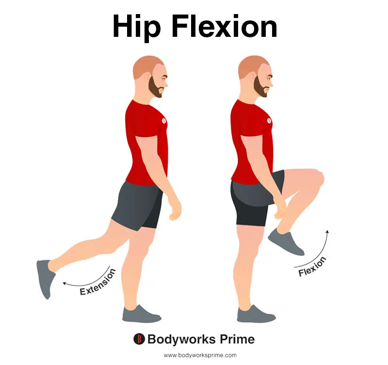
In this image, you can see an example of hip flexion, which is the action of raising your leg up in front of you and causing your hip to bend. If both legs are fixed, the torso will lean forward towards the legs. The opposite movement of hip flexion is hip extension. Only the rectus femoris is able to perform hip flexion.
Rectus Femoris Actions
As the only biarticular muscle in the quadriceps group, the rectus femoris is involved in both hip flexion and knee extension. Biarticular means that it crosses two joints. The rectus femoris is the only quadricep muscle involved in hip flexion because it is the only quadricep muscle that crosses the hip joint [55] [56] [57].
Vastus Lateralis Actions
The vastus lateralis primarily contributes to knee extension [58] [59]. It also plays an important role in stabilising the knee joint, working together with the vastus medialis to maintain proper tracking of the patella and prevent excessive sideways movement [60].
Vastus Medialis Actions
The vastus medialis primarily functions in knee extension [61] [62]. It is mainly active during the terminal range of extension (the end ranges of knee extension) [63]. However, “EMG studies have shown that all components of the quadriceps act in conjunction and that none of the components are predominantly responsible for fully extending the knee” [64]. The lower portion of this muscle, known as the vastus medialis oblique (VMO), helps stabilise the patella during knee extension and prevents patellar tracking issues [65] [66]. Although the vastus medialis does not cross the hip joint itself, it connects to the adductor longus and adductor magnus, which do cross the hip joint. However, the vastus medialis’ contribution to adduction is negligible compared to these direct hip adductors, and it is not a main action of the muscle. The vastus medialis likely plays more of a stabilising role during adduction, rather than directly contributing to the movement [67] [68].
Vastus Intermedius Actions
The vastus intermedius is located beneath the rectus femoris and contributes primarily to knee extension [69]. In addition to knee extension, the vastus intermedius is also involved with maintaining knee stability [70]. As one of the deeper quadricep muscles, the vastus intermedius provides support to the patella and helps distribute force across the knee joint. It has been suggested by Grob et al. (2018) that “substantial parts of the vastus intermedius directly contribute to the medial pull on the patella and help to maintain medial tracking of the patella during knee extension” [71].
Innervation
The quadriceps muscle group is innervated by the femoral nerve, which is a major branch of the lumbar plexus [72] [73]. The femoral nerve originates from the ventral rami of the spinal nerves L2, L3, and L4. The femoral nerve is one of the main nerves which supplies motor and sensory innervation to the muscles of the anterior compartment of the thigh, including the quadriceps muscles [74] [75]. Research has suggested that the VMO part of the vastus medialis is innervated by a distinct branch of the femoral nerve compared to the rest of the muscle [76].

Here we can see the innervation of the quadriceps muscle group, the femoral nerve, which is a major branch of the lumbar plexus. The femoral nerve originates from the spinal nerve roots of L2, L3, and L4.
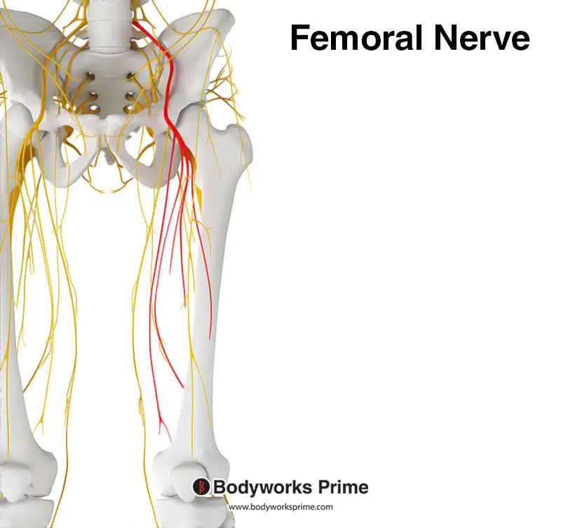
Here we can see the femoral nerve highlighted in red. The femoral nerve innervates the quadriceps muscle group. The femoral nerve originates from the spinal nerve roots of L2, L3, and L4.
Blood Supply
The quadriceps muscle group receives its blood supply from branches of the femoral artery, which is the main arterial supply to the thigh. The femoral artery is a continuation of the external iliac artery, and it enters the thigh deep to the inguinal ligament [77].
Rectus Femoris
The blood supply to the rectus femoris is provided by the lateral circumflex femoral artery, a branch of the femoral artery [78] [79].
Vastus Lateralis
The vastus lateralis is supplied by the lateral circumflex femoral artery and its branches, particularly the descending branch [80] [81].
Vastus Medialis
The blood supply to the vastus medialis is derived from the medial circumflex femoral artery and its branches, which is another branch of the femoral artery [82] [83].
Vastus Intermedius
The vastus intermedius receives its blood supply from perforating branches of the femoral artery, particularly the first and second perforating arteries [84] [85].
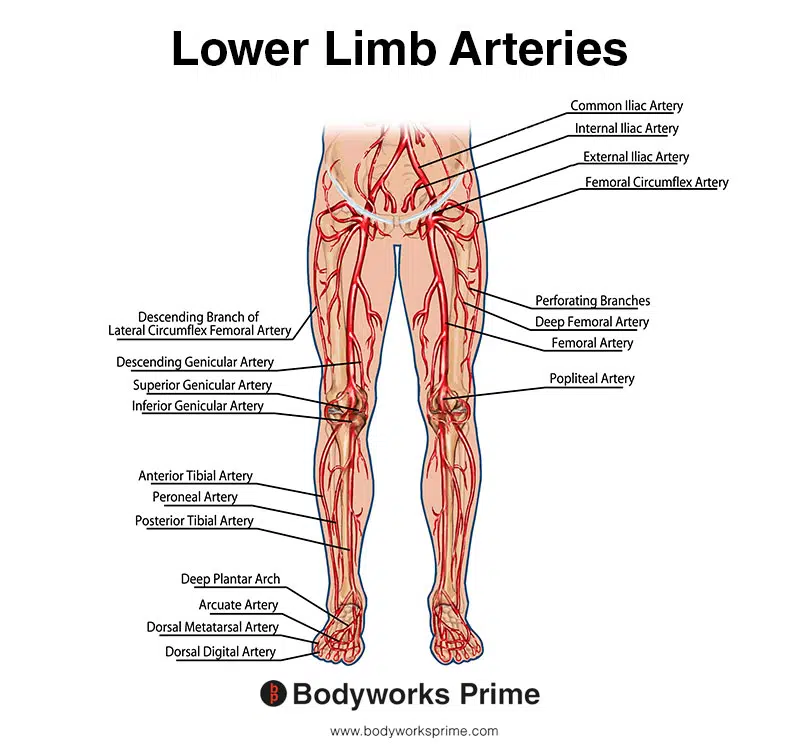
This image shows the arteries of the lower limb. You can see the femoral artery located in the thigh region, labelled on the right leg.
Want some flashcards to help you remember this information? Then click the link below:
Rectus Femoris Flashcards
Vastus Lateralis Flashcards
Vastus Medialis Flashcards
Vastus Intermedius Flashcards
Support Bodyworks Prime
Running a website and YouTube channel can be expensive. Your donation helps support the creation of more content for my website and YouTube channel. All donation proceeds go towards covering expenses only. Every contribution, big or small, makes a difference!
References
| ↑1, ↑22, ↑37, ↑58, ↑61, ↑69 | Standring S. (2015). Gray’s Anatomy: The Anatomical Basis of Clinical Practice, 41st Edn. Amsterdam: Elsevier. |
|---|---|
| ↑2, ↑4, ↑41, ↑51, ↑57, ↑59, ↑62 | Bordoni B, Varacallo M. Anatomy, Bony Pelvis and Lower Limb, Thigh Quadriceps Muscle. 2021 Jul 22. In: StatPearls [Internet]. Treasure Island (FL): StatPearls Publishing; 2021 Jan–. PMID: 30020706. |
| ↑3, ↑6, ↑8, ↑12, ↑21, ↑29, ↑31, ↑43, ↑45, ↑52, ↑73, ↑75 | Moore KL, Agur AMR, Dalley AF. Clinically Oriented Anatomy. 8th ed. Philadelphia: Lippincot Williams & Wilkins; 2017. |
| ↑5, ↑38, ↑40, ↑53, ↑55 | Murdock CJ, Mudreac A, Agyeman K. Anatomy, Abdomen and Pelvis, Rectus Femoris Muscle. [Updated 2022 Nov 4]. In: StatPearls [Internet]. Treasure Island (FL): StatPearls Publishing; 2023 Jan-. Available from: https://www.ncbi.nlm.nih.gov/books/NBK539897/ |
| ↑7, ↑42 | Biondi NL, Varacallo M. Anatomy, Bony Pelvis and Lower Limb, Vastus Lateralis Muscle. [Updated 2021 Aug 13]. In: StatPearls [Internet]. Treasure Island (FL): StatPearls Publishing; 2021 Jan-. Available from: https://www.ncbi.nlm.nih.gov/books/NBK532309/ |
| ↑9 | Nakajima Y, Fujii T, Mukai K, Ishida A, Kato M, Takahashi M, Tsuda M, Hashiba N, Mori N, Yamanaka A, Ozaki N, Nakatani T. Anatomically safe sites for intramuscular injections: a cross-sectional study on young adults and cadavers with a focus on the thigh. Hum Vaccin Immunother. 2020;16(1):189-196. doi: 10.1080/21645515.2019.1646576. Epub 2019 Aug 23. PMID: 31403356; PMCID: PMC7012163. |
| ↑10 | Polania Gutierrez JJ, Munakomi S. Intramuscular Injection. [Updated 2023 Feb 12]. In: StatPearls [Internet]. Treasure Island (FL): StatPearls Publishing; 2023 Jan-. Available from: https://www.ncbi.nlm.nih.gov/books/NBK556121/ |
| ↑11, ↑13, ↑18, ↑48 | Waligora AC, Johanson NA, Hirsch BE. Clinical anatomy of the quadriceps femoris and extensor apparatus of the knee. Clin Orthop Relat Res. 2009 Dec;467(12):3297-306. doi: 10.1007/s11999-009-1052-y. Epub 2009 Aug 19. PMID: 19690926; PMCID: PMC2772911. |
| ↑14 | Hubbard JK, Sampson HW, Elledge JR. Prevalence and morphology of the vastus medialis oblique muscle in human cadavers. Anat Rec. 1997 Sep;249(1):135-42. doi: 10.1002/(SICI)1097-0185(199709)249:1<135::AID-AR16>3.0.CO;2-Q. PMID: 9294658. |
| ↑15 | Lefebvre R, Leroux A, Poumarat G, Galtier B, Guillot M, Vanneuville G, Boucher JP. Vastus medialis: anatomical and functional considerations and implications based upon human and cadaveric studies. J Manipulative Physiol Ther. 2006 Feb;29(2):139-44. doi: 10.1016/j.jmpt.2005.12.006. PMID: 16461173. |
| ↑16 | Peeler J, Cooper J, Porter MM, Thliveris JA, Anderson JE. Structural parameters of the vastus medialis muscle. Clin Anat. 2005 May;18(4):281-9. doi: 10.1002/ca.20110. PMID: 15832351. |
| ↑17 | Smith TO, Nichols R, Harle D, Donell ST. Do the vastus medialis obliquus and vastus medialis longus really exist? A systematic review. Clin Anat. 2009 Mar;22(2):183-99. doi: 10.1002/ca.20737. PMID: 19090000. |
| ↑19 | Smith TO, Bowyer D, Dixon J, Stephenson R, Chester R, Donell ST. Can vastus medialis oblique be preferentially activated? A systematic review of electromyographic studies. Physiother Theory Pract. 2009 Feb;25(2):69-98. doi: 10.1080/09593980802686953. PMID: 19212898. |
| ↑20 | Toumi H, Poumarat G, Benjamin M, Best TM, F’Guyer S, Fairclough J. New insights into the function of the vastus medialis with clinical implications. Med Sci Sports Exerc. 2007 Jul;39(7):1153-9. doi: 10.1249/01.mss.0b013e31804ec08d. Erratum in: Med Sci Sports Exerc. 2008 May;40(5):982. Best, Thomas [corrected to Best, Thomas M]. PMID: 17596784. |
| ↑23 | Willan PL, Mahon M, Golland JA. Morphological variations of the human vastus lateralis muscle. J Anat. 1990 Feb;168:235-9. PMID: 2323995; PMCID: PMC1256904. |
| ↑24 | Becker I, Woodley SJ, Baxter GD. Gross morphology of the vastus lateralis muscle: An anatomical review. Clin Anat. 2009 May;22(4):436-50. doi: 10.1002/ca.20792. PMID: 19306318. |
| ↑25 | Orchard J, Best TM, Verrall GM. Return to play following muscle strains. Clin J Sport Med. 2005 Nov;15(6):436-41. doi: 10.1097/01.jsm.0000188206.54984.65. PMID: 16278548. |
| ↑26, ↑28 | Kary JM. Diagnosis and management of quadriceps strains and contusions. Curr Rev Musculoskelet Med. 2010 Jul 30;3(1-4):26-31. doi: 10.1007/s12178-010-9064-5. PMID: 21063497; PMCID: PMC2941577. |
| ↑27 | Mair SD, Seaber AV, Glisson RR, et al. The role of fatigue in susceptibility to acute muscle strain injury. Am J Sports Med. 1996;24:137–143. doi: 10.1177/036354659602400203. |
| ↑30 | Cook JL, Purdam CR. Is tendon pathology a continuum? A pathology model to explain the clinical presentation of load-induced tendinopathy. Br J Sports Med. 2009 Jun;43(6):409-16. doi: 10.1136/bjsm.2008.051193. Epub 2008 Sep 23. PMID: 18812414. |
| ↑32 | Bloomquist K, Langberg H, Karlsen S, Madsgaard S, Boesen M, Raastad T. Effect of range of motion in heavy load squatting on muscle and tendon adaptations. Eur J Appl Physiol. 2013 Aug;113(8):2133-42. doi: 10.1007/s00421-013-2642-7. Epub 2013 Apr 20. PMID: 23604798. |
| ↑33 | Escamilla RF, Fleisig GS, Zheng N, Lander JE, Barrentine SW, Andrews JR, Bergemann BW, Moorman CT 3rd. Effects of technique variations on knee biomechanics during the squat and leg press. Med Sci Sports Exerc. 2001 Sep;33(9):1552-66. doi: 10.1097/00005768-200109000-00020. PMID: 11528346. |
| ↑34 | Signorile, Joseph F.1; Weber, Brad2; Roll, Brad2; Caruso, John F.3; Lowensteyn, Ilka3; Perry, Arlette C.3. An Electromyographical Comparison of the Squat and Knee Extension Exercises. Journal of Strength and Conditioning Research 8(3):p 178-183, August 1994. |
| ↑35 | Martín-Fuentes I, Oliva-Lozano JM, Muyor JM. Influence of Feet Position and Execution Velocity on Muscle Activation and Kinematic Parameters During the Inclined Leg Press Exercise. Sports Health. 2022 May-Jun;14(3):317-327. doi: 10.1177/19417381211016357. Epub 2021 Jun 4. PMID: 34085847; PMCID: PMC9112713. |
| ↑36 | Wilk KE, Escamilla RF, Fleisig GS, Barrentine SW, Andrews JR, Boyd ML. A comparison of tibiofemoral joint forces and electromyographic activity during open and closed kinetic chain exercises. Am J Sports Med. 1996 Jul-Aug;24(4):518-27. doi: 10.1177/036354659602400418. PMID: 8827313. |
| ↑39 | Kumaravel M, Bawa P, Murai N. Magnetic resonance imaging of muscle injury in elite American football players: Predictors for return to play and performance. Eur J Radiol. 2018 Nov;108:155-164. doi: 10.1016/j.ejrad.2018.09.028. Epub 2018 Sep 27. PMID: 30396649. |
| ↑44 | Hayashi A, Maruyama Y. Lateral intermuscular septum of the thigh and short head of the biceps femoris muscle: an anatomic investigation with new clinical applications. Plast Reconstr Surg. 2001 Nov;108(6):1646-54. doi: 10.1097/00006534-200111000-00032. PMID: 11711941. |
| ↑46, ↑50, ↑63, ↑64, ↑66, ↑71 | Grob K, Manestar M, Filgueira L, Kuster MS, Gilbey H, Ackland T. The interaction between the vastus medialis and vastus intermedius and its influence on the extensor apparatus of the knee joint. Knee Surg Sports Traumatol Arthrosc. 2018 Mar;26(3):727-738. doi: 10.1007/s00167-016-4396-3. Epub 2017 Jan 25. PMID: 28124107. |
| ↑47 | Rajput HB, Rajani SJ, Vaniya VH. Variation in Morphometry of Vastus Medialis Muscle. J Clin Diagn Res. 2017 Sep;11(9):AC01-AC04. doi: 10.7860/JCDR/2017/29162.10527. Epub 2017 Sep 1. PMID: 29207687; PMCID: PMC5713709. |
| ↑49 | Yoshida S, Ichimura K, Sakai T. Structural diversity of the vastus intermedius origin revealed by analysis of isolated muscle specimens. Clin Anat. 2017 Jan;30(1):98-105. doi: 10.1002/ca.22791. Epub 2016 Oct 31. PMID: 27598432. |
| ↑54 | Chumanov ES, Heiderscheit BC, Thelen DG. Hamstring musculotendon dynamics during stance and swing phases of high-speed running. Med Sci Sports Exerc. 2011 Mar;43(3):525-32. doi: 10.1249/MSS.0b013e3181f23fe8. PMID: 20689454; PMCID: PMC3057086. |
| ↑56 | Neumann DA. Kinesiology of the Musculoskeletal System: Foundations for Rehabilitation. 3rd ed. St. Louis, MO: Elsevier; 2017. |
| ↑60 | Powers CM. The influence of altered lower-extremity kinematics on patellofemoral joint dysfunction: a theoretical perspective. J Orthop Sports Phys Ther. 2003 Nov;33(11):639-46. doi: 10.2519/jospt.2003.33.11.639. PMID: 14669959. |
| ↑65 | Philippot R, Boyer B, Testa R, Farizon F, Moyen B. The role of the medial ligamentous structures on patellar tracking during knee flexion. Knee Surg Sports Traumatol Arthrosc. 2012 Feb;20(2):331-6. doi: 10.1007/s00167-011-1598-6. Epub 2011 Jul 12. PMID: 21748394. |
| ↑67 | Han, S-E., & Park, S-K. (2010). The Effect of Hip Adduction on the Activation of the Vastus Medialis Oblique and the Vastus Lateralis. Journal of the Korean Academy of Clinical Electrophysiology, 8(2), 7-12. https://doi.org/10.5627/KACE.2010.8.2.007 |
| ↑68 | Koh, E-K., Lee, K-H., & Jung, D-Y. (2011). The Effect of Isometric Hip Adduction and Abduction on the Muscle Activities of Vastus Medialis Oblique and Vastus Lateralis during Leg Squat Exercises. Korean Journal of Sport Biomechanics, 21(3), 361-368. https://doi.org/10.5103/KJSB.2011.21.3.361 |
| ↑70, ↑76 | Toumi H, Poumarat G, Benjamin M, Best TM, F’Guyer S, Fairclough J. New insights into the function of the vastus medialis with clinical implications. Med Sci Sports Exerc. 2007 Jul;39(7):1153-9. doi: 10.1249/01.mss.0b013e31804ec08d. Erratum in: Med Sci Sports Exerc. 2008 May;40(5):982. Best, Thomas [corrected to Best, Thomas M]. PMID: 17596784. |
| ↑72, ↑74 | Refai NA, Black AC, Tadi P. Anatomy, Bony Pelvis and Lower Limb: Thigh Femoral Nerve. [Updated 2022 Nov 18]. In: StatPearls [Internet]. Treasure Island (FL): StatPearls Publishing; 2023 Jan-. Available from: https://www.ncbi.nlm.nih.gov/books/NBK556065/ |
| ↑77, ↑78, ↑80, ↑82, ↑84 | Swift H, Bordoni B. Anatomy, Bony Pelvis and Lower Limb: Femoral Artery. 2022 Aug 8. In: StatPearls [Internet]. Treasure Island (FL): StatPearls Publishing; 2023 Jan–. PMID: 30855850. |
| ↑79, ↑81, ↑83, ↑85 | Arias DG, Marappa-Ganeshan R. Anatomy, Bony Pelvis and Lower Limb, Arteries. [Updated 2022 Jul 25]. In: StatPearls [Internet]. Treasure Island (FL): StatPearls Publishing; 2023 Jan-. Available from: https://www.ncbi.nlm.nih.gov/books/NBK544319/ |


