| Origin | Ischial tuberosity |
| Insertion | Quadrate tubercle & just below it |
| Action | Hip lateral rotation Hip stabilisation |
| Nerve | Nerve to quadratus femoris (L5, S1) |
| Artery | Inferior gluteal artery (main) Medial circumflex femoral artery (supplemental) |
Location & Overview
The quadratus femoris muscle is a somewhat flat and quadrilateral shaped muscle that is located deep in the gluteal region. The posterior surface of the quadratus femoris is deep to the gluteus maximus muscle. The quadratus femoris also sits inferior to the gemellus muscles and the obturator internus, and it is superior to the adductor magnus. As one of the six deep lateral rotators of the thigh, the primary roles of the quadratus femoris are to laterally rotate and stabilise the hip joint. The six deep primary lateral rotator muscles of the thigh are: piriformis, superior gemellus, obturator internus, inferior gemellus, quadratus femoris, and obturator externus [1] [2].
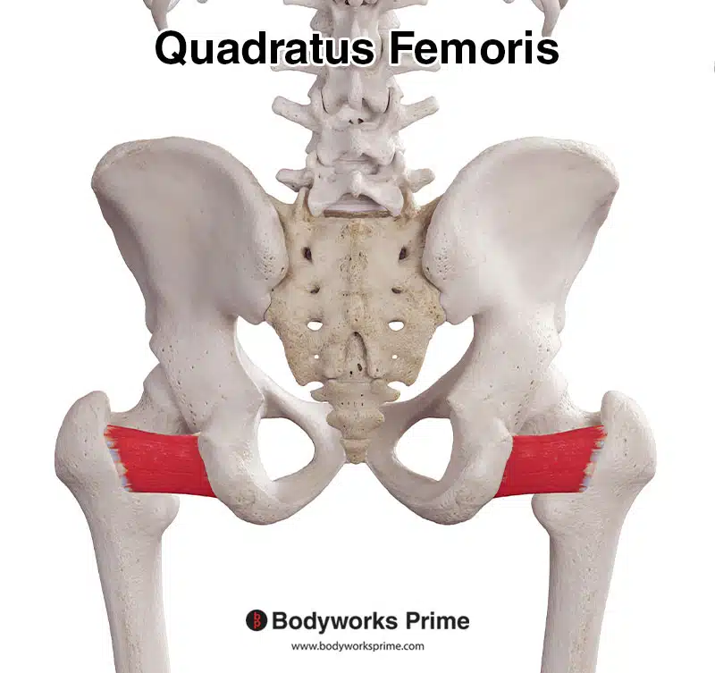
Here we can see the quadratus femoris muscle from a posterior view.
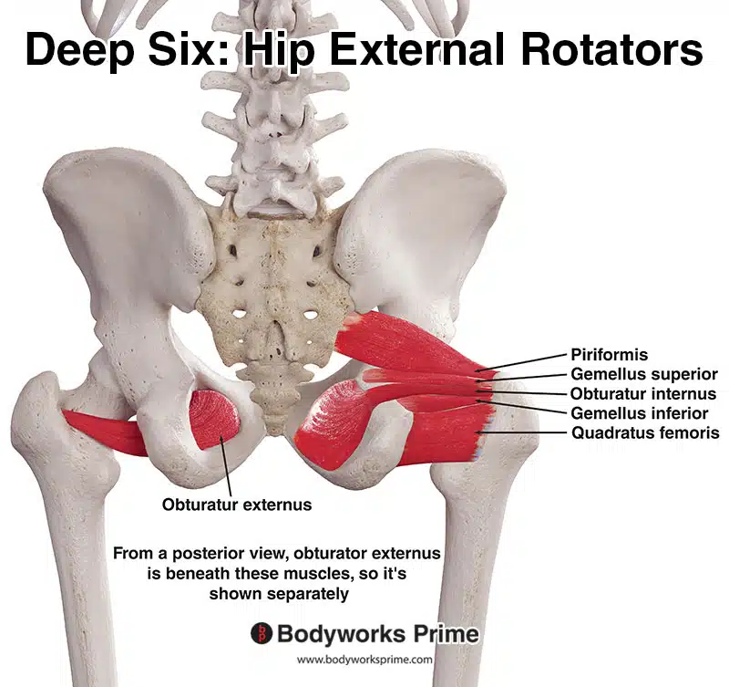
Pictured here are the deep six hip external rotator muscles: the piriformis, gemellus superior, gemellus inferior, obturator internus, obturator externus, and quadratus femoris. These muscles play an important role in stabilising the hip joint and facilitating hip lateral rotation.
Origin & Insertion
The quadratus femoris muscle originates on the lateral aspect of the pelvis and inserts onto the posterior side of the femur. The specific origin point on the pelvis is the ischial tuberosity. The ischial tuberosity is a prominent bony structure that’s felt when sitting on a hard surface (the ischial tuberosity is located on the ischium, one of the three major bones that make up the pelvic bone). Specifically, the quadratus femoris arises from the upper part of the lateral border of the ischial tuberosity. The insertion point on the femur is the quadrate tubercle. The quadrate tubercle is a small raised area located on the intertrochanteric crest. The intertrochanteric crest is a bony ridge on the backside of the femur, connecting the greater and lesser trochanters. Additionally, the muscle also inserts into an area just below the quadrate tubercle on the femur [3] [4].
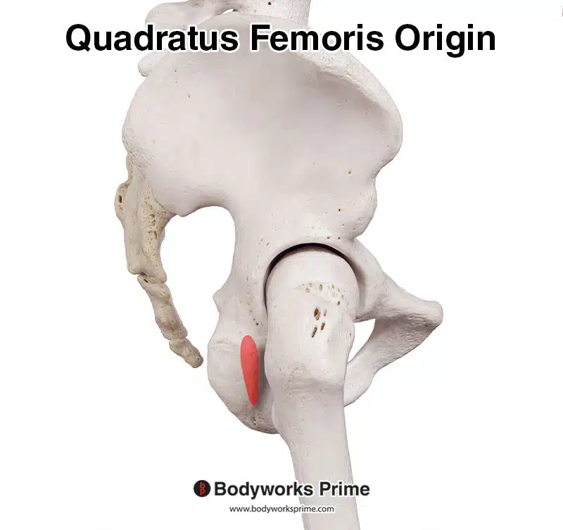
Here we can see the origin of the quadratus femoris muscle marked in red on the ischial tuberosity.
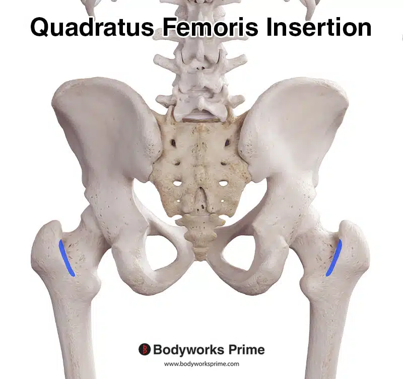
Here we can see the insertion of the quadratus femoris muscle marked in blue on the quadrate tubercle & just below it.
Actions
Contraction of the quadratus femoris produces lateral (external) rotation of the thigh at the hip joint. This action rotates the leg so the knee turns outwards. In addition to rotation, the quadratus femoris assists in stabilising the hip joint as one of the deep lateral rotators of the thigh [5] [6].
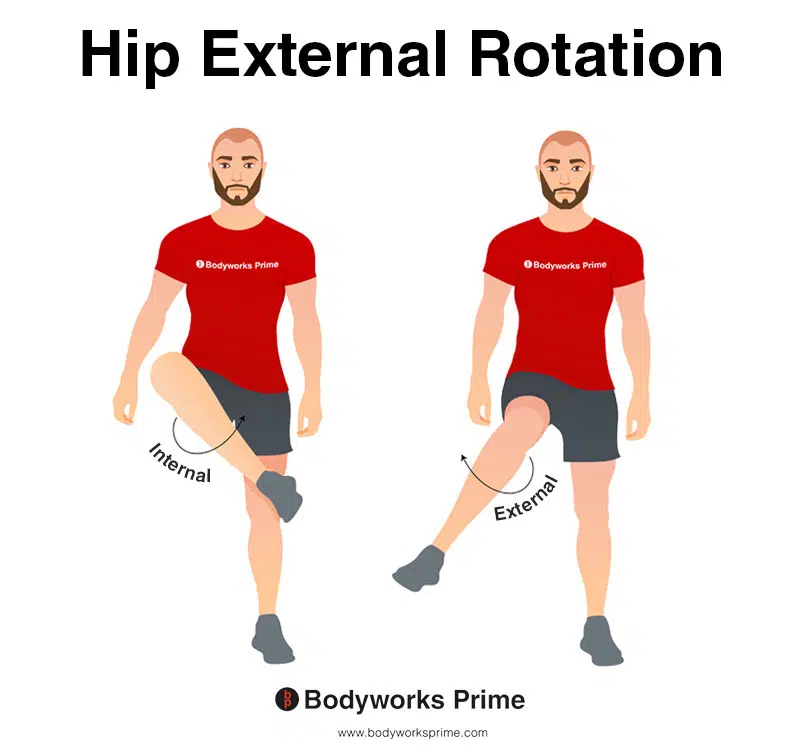
This image demonstrates hip external rotation, where the leg rotates outwards (laterally) from the hip joint. External rotation is also referred to as lateral rotation. The opposite of external rotation is internal rotation (medial rotation). External rotation of the hip joint is the primary action of the quadratus femoris muscle and it also assists with stabilisation of the hip joint.
Innervation
The quadratus femoris is innervated by the nerve to quadratus femoris, which originates from the L5 and S1 spinal nerves. This nerve leaves the pelvis via the greater sciatic foramen. The nerve to quadratus femoris also passes over the posterior surface of the hip joint, anterior to the sciatic nerve and obturator internus muscle, to reach the deep surface of the quadratus femoris muscle. The nerve to quadratus femoris also supplies the gemellus inferior muscle [7] [8].
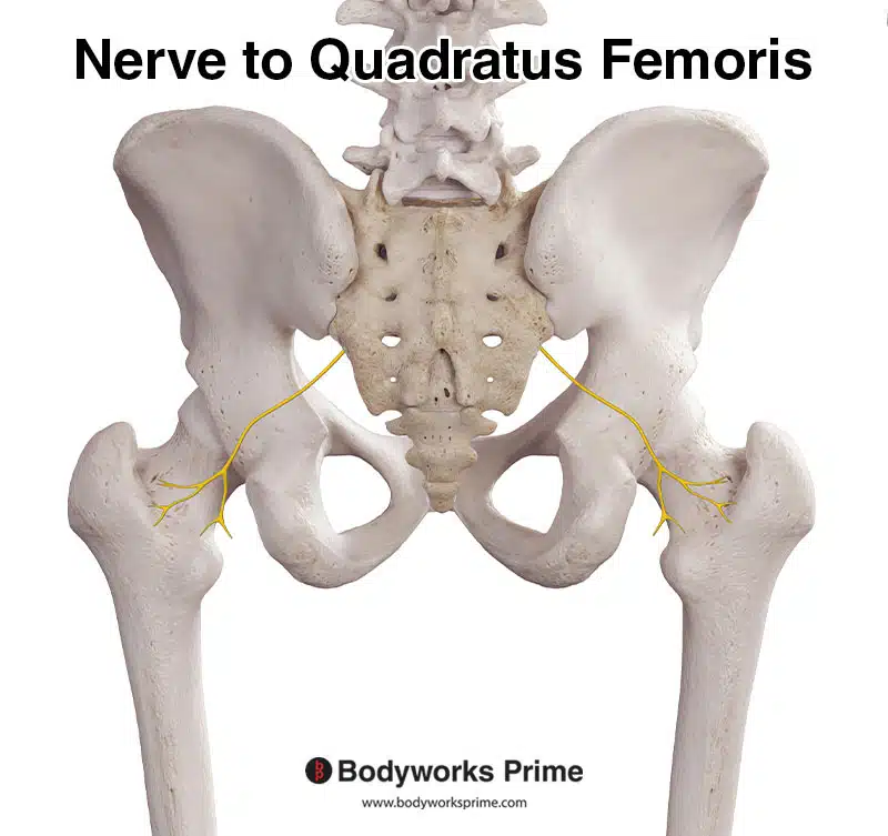
This image shows the nerve to quadratus femoris which innervates the quadratus femoris muscle.
Blood Supply
The main arterial supply to the quadratus femoris is via the inferior gluteal artery, which exits the pelvis through the greater sciatic foramen inferior to the piriformis muscle. Additionally, small branches from the medial circumflex femoral artery reach the superficial surface of the muscle [9] [10].
Want some flashcards to help you remember this information? Then click the link below:
Quadratus Femoris Flashcards
Support Bodyworks Prime
Running a website and YouTube channel can be expensive. Your donation helps support the creation of more content for my website and YouTube channel. All donation proceeds go towards covering expenses only. Every contribution, big or small, makes a difference!


