| Origin | Pelvic surfaces of the ilium & ischium Obturator membrane |
| Insertion | Medial surface of the greater trochanter of the femur, specifically the trochanteric fossa |
| Action | Laterally rotates the hip joint Stabilises the hip joint Assists with abduction of the hip when flexed |
| Nerve | Nerve to obturator internus (L5 & S1) |
| Artery | Obturator Artery Inferior Gluteal Artery Pudendal Artery |
Location & Overview
The obturator internus muscle is primarily situated inside the pelvic area, where it plays a key role in supporting the lateral wall. It originates within the lesser pelvis, acting as a protective pad for most of its lateral aspects. As the muscle fibers progress, they converge posteriorly and laterally and becoming tendinous. This tendinous segment departs the pelvis through an opening/passage known as the lesser sciatic foramen. Once outside the pelvis, the muscle attaches itself to the greater trochanter of the femur [1] [2].
The obturator internus muscle is mainly positioned medial to the gemelli muscles. These gemelli muscles consist of two parts: the superior gemellus and the inferior gemellus. As the obturator internus extends laterally toward the femur, its tendon passes between and merges with the tendons of the gemelli muscles. Collectively, the tendons of the obturator internus, superior gemellus, and inferior gemellus join to form a single tendon. This common tendon then attaches to the greater trochanter of the femur [3] [4].
Moreover, aside from its anatomical structure, the obturator internus doesn’t function in isolation either; it collaborates with the superior gemellus and inferior gemellus, to form a muscle group called the ‘triceps coxae’. This muscle group is located between two other muscles: the piriformis and the quadratus femoris. The triceps coxae play a role in hip stabilisation and movement, especially in terms of lateral rotation of the thigh [5] [6].
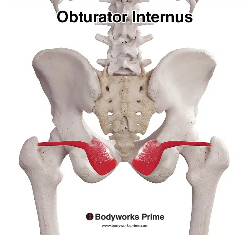
Pictured here we can see the obturator internus muscle from a posterior view.
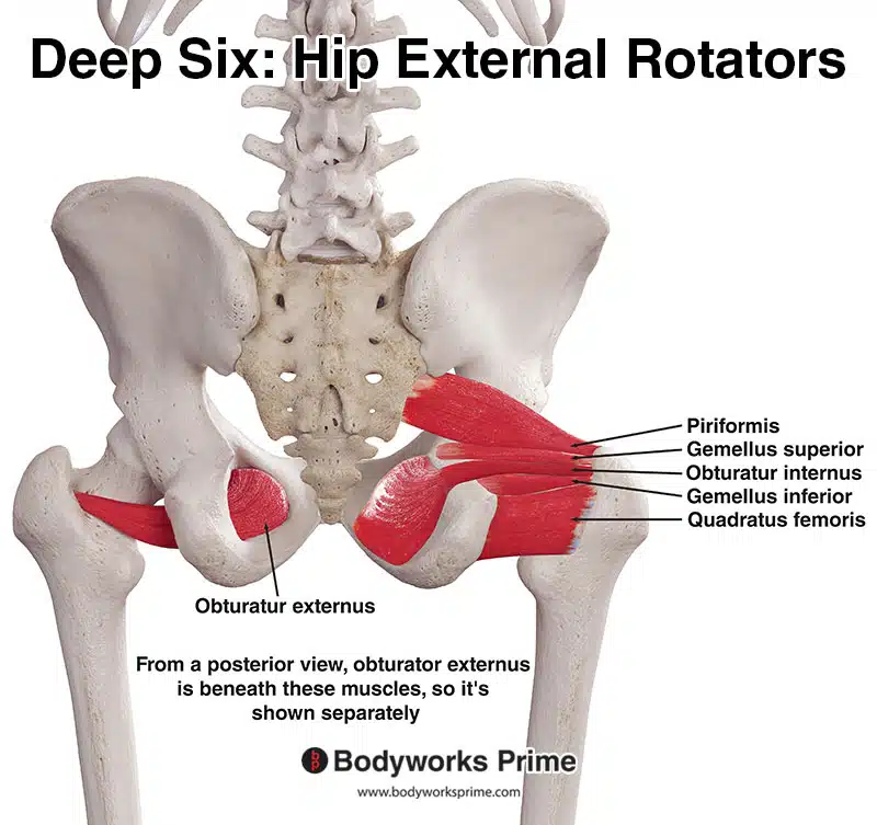
Pictured here are the deep six hip external rotator muscles: the piriformis, gemellus superior, gemellus inferior, obturator internus, obturator externus, and quadratus femoris. These muscles play an important role in stabilising the hip joint and facilitating hip lateral rotation.
Origin & Insertion
The obturator internus originates from the pelvic surface of the obturator membrane, which is a thin, fibrous sheet that covers the obturator foramen (a large opening in the pelvic bone). Additionally, it originates from the surrounding bones, particularly the pelvic surfaces of the ilium, which is the broad, fan-shaped upper part of the hip bone, and the ischium, which is the lower and posterior part of the hip bone. The muscle then inserts onto the medial surface of the greater trochanter, a prominent bony projection located at the upper and outer part of the femur (thigh bone). Specifically, it attaches to the trochanteric fossa, which is a shallow depression on the medial aspect of the greater trochanter of the femur [7] [8].
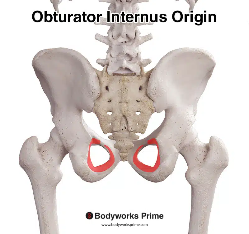
Here we can see the origin of the obturator internus muscle marked out in red on the pelvic surfaces of the ilium & ischium. The obturator internus also originates from the obturator membrane, shown below.
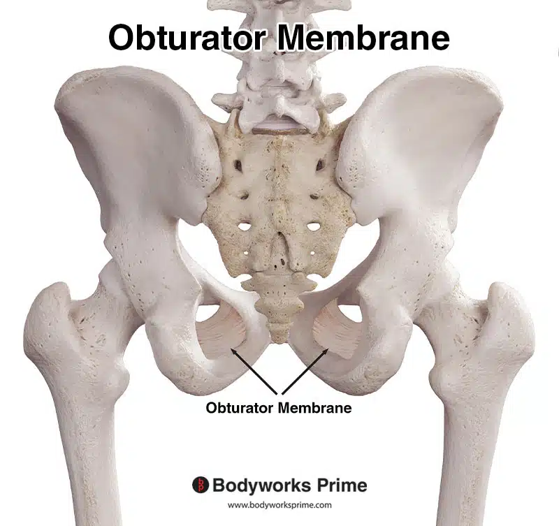
Here the can see the obturator membrane, another origin point of the obturator internus muscle.
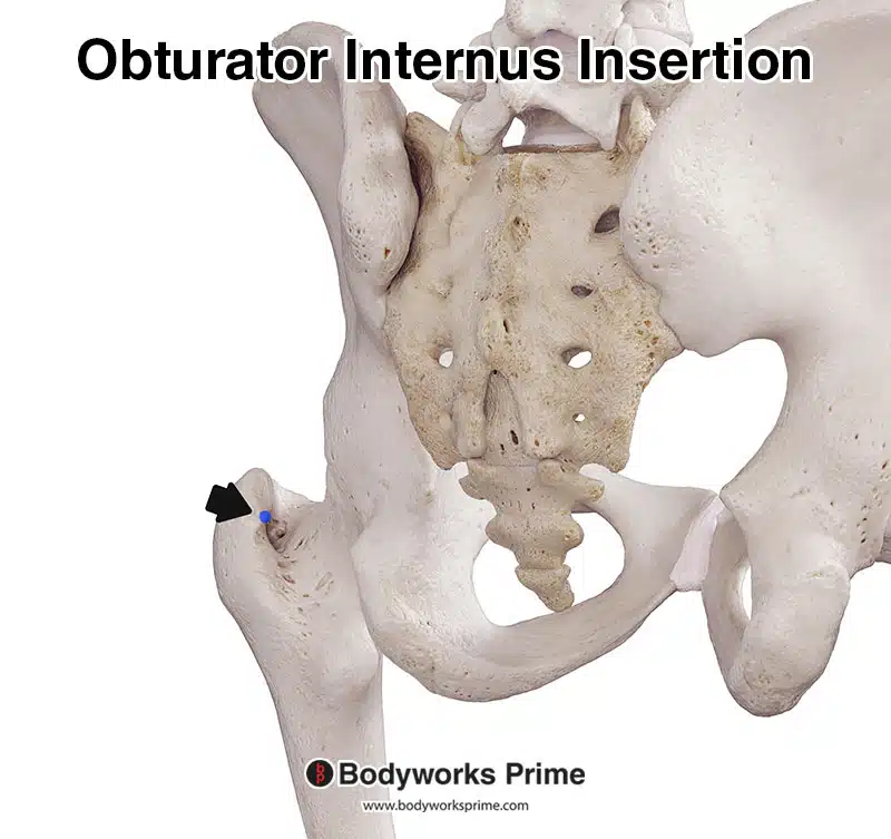
Here we can see the insertion of the obturator internus muscle marked in blue on the medial surface of the greater trochanter of the femur, specifically the trochanteric fossa.
Actions
The obturator internus primarily functions to laterally (or externally) rotate the hip joint. This means it helps to turn the thigh outward, away from the midline of the body. This action is particularly emphasised when the hip is in an extended position. Additionally, when the hip is flexed, the obturator internus weakly assists in abduction, which is the movement of the thigh away from the body’s midline [9] [10].
Beyond these movements, an important function of the obturator internus muscle is the stabilisation of the hip joint. Furthermore, the obturator internus plays a significant postural role by holding the head of the femur securely within the acetabulum, which is the cup-shaped socket of the hip bone where the ball-shaped head of the femur fits. This ensures stability and integrity of the hip joint, especially during movement [11] [12].
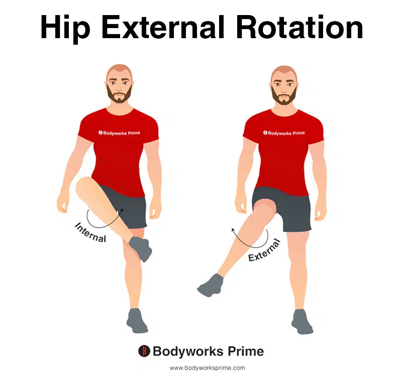
This image shows an example of hip external rotation, which involves rotating the leg from the hip joint outwards (laterally). External rotation is also referred to as lateral rotation. The opposite of external rotation (lateral rotation) is internal rotation (medial rotation). External rotation of the hip joint is the primary action of the obturator internus (alongside stabilising the hip joint).
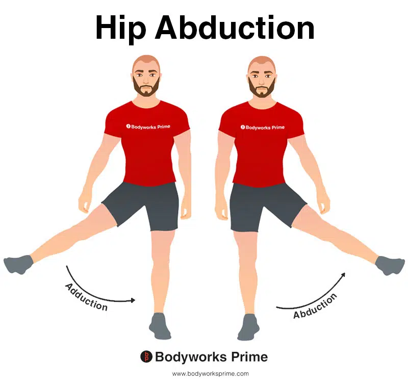
This image shows an example of hip abduction, which involves moving the leg out to the side (laterally). The opposite of hip abduction is hip adduction. The obturator internus can weakly assist with hip abduction, but only when the hip is flexed.
Innervation
The obturator internus muscle is innervated by the nerve to the obturator internus. This nerve arises from the anterior divisions of the anterior rami of the spinal nerves, specifically from the levels L5 and S1 [13] [14].
The course of the nerve to the obturator internus exits the pelvis via the greater sciatic foramen, which is a large opening in the posterior pelvis formed by the sacrum and the ilium. This foramen is positioned inferior to the piriformis muscle [15] [16].
Subsequently, the nerve descends behind the sacrospinous ligament, a thin ligament that extends from the sacrum to the ischial spine, helping to convert the greater and lesser sciatic notches into foramina [17] [18].
During its journey, the nerve supplies the superior gemellus muscle as it passes around the base of the ischial spine, which is a pointed bony projection from the posterior part of the ischium, serving as an attachment point for several ligaments and muscles [19] [20].
Upon entering the perineum, which is the region located below the pelvic diaphragm and between the thighs, encompassing the external genitalia and the anus, the nerve passes through the lesser sciatic foramen. This is a smaller opening formed by the ischium and the sacrum, situated below the greater sciatic foramen. Following this path, the nerve provides innervation to the obturator internus muscle [21] [22].
The nerve to the obturator internus also innervates the superior gemellus muscle. However, there is some variability in this innervation. In a study conducted by Htar Htar Aung et al. (2001), it was shown that the superior gemellus muscle received dual innervation from both the nerve to the obturator internus and the nerve to the quadratus femoris approximately 60.4% of the time. Htar Htar Aung et al. (2001) also confirmed that these two nerves are distinct, as they follow different pathways and do not communicate with each other [23].
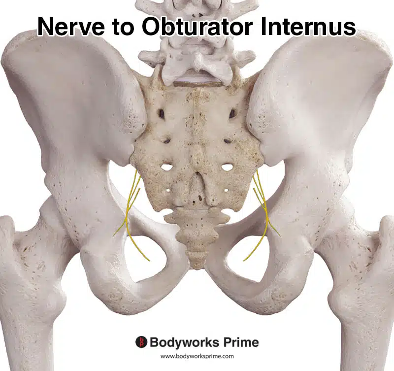
This image shows the nerve to the obturator internus which innervates the obturator internus and the superior gemellus muscles.
Blood Supply
The obturator internus muscle receives its blood supply from multiple sources. Primarily, it receives blood from branches of the obturator artery. Additionally, the muscle benefits from the muscular branches of the inferior gluteal artery. Furthermore, the gemellar and inferior rectal branches of the pudendal artery are responsible for the vascularisation of the muscle’s lateral section [24].
Want some flashcards to help you remember this information? Then click the link below:
Obturator Internus Flashcards
Support Bodyworks Prime
Running a website and YouTube channel can be expensive. Your donation helps support the creation of more content for my website and YouTube channel. All donation proceeds go towards covering expenses only. Every contribution, big or small, makes a difference!
References
| ↑1, ↑3, ↑5, ↑7, ↑9, ↑11, ↑13, ↑15, ↑17, ↑19, ↑21 | Dalley AF II, Agur AMR. Moore’s Clinically Oriented Anatomy. 9th ed. Wolters Kluwer Health; 2022. |
|---|---|
| ↑2, ↑4, ↑6, ↑8, ↑10, ↑12, ↑14, ↑16, ↑18, ↑20, ↑22, ↑24 | Larson MR, Ryan W. Anatomy, Abdomen and Pelvis, Obturator Muscles. [Updated 2023 Jan 17]. In: StatPearls [Internet]. Treasure Island (FL): StatPearls Publishing; 2023 Jan-. Available from: https://www.ncbi.nlm.nih.gov/books/NBK589636/ |
| ↑23 | Aung HH, Sakamoto H, Akita K, Sato T. Anatomical study of the obturator internus, gemelli and quadratus femoris muscles with special reference to their innervation. Anat Rec. 2001 May 1;263(1):41-52. doi: 10.1002/ar.1075. PMID: 11331970. |


