| Origin | Pectineal line of pubis (on the superior pubic ramus) |
| Insertion | Pectineal line of femur (just inferior to lesser trochanter) |
| Action | Adduction of hip joint Flexion of hip joint May contribute to internal/external rotation of the hip but more likely plays a dynamic stability role during rotation |
| Nerve | Femoral nerve (L2, L3) May also receive a branch from the obturator nerve |
| Artery | Obturator artery Medial circumflex femoral artery |
Location & Overview
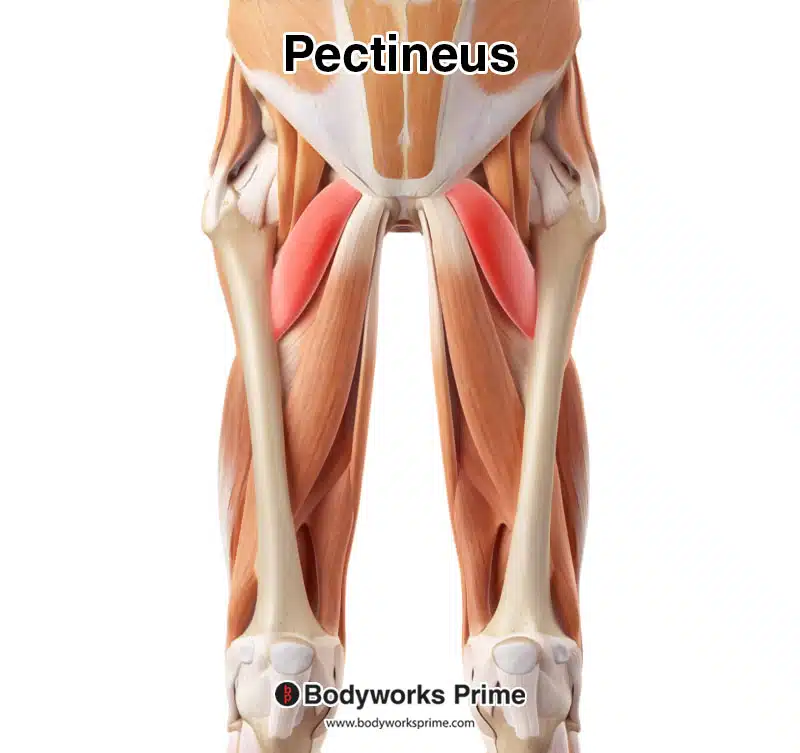
The pectineus muscle is somewhat small, flat, and quadrangular shaped. It’s located in the medial (inner) portion of the superior (upper) thigh. This muscle belongs to the hip adductor group of muscles, which also includes: the adductor brevis, adductor longus, adductor magnus, and the gracilis. The pectineus muscle is unique, because it is the most superior of the adductor muscles. It is also one of the few muscles in the body that is is innervated by two different nerves [1] [2].
The pectineus muscle is located between the iliopsoas and adductor longus muscles anteriorly, and the obturator externus and adductor brevis muscles posteriorly. It’s also deep and medial to the sartorius muscle. This location puts the pectineus muscle in a prime position to contribute to the complex functions of the hip joint [3].
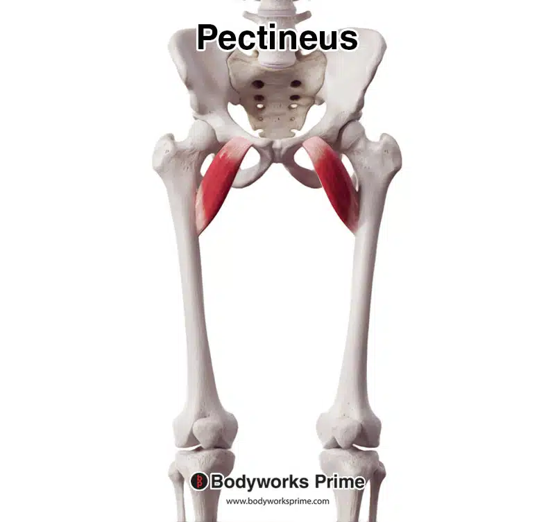
Here we can see the pectineus muscle, seen from an anterior view.
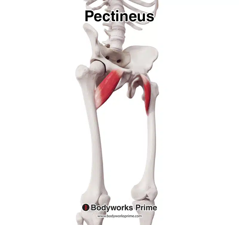
Here we can see the pectineus muscle, seen from an anterolateral view.
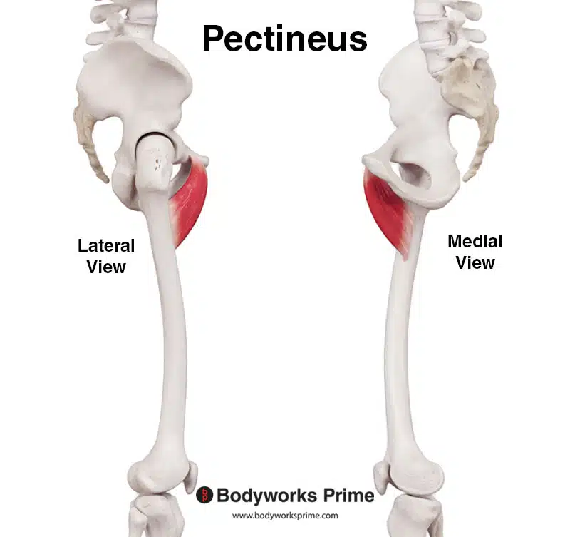
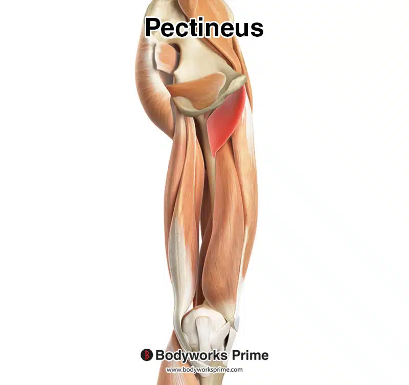
Here we can see the pectineus muscle, seen from a lateral and medial view.
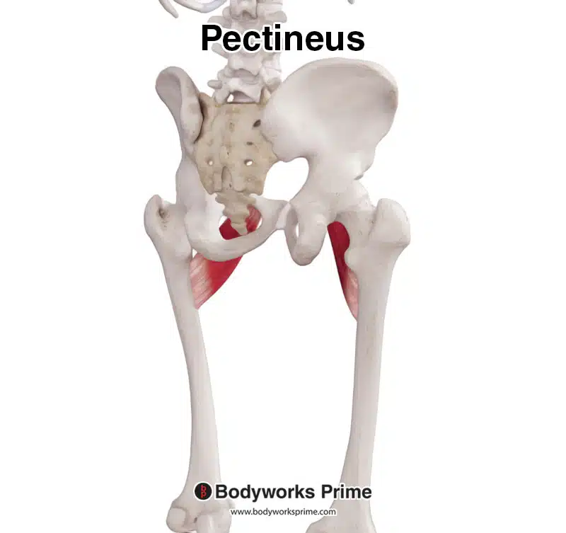
Here we can see the pectineus muscle, seen from a posterior view.
Here we can see the pectineus muscle highlighted in red amongst the other muscles of the body, seen from an anterior view. Superficial muscles have been removed to reveal the pectineus.
Here we can see the pectineus muscle highlighted in red amongst the other muscles of the body, seen from a medial view. Superficial muscles have been removed to reveal the pectineus.
Origin & Insertion
The pectineus muscle originates from the pectineal line of the pubic bone. The pectineal line of the pubic bone is a ridge located anteriorly on a section of the pubic bone called the superior ramus. The superior ramus is above the large O shaped opening on the hip bone, this opening is called the obturator foramen. The picture below shows the origin on the pectineal line of the pubic bone [4] [5] [6].
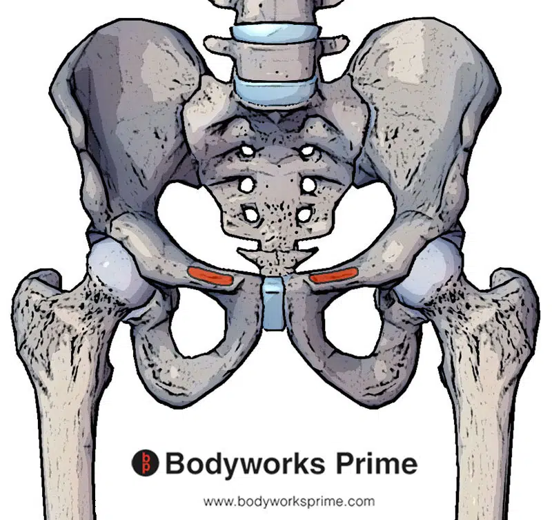
Here we can see the origin of the pectineus muscle highlighted in red.
From the pectineal line of the pubic bone, the pectineus’ muscle fibers traverse diagonally and laterally, extending towards the femur. It then reaches its insertion, located on the pectineal line of the femur. The pectineal line of the femur is a bony ridge that runs obliquely along the upper part of the medial surface of the femur, just inferior to the lesser trochanter. The lesser trochanter is a small, bony prominence on the posterior aspect of the femur, specifically at the junction of the neck and the shaft of the femur. The picture below shows the insertion point on the pectineal line of the femur [7] [8] [9].
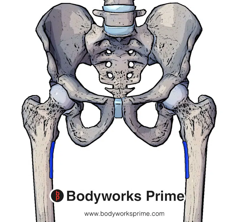
Here we can see the insertion of the pectineus muscle highlighted in blue.
Actions
The pectineus muscle serves an important role in movements involving the hip joint. The pectineus’ primary actions are to facilitate the adduction and flexion of the hip joint [10] [11] [12].
To visualise these movements, consider the action of bringing your leg inward towards the centre of your body; this is hip adduction. Now imagine the action of lifting your knee towards your chest while standing; this is hip flexion. Contraction of the pectineus muscle contributes to both of these actions.
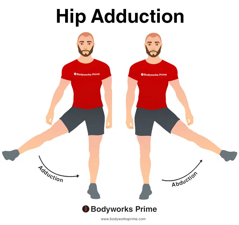
This image shows an example of hip adduction, which involves moving the leg towards the body (medially). The opposite of hip adduction is hip abduction. The pectineus can adduct the hip joint.
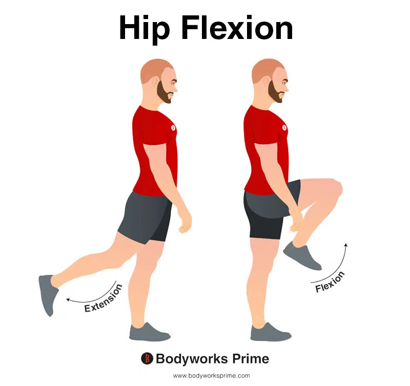
In this image, you can see an example of hip flexion, which is the action of raising your leg up in front of you and causing your hip to bend. If both legs are fixed, the torso will lean forward towards the legs. The opposite movement of hip flexion is hip extension. The pectineus can flex the hip joint.
In addition to hip adduction and flexion, there is ongoing research into the role of the pectineus and rotation of the femur [13] [14]. Studies suggest that pectineus may provide a supportive role in maintaining dynamic stability during rotation. Furthermore, due to the muscle’s attachment on the medial side of the femur, it is hypothesised that the pectineus may provide a small degree of internal rotation when the femur is externally rotated, or provide a small degree of external rotation when the femur is internally rotated. However, more research is needed to conclusively determine if the pectineus directly contributes to rotation of the femur. If it does contribute to rotation, its contribution would be very minimal comparative to other primary external and internal rotators of the hip joint. Presently, the consensus points more towards the pectineus acting as more of a stabiliser during rotational movements of the hip joint, rather than directly causing the rotation.
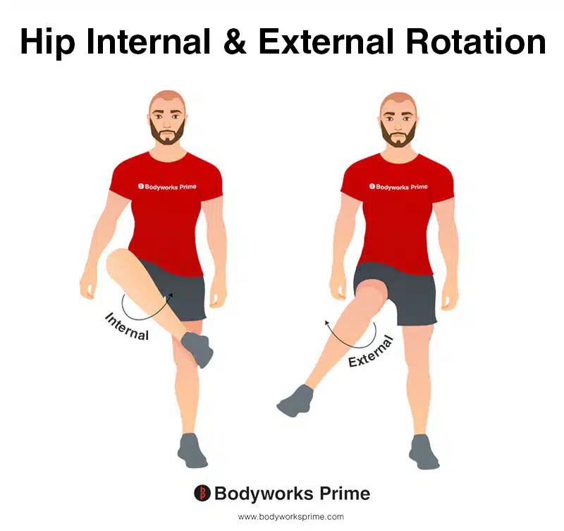
This image shows an example of hip internal (medial) rotation and external (lateral) rotation. Internal rotation involves rotating the leg from the hip joint inwards (medially). The opposite of internal rotation is external rotation, which involves rotating the leg from the hip joint outwards (laterally). The pectineus may assist weakly with rotation of the femur. More research is needed to conclusively determine if the pectineus directly contributes to rotation of the femur. Presently, the consensus points more towards the pectineus acting as more of a stabiliser during rotational movements of the hip joint, rather than directly causing the rotation.
Innervation
The pectineus muscle is primarily innervated by the femoral nerve, with occasional contributions from the obturator nerve [15].
The femoral nerve, the largest branch of the lumbar plexus, originates from the dorsal divisions of the second, third, and fourth lumbar nerves (L2, L3, L4). It descends posterior to the psoas major, running between this muscle and the iliacus. It then passes beneath the inguinal ligament and enters the femoral triangle, an area in the upper thigh. Here, it divides into two main branches: anterior and posterior. The anterior division provides nerve supply to the pectineus and iliacus muscles among others [16] [17].
Occasionally, the obturator nerve also provides innervation to the pectineus. This nerve begins at the lumbar plexus and descends alongside the iliopectineal line, traversing the fibers of the psoas major muscle. Upon emerging from the medial border of the psoas major near the pelvic brim, it continues posterior to the common iliac arteries, descending towards the obturator canal. The obturator nerve then exits the pelvis through the obturator canal, making its way towards the medial thigh where it can innervate parts of the pectineus [18] [19].
It’s important to note that the pattern of innervation can vary among individuals, with some having slightly different configurations than the typical pattern described here [20].
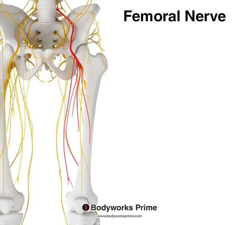
Here we can see the femoral nerve highlighted in red. The pectineus muscle is primarily innervated by the femoral nerve.
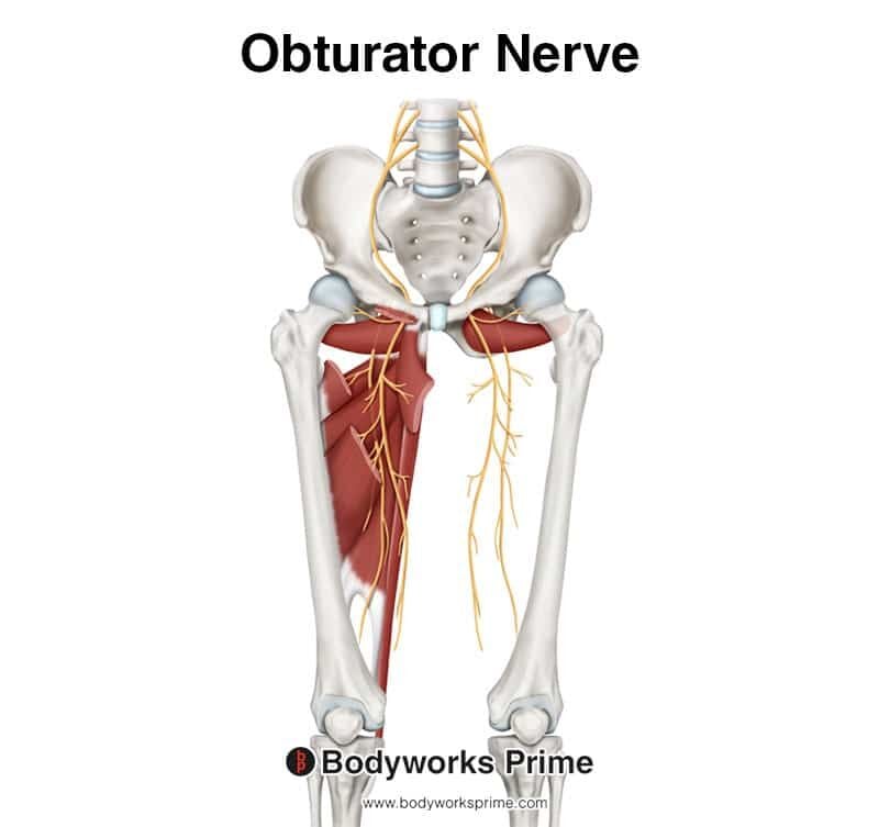
This image shows the obturator nerve arising from the lumbar plexus. The pectineus occasionally receives contributions from the obturator nerve.
Blood Supply
The pectineus muscle receives its blood supply primarily from the obturator artery and occasionally the medial circumflex femoral artery [21] [22] [23].
The obturator artery usually originates from the anterior division of the internal iliac artery. This is located in the lower pelvic region. The obturator artery then travels towards the obturator canal, which is an opening in the pelvic bone, to reach the inner thigh area. The obturator artery provides blood to a number of muscles in this region, including the pectineus [24] [25].
Additionally, the pectineus muscle can receive blood supply from the medial circumflex femoral artery. This artery originates from the deep femoral artery (aka profunda femoris) which is a major branch of the femoral artery in the thigh. The medial circumflex femoral artery curves around the posterior aspect of the femur, where it supplies blood to various muscles of the hip and thigh, including potentially the pectineus muscle [26] [27].
It’s important to bear in mind that the exact pattern of blood supply can vary among individuals. Some people have slightly different vascular configurations than the pattern described above.
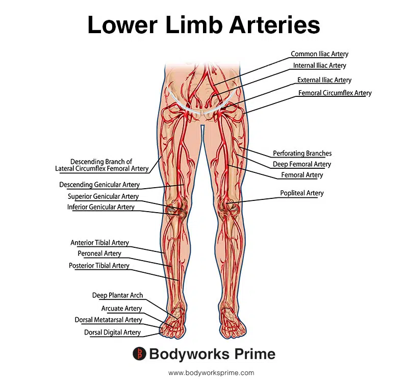
This image shows the arteries of the lower limb.
Want some flashcards to help you remember this information? Then click the link below:
Pectineus Muscle Flashcards
Support Bodyworks Prime
Running a website and YouTube channel can be expensive. Your donation helps support the creation of more content for my website and YouTube channel. All donation proceeds go towards covering expenses only. Every contribution, big or small, makes a difference!
References
| ↑1, ↑15, ↑20 | Kim H, Nam YS. Variation of pectineus muscle forming a hiatus. Anat Sci Int. 2021;96(3):481-484. doi:10.1007/s12565-020-00593-5 |
|---|---|
| ↑2, ↑3, ↑5, ↑8, ↑16, ↑18, ↑22, ↑24, ↑26 | Standring S. (2015). Gray’s Anatomy: The Anatomical Basis of Clinical Practice, 41st Edn. Amsterdam: Elsevier. |
| ↑4, ↑7, ↑11, ↑17, ↑19, ↑23, ↑25, ↑27 | Moore KL, Agur AMR, Dalley AF. Clinically Oriented Anatomy. 8th ed. Philadelphia: Lippincot Williams & Wilkins; 2017. |
| ↑6, ↑9, ↑12 | Kiel J, Kaiser K. Adductor Strain. [Updated 2021 Jul 25]. In: StatPearls [Internet]. Treasure Island (FL): StatPearls Publishing; 2021 Jan-. Available from: https://www.ncbi.nlm.nih.gov/books/NBK493166/ |
| ↑10 | Ransom AL, Sinkler MA, Nallamothu SV. Anatomy, Bony Pelvis and Lower Limb, Femoral Muscles. [Updated 2021 Oct 6]. In: StatPearls [Internet]. Treasure Island (FL): StatPearls Publishing; 2021 Jan-. Available from: https://www.ncbi.nlm.nih.gov/books/NBK500008/ |
| ↑13 | Decker MJ, Krong J, Hageman LR, Torry MR, Philippon MJ, Steadman. Annual Meeting of the Orthopaedic Research Society, 2010. Hip Rotation Function of the Pectineus Muscle. [online] (Poster No. 1607). Available from: http://www.ors.org/Transactions/56/1607.pdf [Accessed 15 June 2022]. |
| ↑14 | Giphart JE, Stull JD, Laprade RF, Wahoff MS, Philippon MJ. Recruitment and activity of the pectineus and piriformis muscles during hip rehabilitation exercises: an electromyography study. Am J Sports Med. 2012 Jul;40(7):1654-63. doi: 10.1177/0363546512443812. Epub 2012 Apr 20. PMID: 22523373. |
| ↑21 | Arias DG, Marappa-Ganeshan R. Anatomy, Bony Pelvis and Lower Limb, Arteries. [Updated 2021 Jul 26]. In: StatPearls [Internet]. Treasure Island (FL): StatPearls Publishing; 2021 Jan-. Available from: https://www.ncbi.nlm.nih.gov/books/NBK544319/ |


