| Origin | Obturator foramen & membrane |
| Insertion | Trochanteric fossa of the greater trochanter |
| Action | Hip lateral rotation Hip stabilisation Potentially assists with adduction when hip is flexed |
| Nerve | Posterior division of obturator nerve (L3, L4) |
| Artery | Anterior branch of obturator artery & medial circumflex femoral artery |
Location & Overview
The obturator externus is a flat, fan-shaped muscle located deep in the medial compartment of the thigh. It originates on the anterior side of the hip’s obturator foramen. As it extends laterally, it passes posteriorly beneath the neck of the femur, lying deep to the quadratus femoris muscle
[1] [2].
The primary function of the obturator externus is to act as a lateral rotator of the hip joint and also stabilising the femoral head within the acetabulum. It is one of the short and deep lateral rotators of the hip, often referred to as the “deep six”. This group comprises the piriformis, superior gemellus, obturator internus, inferior gemellus, quadratus femoris, and the obturator externus [3] [4].
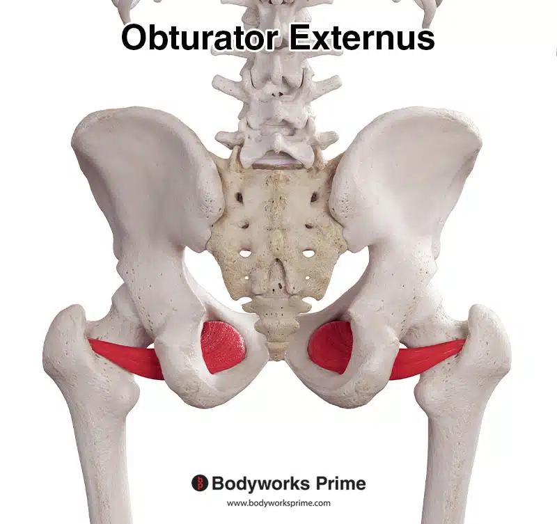
Pictured here we can see the obturator externus muscle from a posterior view.
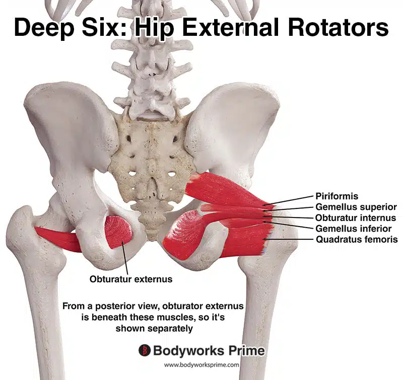
Pictured here are the deep six hip external rotator muscles: the piriformis, gemellus superior, gemellus inferior, obturator internus, obturator externus, and quadratus femoris. These muscles play an important role in stabilising the hip joint and facilitating hip lateral rotation.
Origin & Insertion
The obturator externus originates from the bony margins of the obturator foramen, which is a large opening in the pelvis situated between the pubis and ischium bones. This foramen is covered by the obturator membrane, a thin sheet of connective tissue that stretches across the opening, giving an additional area for the obturator externus to originate from. The muscle fibers of the obturator externus then converge into a tendon that courses inferomedially beneath the neck of the femur. This tendon attaches onto the trochanteric fossa, a shallow depression on the medial aspect of the greater trochanter. The greater trochanter is a prominent bony projection located at the upper part of the femur, serving as a major attachment point for several muscles [5] [6] [7].
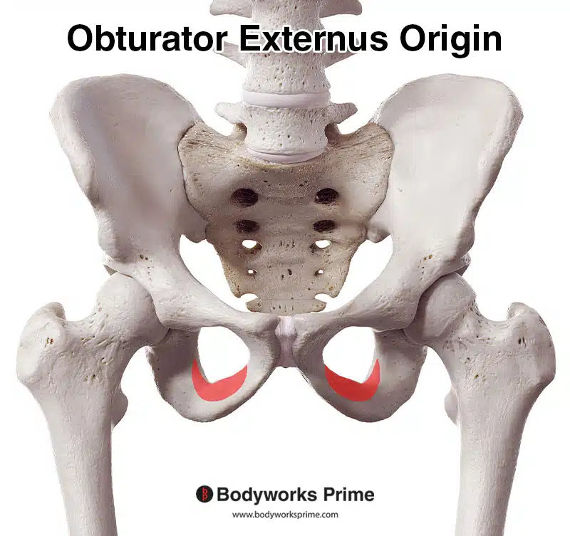
Here we can see the obturator externus origin marked in red on the obturator foramen. The obturator externus also originates on the obturator membrane, displayed below.
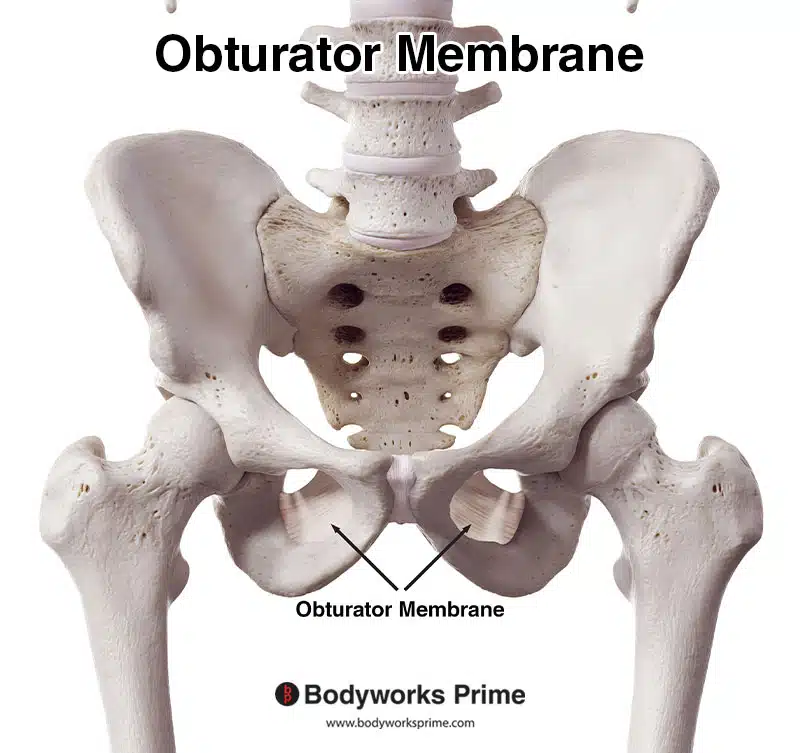
Here we can see the obturator membrane, another origin point of the obturator externus muscle.
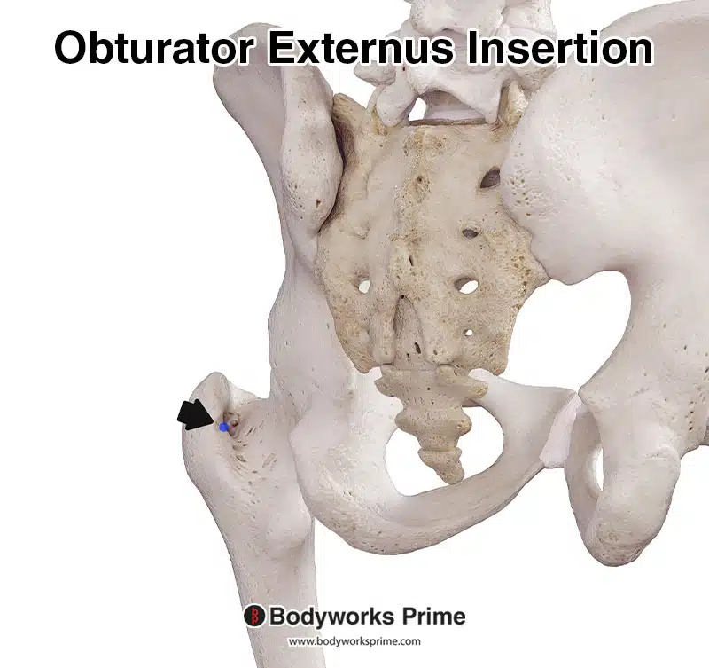
Here we can see the obturator externus insertion marked in blue on the trochanteric fossa of the greater trochanter.
Actions
Contraction of the obturator externus primarily produces lateral (external) rotation of the thigh at the hip joint, causing the leg to rotate so that the knee turns outwards. Additionally, when the hip is flexed, the obturator externus potentially contributes to adduction, drawing the thigh toward the midline of the body (there is conflicting information regarding its adduction ability in the literature). The obturator externus also plays an important role in stabilising the hip joint, ensuring the femoral head remains anchored securely within the acetabulum [8] [9] [10].
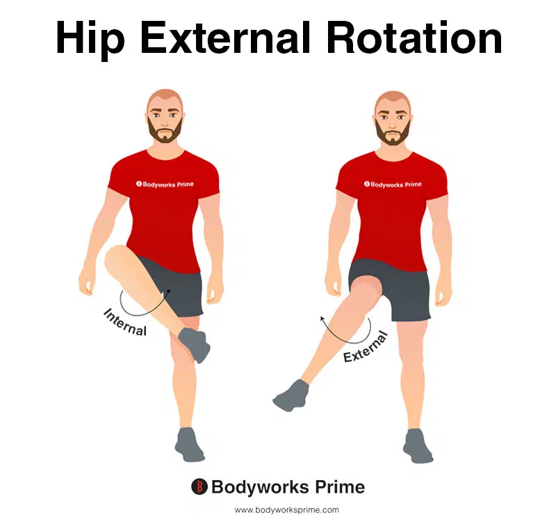
This image shows an example of hip external rotation, which involves rotating the leg from the hip joint outwards (laterally). External rotation is also referred to as lateral rotation. The opposite of external rotation is internal rotation (medial rotation). External rotation of the hip joint is the primary action of the obturator externus (alongside hip stabilisation).
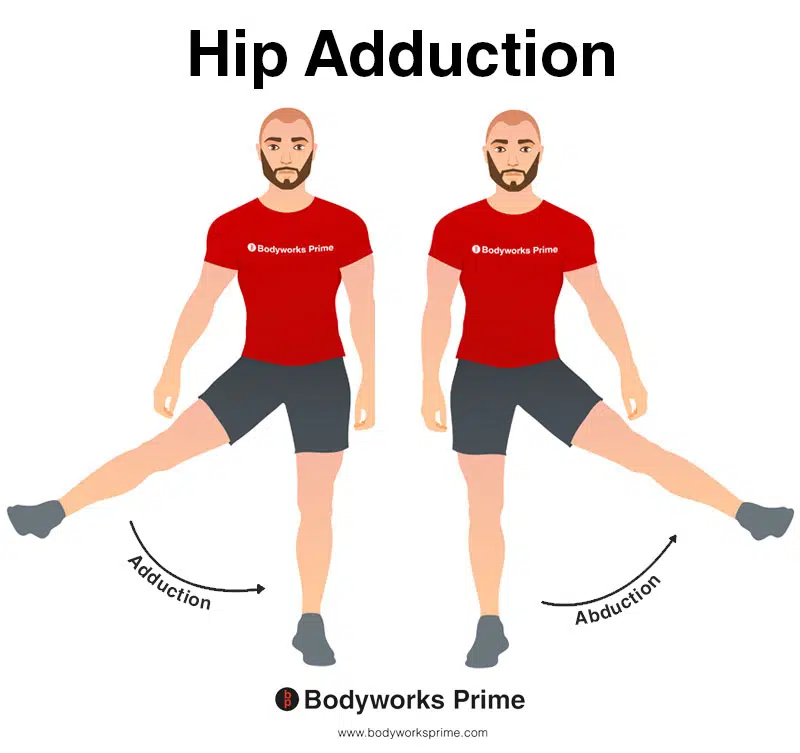
This image shows an example of hip adduction, which involves moving the leg towards the body’s midline. The opposite of hip adduction is hip abduction. The obturator externus can weakly assist with hip adduction, but only when the hip is flexed.
Innervation
The obturator externus is innervated by the posterior division of the obturator nerve, specifically the spinal roots L3 and L4. Originating from the lumbar plexus, the obturator nerve divides into anterior and posterior branches near the obturator foramen [11] [12] [13].
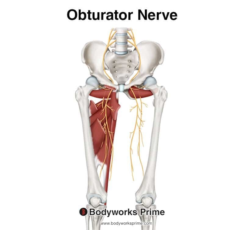
Here we can see the obturator nerve which innervates the obturator externus muscle.
Blood Supply
Arterial supply to the obturator externus is primarily via the anterior branch of the obturator artery. Additional vascularisation comes from the medial circumflex femoral artery, an offshoot of the femoral artery. These vessels join together near the obturator externus muscle [14].
Want some flashcards to help you remember this information? Then click the link below:
Obturator Externus Flashcards
Support Bodyworks Prime
Running a website and YouTube channel can be expensive. Your donation helps support the creation of more content for my website and YouTube channel. All donation proceeds go towards covering expenses only. Every contribution, big or small, makes a difference!
References
| ↑1, ↑3, ↑5, ↑8, ↑12 | Dalley AF II, Agur AMR. Moore’s Clinically Oriented Anatomy. 9th ed. Wolters Kluwer Health; 2022. |
|---|---|
| ↑2, ↑4, ↑6, ↑9, ↑13 | Standring S. (2015). Gray’s Anatomy: The Anatomical Basis of Clinical Practice, 41st Edn. Amsterdam: Elsevier |
| ↑7, ↑10, ↑11, ↑14 | Larson MR, Ryan W. Anatomy, Abdomen and Pelvis, Obturator Muscles. [Updated 2023 Jan 17]. In: StatPearls [Internet]. Treasure Island (FL): StatPearls Publishing; 2023 Jan-. Available from: https://www.ncbi.nlm.nih.gov/books/NBK589636/ |


