| Origin | Superior two-thirds of the lateral surface of the fibula Fibular Head |
| Insertion | Base of the first metatarsal Medial cuneiform |
| Action | Eversion of the foot Assists with plantar flexion |
| Nerve | Superficial fibular nerve (peroneal nerve), L5 & S1 |
| Main Artery | Anterior tibial artery Fibular artery (peroneal artery) |
Location & Overview
The fibularis longus, which was previously named the peroneus longus, is a slender and elongated fusiform muscle located on the lateral aspect of the leg. This muscle is one of three other fibularis muscles, alongside the fibularis tertius and fibularis brevis. At its proximal end, it is superficial to part of the fibularis brevis muscle. Moreover, the tendons of fibularis longus and brevis run parallel to each other towards the ankle joint, with the longus being more posteriorly placed. However, an intriguing divergence happens at the muscle’s distal end. Here, the longus tendon continues beneath the foot, terminating at the base of the first metatarsal. This pathway is in contrast to the brevis, which attaches to the fifth metatarsal. Its positioning is demonstrated in the images below [1] [2] [3].
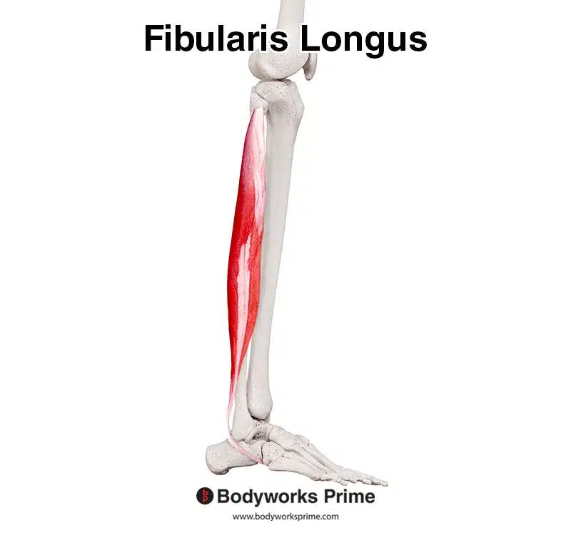
Pictured here, we can see the fibularis longus muscle from a lateral view.
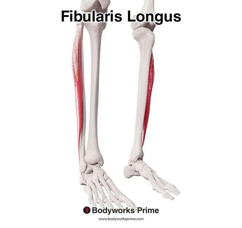
Pictured here, we can see the fibularis longus muscle from an anterolateral view.
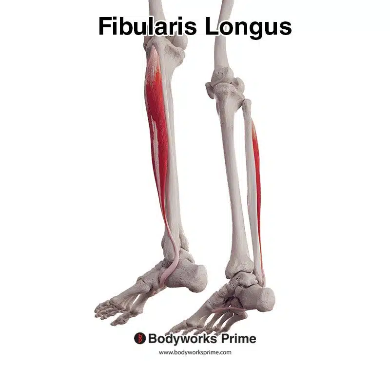
Pictured here, we can see the fibularis longus muscle from a posterior and somewhat inferior view.
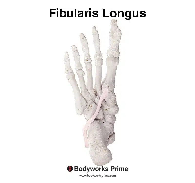
Pictured here, we can see the fibularis longus muscle’s distal tendon underneath the foot.
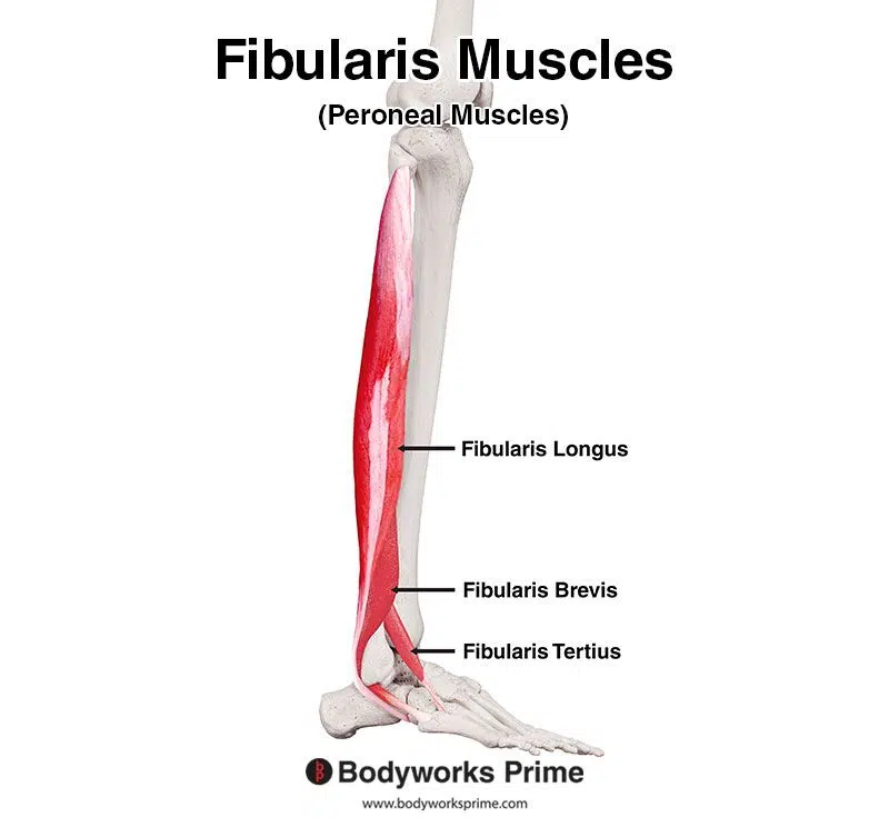
Pictured here, we can see all three of the fibularis muscles: the fibularis longus, fibularis brevis, and fibularis tertius.
Origin & Insertion
The origin of the fibularis longus is notably expansive, encompassing the head and the superior two-thirds of the lateral surface of the fibula, and extending to the intermuscular septa that separates it from adjacent muscles. This broad origin provides a robust anchorage for the muscle. Progressing distally, the muscle’s tendon passes posterior to the lateral malleolus continuing past the calcaneus where it directs itself underneath the foot, inserting onto the base of the first metatarsal and the medial cuneiform. This distinctive pathway not only facilitates its mechanical leverage but also influences its functional roles in foot dynamics [4] [5] [6].

Pictured here, we can see the fibularis longus muscle’s origin highlighted in red on the superior two-thirds of the lateral surface of the fibula.
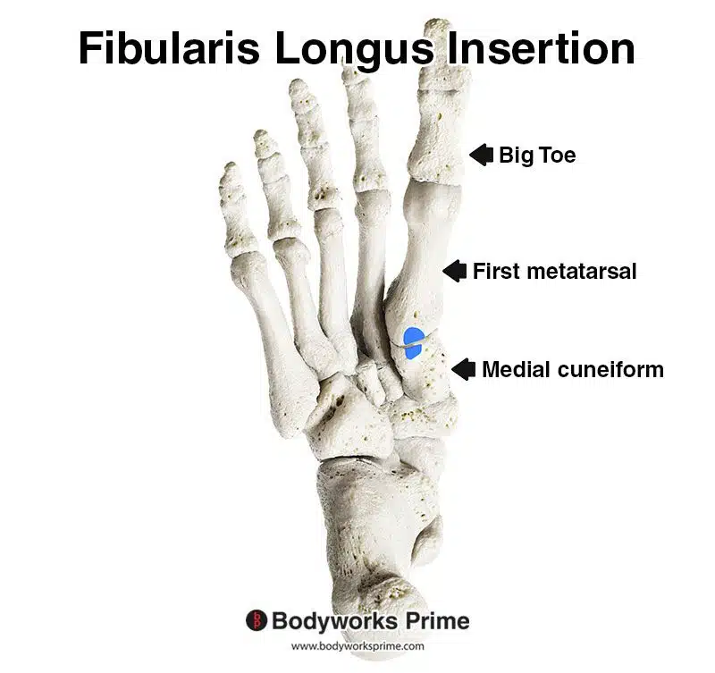
Pictured here, we can see the fibularis longus muscle’s insertion highlighted in blue on the base of the first metatarsal and the medial cuneiform.
Actions
The fibularis longus plays an important role in the eversion of the foot, where it helps move the foot outward in a lateral direction (noticeable when you try to view the sole of your foot by turning it outward). Additionally, it assists weakly with plantarflexion of the ankle, a movement that involves pointing the toes and foot downward, as seen when standing on your tiptoes [7] [8] [9].
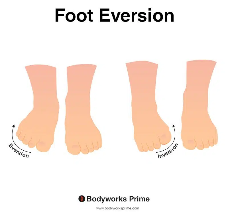
This picture demonstrates foot eversion, which means rotating the foot outwards, causing the sole of the foot to turn away from the body. The opposite of eversion is inversion. Eversion is the primary action of the fibularis longus.
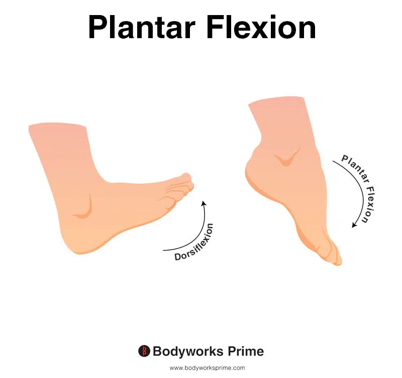
This image shows an example of foot plantar flexion, which involves extending the ankle. The opposite of plantar flexion is dorsiflexion. Plantar flexion is a secondary action of the fibularis longus
Innervation
The fibularis longus is innervated by the superficial fibular (peroneal) nerve, with root values of L5 and S1. The superficial fibular nerve is a terminal branch of the common fibular nerve. The common fibular nerve is a branch of the sciatic nerve [10] [11] [12].
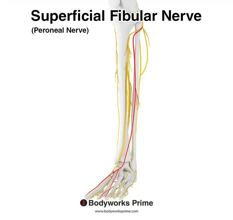
Pictured here, we can see the superficial fibular nerve highlighted in red. The superficial fibular nerve innervates the fibularis longus from the nerve roots of L5 and S1. The superficial fibular nerve was previously known as the peroneal nerve.
Blood Supply
Vascularization of the fibularis longus is quite extensive, derived from numerous arteries. Proximally, its muscular structure receives blood primarily from the anterior tibial artery and the fibular artery (aka peroneal artery). It may also receive contributions from the circumflex fibular branch of the posterior tibial artery and the artery accompanying the common fibular nerve (sural communicating artery). Distally, the tendons obtain vascular supply from an assortment of sources, encompassing the fibular perforating, anterior lateral malleolar, lateral calcaneal, lateral tarsal, arcuate, and both lateral and medial plantar arteries. However, the main two arteries to remember with regards to supplying the fibularis longus (especially in the proximal and mid-portions) are the anterior tibial artery and the fibular artery [13] [14] [15].
Want some flashcards to help you remember this information? Then click the link below:
Fibularis Longus Flashcards
Support Bodyworks Prime
Running a website and YouTube channel can be expensive. Your donation helps support the creation of more content for my website and YouTube channel. All donation proceeds go towards covering expenses only. Every contribution, big or small, makes a difference!
References
| ↑1, ↑4, ↑7, ↑10 | Dalley AF II, Agur AMR. Moore’s Clinically Oriented Anatomy. 9th ed. Wolters Kluwer Health; 2022. |
|---|---|
| ↑2, ↑5, ↑8, ↑11, ↑13 | Standring S. (2015). Gray’s Anatomy: The Anatomical Basis of Clinical Practice, 41st Edn. Amsterdam: Elsevier |
| ↑3, ↑6, ↑9, ↑12 | Palastanga N, Soames RW. Anatomy and Human Movement: Structure and Function (Physiotherapy Essentials), 6th ed. Elsevier; 2011. ISBN-13: 978-0702035531. |
| ↑14 | Lezak B, Varacallo M. Anatomy, Bony Pelvis and Lower Limb: Calf Peroneus Longus Muscle. [Updated 2022 Aug 25]. In: StatPearls [Internet]. Treasure Island (FL): StatPearls Publishing; 2023 Jan-. Available from: https://www.ncbi.nlm.nih.gov/books/NBK546650/ |
| ↑15 | Khan IA, Mahabadi N, D’Abarno A, et al. Anatomy, Bony Pelvis and Lower Limb: Leg Lateral Compartment. [Updated 2023 Aug 14]. In: StatPearls [Internet]. Treasure Island (FL): StatPearls Publishing; 2023 Jan-. Available from: https://www.ncbi.nlm.nih.gov/books/NBK519526/ |


