| Origin | Ischiopubic ramus |
| Insertion | Medial surface of proximal tibia (pes anserinus) |
| Action | Hip adduction Knee flexion Knee internal rotation (i.e medial rotation of tibia) Hip flexion Hip internal rotation |
| Nerve | Anterior branch of obturator nerve (L2, L3, L4) |
| Artery | Medial circumflex femoral artery Superficial femoral artery Deep femoral artery Descending genicular artery Anterior branch of the obturator artery |
Location & Overview
The gracilis muscle, is name so due to its long and slender shape, from the Latin ‘gracilis’, which means thin or slender. The gracilis is located in the medial compartment of the thigh. As a superficial muscle, it is located close to the skin’s surface which makes it easy to palpate [1] [2].
The gracilis is one of the adductor muscles of the hip (its other actions are covered in the actions section below). It is situated immediately adjacent to the adductor longus muscle and beneath the adductor magnus muscle. Its position in the medial compartment of the thigh places it closer to the body’s midline. This gives it a strategic role in the movements of the lower limb, such as adduction [3] [4].
The gracilis is part of the pes anserinus, which if formed by a collection of tendons. The term is derived from Latin which translates to ‘goose foot’, due to its three-pronged structure of tendons, similar to the foot of a goose. This tendons involved in the pes anserinus consist of three muscles: the gracilis, the semitendinosus, and the sartorius. These tendons converge at their insertion point on the proximal tibia, forming a robust structure that plays a significant role in stabilising the medial side of the knee joint [5] [6].
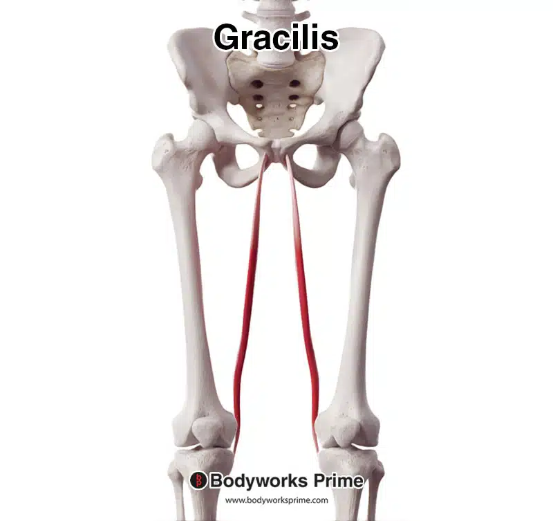
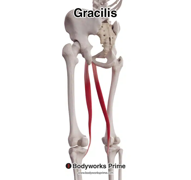
Here we can see the gracilis muscle, seen from an anterior view.
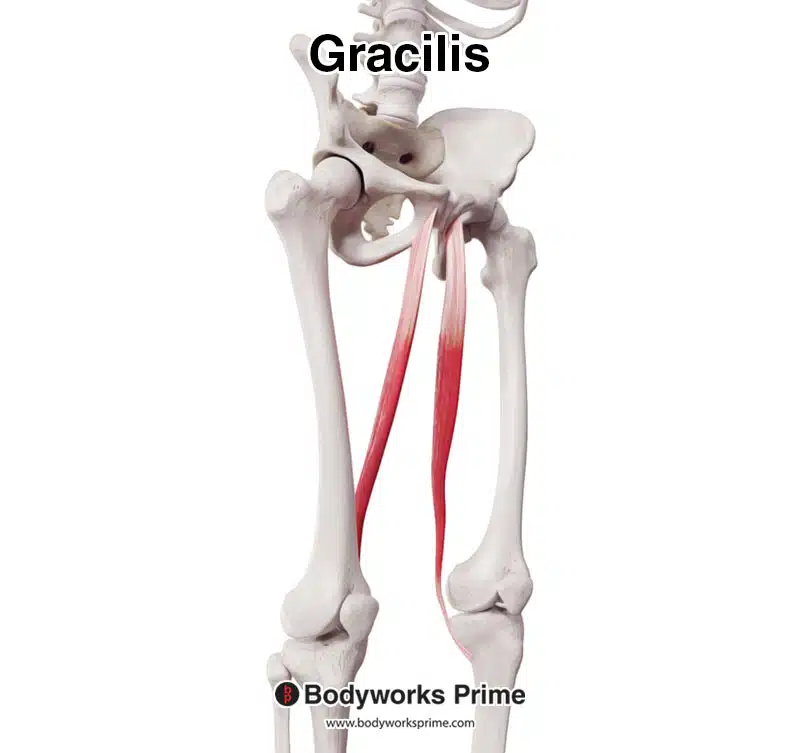
Here we can see the gracilis muscle, seen from an anterolateral view.
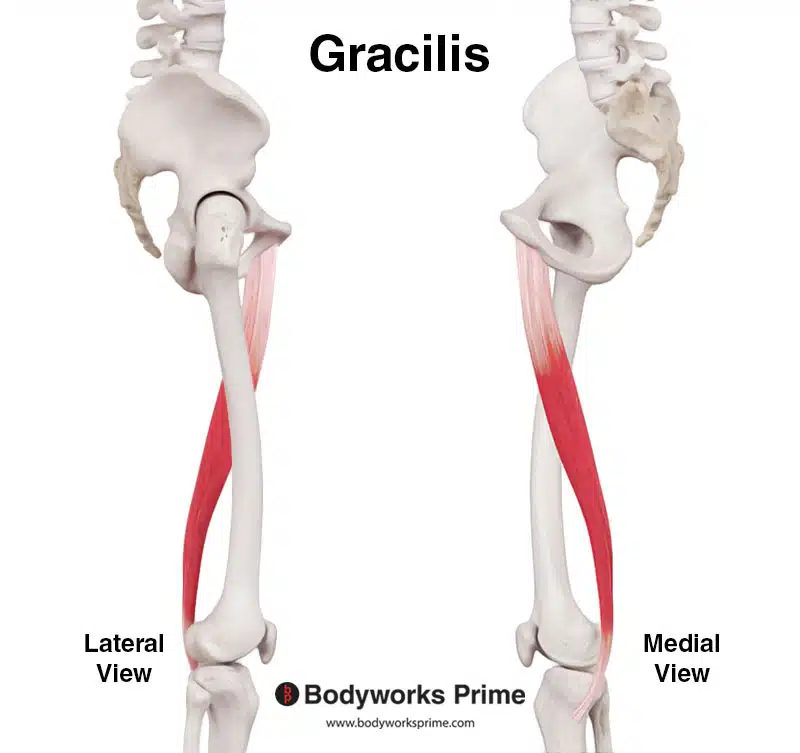
Here we can see the gracilis muscle, seen from a lateral view.
Here we can see the gracilis muscle, seen from a posterolateral view.
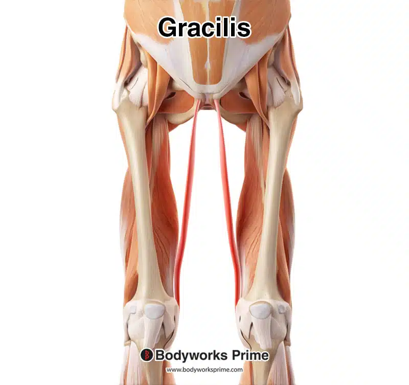
Here we can see the gracilis highlighted in red amongst the other muscles of the body, seen from an anterior view. Nearby muscles have been removed to better show the gracilis from this view.
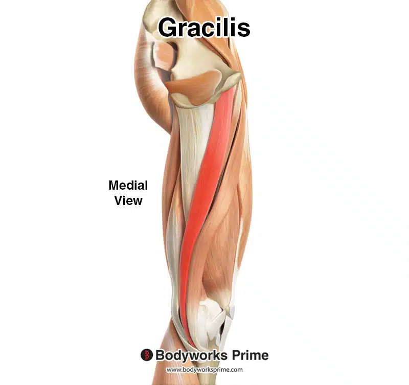
Here we can see the gracilis highlighted in red amongst the other muscles of the body, seen from a medial view.
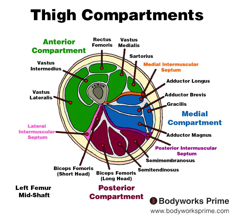
Here we can see an image of the compartments of the thigh. We can see the gracilis in the medial compartment, the section coloured in blue.
Origin & Insertion
The gracilis muscle originates on the lower half of the hip, more specifically on a structure called the ischiopubic ramus [7] [8] The ischiopubic ramus is a bony structure formed by the fusion of the ischium and the pubis, two of the three bones that compose the hip. This structure creates a kind of bridge at the lower anterior part of the hip, contributing to the formation of the obturator foramen, which is a large opening in the hip bone. The gracilis muscle takes its origin from the anterior aspect of this bridge-like structure. Its origin is on the of the anterior aspect ischiopubic ramus [9] [10].
The gracilis then extends down the inner part of the thigh in a relatively straight path, due to being the most medial of the thigh muscles. It runs almost parallel to the femur providing a clear path of the direction for the muscle [11].
The muscle terminates at its insertion point located on the medial surface of the proximal tibia. To be more precise, the gracilis inserts just below the knee, attaching to an area on the tibia known as the pes anserinus [12]. This insertion is just medial to the tibial tuberosity, where the quadriceps insert via the patella tendon. The common tendinous insertion of the gracilis is shared with two other muscles, the sartorius and semitendinosus. The insertion of these tendons forms a ‘goose foot’ pattern known as the pes anserinus, on the inner side of the upper tibia [13] [14].
This anatomical path, from the hip bone to the tibia, allows the gracilis muscle to span two joints: the hip and the knee. Thus, it is capable of influencing the movements of both these joints [15] [16], which will be discussed in more detail in the actions section next.
The origin of the gracilis is highlighted in red at the ischiopubic ramus.
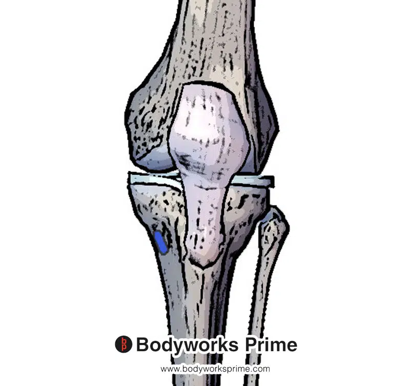
The insertion of the gracilis is highlighted in blue at the medial surface of proximal tibia and is one of the three muscles of the pes anserinus.
Actions
The gracilis muscle performs several important actions in the lower body. Its positioning and origin and insertion enable it to influence movements at both the hip and the knee joints [17] [18]. These actions, with accompanying illustrations, are outlined below.
Hip adduction: This is the primary action of the gracilis muscle. Adduction of the hip joint refers to the movement that brings the leg towards to the midline of the body. For example, if you were standing with your legs apart, the act of bringing your feet together would be accomplished through hip adduction. The gracilis muscle works together with the other adductor muscles to accomplish this (such as the adductor longus, adductor brevis, adductor magnus and the pectineus) [19] [20].
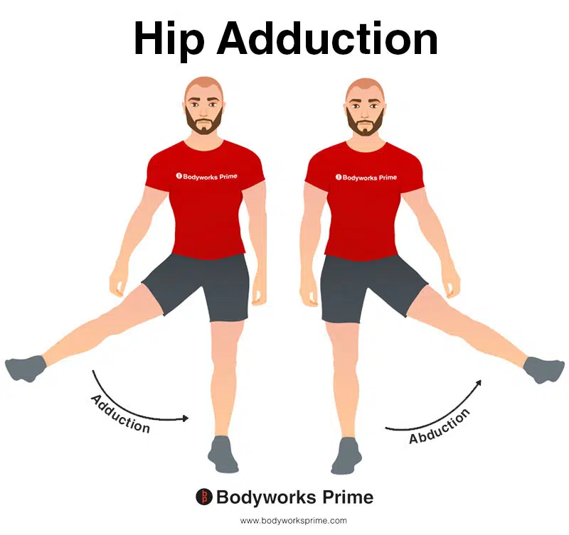
This image shows an example of hip adduction, which involves moving the towards the body (medially). The opposite of hip adduction is hip abduction. Hip adduction is the primary action of the gracilis muscle.
Knee flexion: The gracilis muscle also contributes to flexion of the knee joint, an action that involves bending the knee to decrease the angle between the thigh and the lower leg [21].
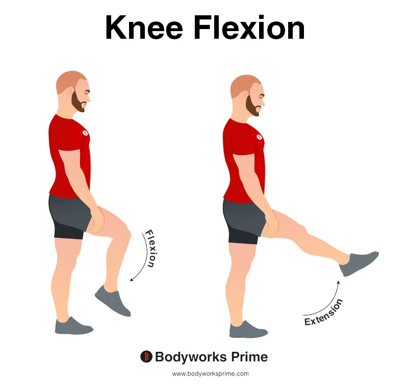
In this image, you can see an example of knee flexion, which is the action of bending your knee. The opposite movement of knee flexion is knee extension. The gracilis muscle contributes to flexion.
Tibia medial rotation (i.e knee internal rotation): When the knee is flexed, the gracilis can contribute to internal rotation of the tibia (also known as medial rotation of the tibia). This action involves turning the tibia inward, towards the midline of the body, which would result in the foot also turning inward if the foot was off the ground [22].
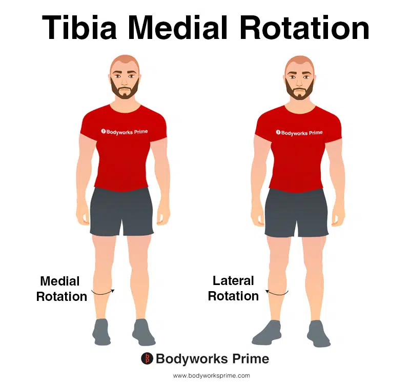
In this image, you can see an example of medial rotation of the tibia. Medial rotation of the tibia involves inward rotation of the shinbone towards the midline of the body. The opposite movement of medial rotation of the tibia is lateral rotation of the tibia. When the knee is flexed, the gracilis can contribute to medial rotation of the tibia.
Hip flexion: The gracilis muscle assists in flexion of the hip joint. This action involves lifting the thigh or moving it in the direction towards the abdomen [23] [24].
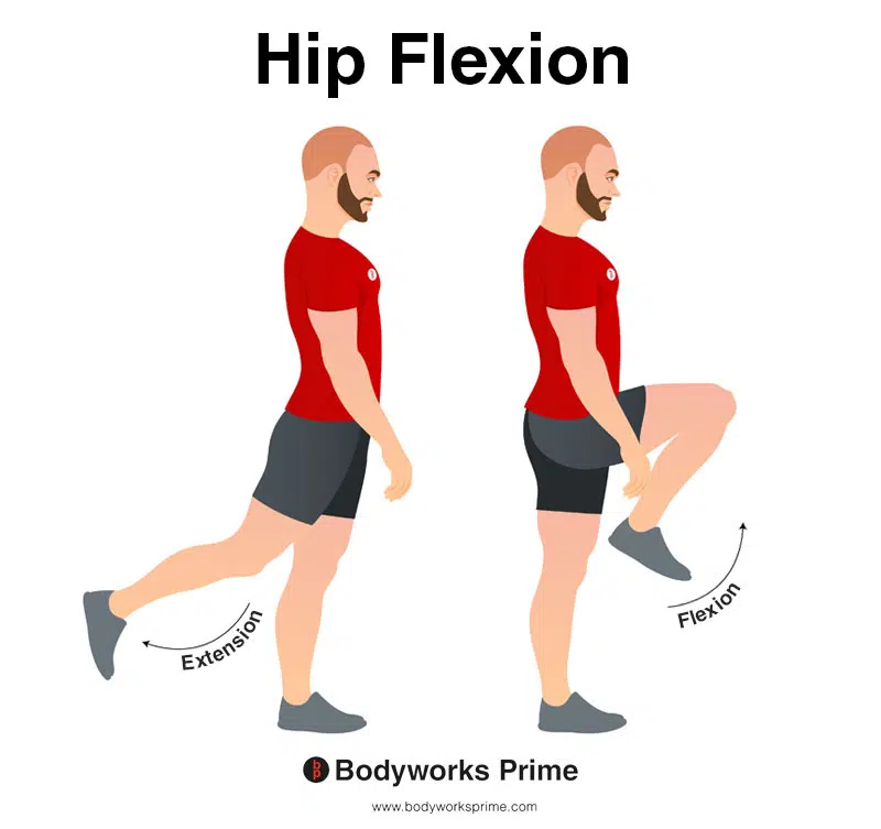
In this image, you can see an example of hip flexion, which is the action of raising your leg up in front of you and causing your hip to bend. If both legs are fixed, the torso will lean forward towards the legs. The opposite movement of hip flexion is hip extension. The gracilis muscle assists in flexion of the hip joint.
Hip internal rotation: When the hip is flexed, the gracilis can contribute to the internal rotation of the hip. This is a movement that turns the femur and vicariously the leg and foot inward, towards the midline of the body [25].
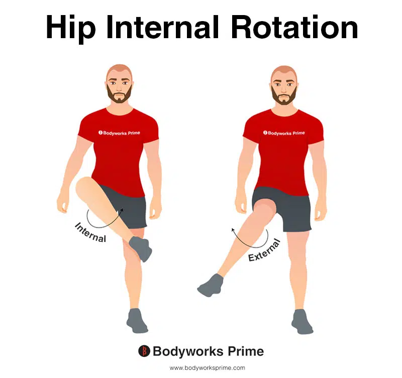
This image depicts an example of hip internal rotation, which involves rotating the leg from the hip joint inwards (medially). Internal rotation is also referred to as medial rotation. The opposite of internal rotation or medial rotation is external rotation or lateral rotation. When the hip is flexed, the gracilis can contribute to the internal rotation of the hip.
Innervation
The gracilis muscle which is located in the medial compartment of the thigh, is primarily innervated by the anterior branch of the obturator nerve. The obturator nerve originates from the L2, L3, and L4 nerve roots of the lumbar plexus [26] [27].
The formation of the obturator nerve begins at the lumbar plexus, and it subsequently descends alongside the iliopectineal line, traversing the fibers of the psoas major muscle. Upon emerging from the medial border of the psoas major near the pelvic brim, it continues posteriorly to the common iliac arteries. The obturator nerve continues along the lateral side of the internal iliac artery, descending towards the obturator canal. The obturator nerve then exits the pelvis through the obturator canal, making its way towards the medial thigh. Here, it splits into anterior and posterior divisions [28] [29].
The anterior branch of the obturator nerve, which innervates the gracilis muscle, continues its journey between the adductor longus and adductor brevis. It also provides sensory innervation to the skin of the medial upper thigh, and motor innervation to other adductor muscles of the lower leg, such as the adductor longus, external obturator, adductor magnus, and adductor brevis [30] [31].
The point of division of the obturator nerve into the anterior and posterior branches can be intrapelvic, within the obturator canal, or in the thigh. Typically, the anterior branch, which innervates the gracilis, divides into three major muscular branches that innervate the adductor longus, adductor brevis, and gracilis muscles [32] [33].
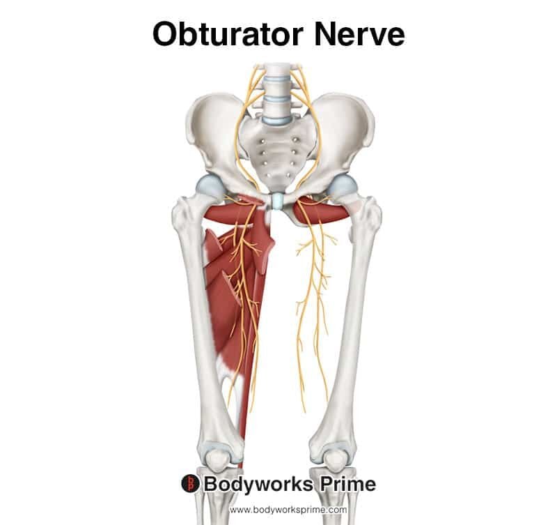
This image shows the obturator nerve arising from the lumbar plexus with nerve roots from L2-L4. The gracilis muscle is innervated by the anterior branch of the obturator nerve.
Blood Supply
The gracilis muscle gets its blood supply from the medial circumflex femoral artery, superficial femoral artery, deep femoral artery, descending genicular artery, and the anterior branch of the obturator artery [34].
The gracilis muscle’s primary blood supply is the medial circumflex femoral artery. This artery originates from the deep femoral artery, which is a major branch of the femoral artery which is responsible for supplying most of the thigh muscles. The medial circumflex femoral artery, after its origin, courses around the posterior aspect of the femur [35].
In addition to the medial circumflex femoral artery, the superficial femoral artery also plays a significant role in the providing blood to the gracilis muscle. This artery is a continuation of the common femoral artery which gives off to the deep femoral artery. The superficial femoral artery travels down the thigh’s anterior compartment, providing blood supply to the various muscles it encounters, including the gracilis [36].
The deep femoral artery, also known as the profunda femoris artery also contributes to the blood supply of the gracilis muscle as well. After its emergence from the common femoral artery, it dives deep into the posterior compartment of the thigh sending numerous branches to the muscles, including the gracilis [37].
The descending genicular artery, a branch of the femoral artery, is another artery which provides blood to the gracilis muscle. After its origin, it travels towards the knee, giving off articular and muscular branches which supply the knee joint and adjacent muscles, including the gracilis [38].
The last artery supplying blood the gracilis is the anterior branch of the obturator artery also. The obturator artery originates from the internal iliac artery, where it then travels through the obturator canal to reach the medial compartment of the thigh. Its anterior branch follows the obturator nerve, supplying blood to the adductor muscles, including the gracilis [39].
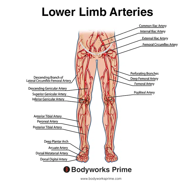
This image shows the arteries of the lower limb.
Want some flashcards to help you remember this information? Then click the link below:
Gracilis flashcards
Support Bodyworks Prime
Running a website and YouTube channel can be expensive. Your donation helps support the creation of more content for my website and YouTube channel. All donation proceeds go towards covering expenses only. Every contribution, big or small, makes a difference!
References
| ↑1, ↑8, ↑10, ↑11, ↑14, ↑16, ↑18 | Standring S. (2015). Gray’s Anatomy: The Anatomical Basis of Clinical Practice, 41st Edn. Amsterdam: Elsevier. |
|---|---|
| ↑2, ↑4, ↑7, ↑12, ↑20, ↑21, ↑22, ↑24, ↑25, ↑34, ↑35, ↑36, ↑37, ↑38, ↑39 | Khan IA, Bordoni B, Varacallo M. Anatomy, Bony Pelvis and Lower Limb, Thigh Gracilis Muscle. [Updated 2022 Apr 15]. In: StatPearls [Internet]. Treasure Island (FL): StatPearls Publishing; 2023 Jan-. Available from: https://www.ncbi.nlm.nih.gov/books/NBK538229/ |
| ↑3, ↑19, ↑23 | Neumann DA. Kinesiology of the hip: a focus on muscular actions. J Orthop Sports Phys Ther. 2010 Feb;40(2):82-94. doi: 10.2519/jospt.2010.3025. PMID: 20118525. |
| ↑5, ↑9, ↑13, ↑15, ↑17 | Moore KL, Agur AMR, Dalley AF. Clinically Oriented Anatomy. 8th ed. Philadelphia: Lippincot Williams & Wilkins; 2017. |
| ↑6 | Mohseni M, Mabrouk A, Graham C. Pes Anserine Bursitis. [Updated 2023 Jan 16]. In: StatPearls [Internet]. Treasure Island (FL): StatPearls Publishing; 2023 Jan-. Available from: https://www.ncbi.nlm.nih.gov/books/NBK532941/ |
| ↑26, ↑28, ↑30, ↑32 | Koh M, Markovich B. Anatomy, Abdomen and Pelvis, Obturator Nerve. [Updated 2022 Jul 25]. In: StatPearls [Internet]. Treasure Island (FL): StatPearls Publishing; 2023 Jan-. Available from: https://www.ncbi.nlm.nih.gov/books/NBK551640/ |
| ↑27, ↑29, ↑31, ↑33 | Anagnostopoulou S, Kostopanagiotou G, Paraskeuopoulos T, Chantzi C, Lolis E, Saranteas T. Anatomic variations of the obturator nerve in the inguinal region: implications in conventional and ultrasound regional anesthesia techniques. Reg Anesth Pain Med. 2009 Jan-Feb;34(1):33-9. doi: 10.1097/AAP.0b013e3181933b51. PMID: 19258986. |


