| Origin | Inferior angle of the medial border of the scapula |
| Insertion | Medial crest of the intertubercular groove (bicipital groove) |
| Action | Shoulder adduction Shoulder extension Shoulder internal rotation |
| Nerve | Lower subscapular nerve (C5, C6, C7) |
| Artery | Posterior circumflex humeral artery Thoracodorsal branch of the subscapular artery |
Location & Overview
The teres major is a muscle characterised by its thick, rectangular, and somewhat flat shape. It is situated in the shoulder region, extending from the scapula to the humerus. This muscle is found just below the infraspinatus and teres minor muscles, both of which are parts of the rotator cuff. However, despite its proximity, the teres major is not considered a component of the rotator cuff. Despite this, it still plays an important role in the movements and stability of the shoulder joint [1] [2] [3].
The teres major is located just superior to the latissimus dorsi muscle, and these two muscles often work synergistically in extending, adducting, and internally rotating the humerus [4]. Therefore, the teres major is occasionally referred to as the “lats little helper”.
In the broader context of the muscular system, the teres major is one of seven scapulohumeral muscles. These muscles, which include the teres major, are closely associated with the movement and stabilisation of the shoulder joint. The other scapulohumeral muscles are the deltoid, coracobrachialis, infraspinatus, subscapularis, supraspinatus, and teres minor [5] [6].
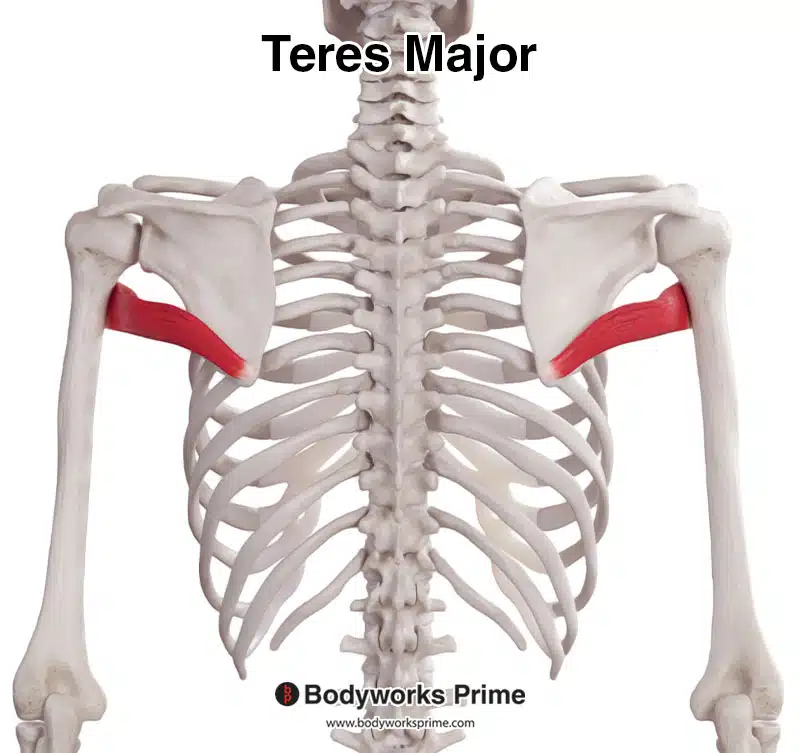
Here we can see the teres major from a posterior view.
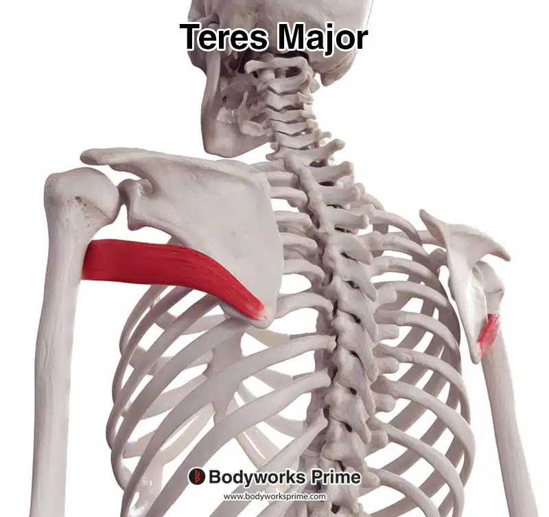
Here we can see the teres major from a posterolateral view.
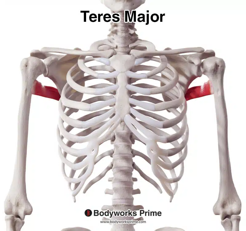
Here we can see the teres major from an anterior view.
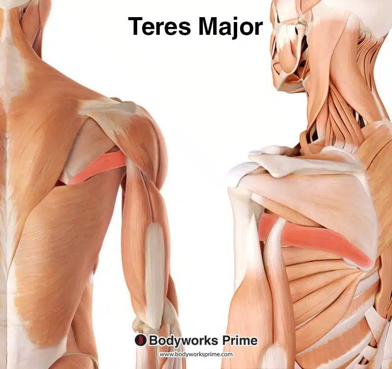
Here we can see the teres major highlighted in red amongst the other muscles of the body. The left shows a superficial view whereas the right shows a deep view.
Origin & Insertion
The teres major muscle originates on the posterior surface of the inferior angle of the scapula, which is the lowermost point of the scapula towards (or shoulder blade) [7] [8].
The teres major inserts at the medial lip of the intertubercular groove of the humerus. The muscle travels anterior to the triceps long head before attaching to the humerus [9] [10]. The intertubercular groove is a prominent groove that runs vertically down the upper part of the humerus bone. This groove is also known as the ‘bicipital groove and it is where the long tendon of the biceps brachii tendon passes through [11].
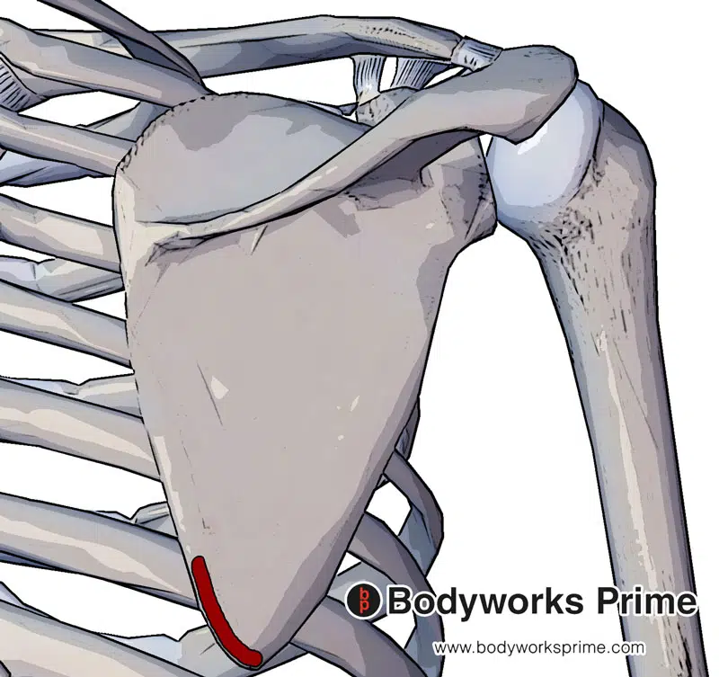
The teres major originates from the posterior side of the scapula. Specifically, the inferior aspect of the medial border of the scapula.
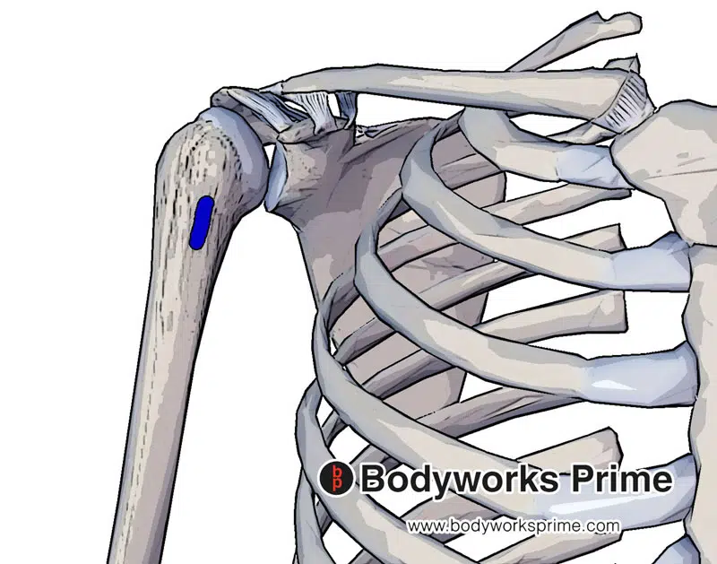
The teres major inserts on the medial crest of the intertubercular groove. Also known as the bicipital groove.
Actions
The teres major performs several actions at the shoulder. Its primary roles include extending, medially rotating, and adducting the humerus, or in simpler terms, moving the arm back, rotating it inward, and pulling it towards the body’s midline [12] [13].
The teres major also plays an important role in stabilising the humeral head, preventing its subluxation or partial dislocation. In carrying out these actions, the teres major often works in synergy with the latissimus dorsi muscle [14] [15].
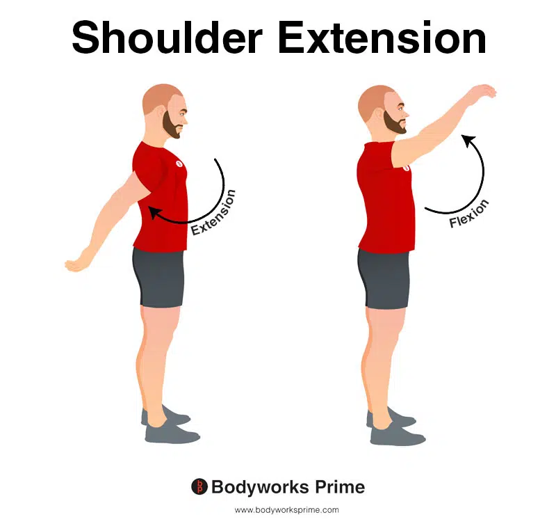
This image shows shoulder extension. Extension is where the arm is moved backward in a posterior direction (towards the back of the body), increasing the angle between the arm and the torso in the sagittal plane. The opposite of this is shoulder flexion which occurs as the arm is moved forward and upward in an anterior direction. The teres major contributes to shoulder extension.
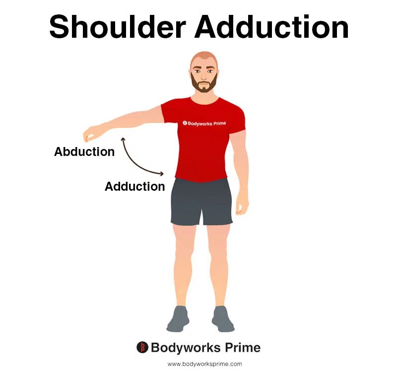
This image shows shoulder adduction. Adduction is where the arm is brought towards the midline of the body, reducing the angle between the arm and the torso. The opposite of adduction is abduction. Abduction occurs as the arm is moved away from the midline, increasing the angle between the arm and the torso. The teres major contributes to shoulder adduction.

This image shows shoulder internal rotation. Internal rotation is when the arm is rotated inward toward the centre of the body, causing the hand and forearm to move towards the body’s midline. The opposite of this is external rotation, which is when arm is rotated outward away from the centre of the body, moving the hand and forearm away from the body’s midline. Internal rotation is also known as medial rotation and external rotation is also known as lateral rotation. The teres major contributes to shoulder internal rotation.
Innervation
The teres major is innervated by the lower subscapular nerve, which originates from the nerve roots C5, C6, and C7. This nerve branches from the brachial plexus and is responsible for transmitting nerve signals between the teres major and the central nervous system, enabling the muscle to contract. The lower subscapular nerve also innervates the subscapularis muscle [16] [17] [18].
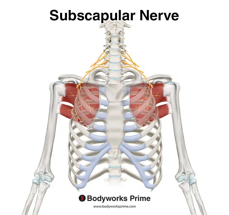
Here we can see the subscapular nerve, which innervates the teres major from the nerve roots of C5, C6, and C7.
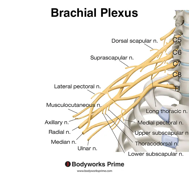
Here we can see the nerves of the brachial plexus. The subscapular nerve is a part of the brachial plexus.
Blood Supply
The teres major’s blood supply is primarily provided by the posterior circumflex humeral artery and the thoracodorsal branch of the subscapular artery. The posterior circumflex humeral artery arises from the third most distal portion of the axillary artery, travels posteriorly through the quadrangular space, and supplies blood to the teres major, teres minor, deltoid, and long head of the triceps [19] [20].
The subscapular artery, a large branch of the axillary artery, gives rise to the thoracodorsal artery. This artery supplies blood to the teres major, among other muscles like the subscapularis and latissimus dorsi [21] [22]. These arteries ensure the teres major muscle receives the necessary oxygen and nutrients to function effectively.
Want some flashcards to help you remember this information? Then click the link below:
Teres Major Flashcards
Support Bodyworks Prime
Running a website and YouTube channel can be expensive. Your donation helps support the creation of more content for my website and YouTube channel. All donation proceeds go towards covering expenses only. Every contribution, big or small, makes a difference!
References
| ↑1, ↑5, ↑9 | Moore KL, Agur AMR, Dalley AF. Clinically Oriented Anatomy. 8th ed. Philadelphia: Lippincot Williams & Wilkins; 2017. |
|---|---|
| ↑2 | Maruvada S, Madrazo-Ibarra A, Varacallo M. Anatomy, Rotator Cuff. [Updated 2023 Mar 27]. In: StatPearls [Internet]. Treasure Island (FL): StatPearls Publishing; 2023 Jan-. Available from: https://www.ncbi.nlm.nih.gov/books/NBK441844/ |
| ↑3, ↑6 | Wu JG, Bordoni B. Anatomy, Shoulder and Upper Limb, Scapulohumeral Muscles. [Updated 2023 May 19]. In: StatPearls [Internet]. Treasure Island (FL): StatPearls Publishing; 2023 Jan-. Available from: https://www.ncbi.nlm.nih.gov/books/NBK546633/ |
| ↑4, ↑8, ↑11, ↑16, ↑19, ↑21 | Standring S. (2015). Gray’s Anatomy: The Anatomical Basis of Clinical Practice, 41st Edn. Amsterdam: Elsevier. |
| ↑7, ↑10, ↑12, ↑14 | Donohue BF, Lubitz MG, Kremchek TE. Sports Injuries to the Latissimus Dorsi and Teres Major. Am J Sports Med. 2017 Aug;45(10):2428-2435. doi: 10.1177/0363546516676062. Epub 2016 Dec 20. PMID: 28125914. |
| ↑13, ↑15, ↑18, ↑20, ↑22 | Syros A, Rizzo MG. Anatomy, Shoulder and Upper Limb, Teres Major Muscle. [Updated 2023 Apr 17]. In: StatPearls [Internet]. Treasure Island (FL): StatPearls Publishing; 2023 Jan-. Available from: https://www.ncbi.nlm.nih.gov/books/NBK580487/ |
| ↑17 | Dancker M, Lambert S, Brenner E. The neurovascular anatomy of the teres major muscle. J Shoulder Elbow Surg. 2015 Mar;24(3):e57-67. doi: 10.1016/j.jse.2014.07.001. Epub 2014 Sep 17. PMID: 25240812. |


