| Origin | Short head: coracoid process of the scapula Long head: supraglenoid tubercle & superior glenoid labrum |
| Insertion | Radial tuberosity Bicipital aponeurosis (forearm fascia) |
| Action | Elbow flexion Forearm supination Shoulder dynamic stability Very weak shoulder flexion |
| Nerve | Musculocutaneous nerve (C5, C6) |
| Artery | Brachial artery and its branches, particularly the ulnar collateral artery |
Location & Overview
The biceps brachii muscle, commonly termed ‘the biceps’, is a prominent superficial muscle of the upper arm, situated in its anterior compartment (front of arm). This anterior compartment also houses the brachialis and coracobrachialis muscles, both deep to the biceps brachii [1] [2].
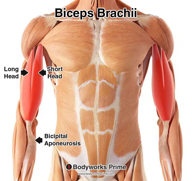
The biceps brachii is a two headed muscle, typically consisting of a short head (caput breve) and a long head (caput longum). The long head is biarticular, crossing both the elbow joint and shoulder joint, whereas the short head only crosses the elbow joint [3] [4]. The deltoid muscle covers the proximal part of the long head, enveloping the bicipital groove where it resides. Despite being part of the same muscle, these two heads have distinct locations and contribute to the muscle’s characteristic curved appearance [5].
Located in opposition to the biceps brachii, in the posterior compartment of the arm, is its antagonist muscle – the triceps brachii. Antagonist muscles work in contrast to each other. In this scenario, while the biceps brachii contributes to flexing the arm, the triceps brachii is involved in extending the arm [6] [7].
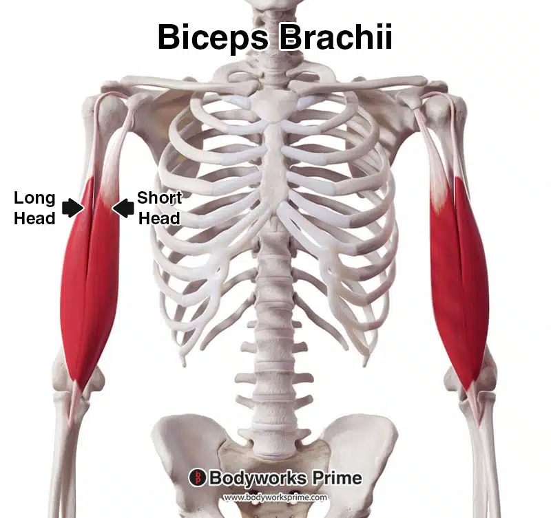
Here we can see the biceps brachii from an anterior view.

Here we can see the biceps brachii from an anterolateral view.

Here we can see the biceps brachii from another anterolateral view.

Here we can see the biceps brachii from a lateral view.
Here we can see the biceps brachii highlighted in red, amongst the other muscles of the body, seen from an anterior view.
Origin & Insertion
The biceps brachii is comprised of two heads – a long head and a short head. The long head originates from a bony prominence on the scapula known as the supraglenoid tubercle, which is a small, rough projection superior to the glenoid cavity. The long head also originates from the superior glenoid labrum, which is a fibrocartilaginous complex attaching like a rim to the articular cartilage of the glenoid fossa. The short head of the biceps brachii originates from the tip of the coracoid process, another prominent structure on the scapula which is a hook-like structure on the lateral edge of the superior and anterior portion of the scapula [8] [9] [10] [11]. These origins allow the two heads to span a considerable distance.
Following their respective origins on the scapula, both heads journey down the arm. The long head crosses obliquely through a ridge in the upper arm known as the bicipital groove. Upon convergence, the two heads form a unified muscle belly around the mid-upper arm region. This single muscle belly then narrows and passes across the elbow’s anterior aspect to insert at two sites: the radial tuberosity, a small prominence on the proximal radius, and the fascia of the forearm. This connection to the fascia is facilitated by a broad, flat tendon known as the bicipital aponeurosis [12] [13] [14] [15].
An interesting point to note is the existence of anatomical variations in the biceps brachii. Studies have reported that the muscle can sometimes display additional heads originating from the humerus – these are termed ‘supernumerary heads’ and can number anywhere from 3 to 7. In addition, the distal tendon of the biceps brachii (the part that inserts into the radius) can sometimes bifurcate, forming two distinct tendons. It has also been observed that the biceps brachii can insert via a single tendon (most common), double tendon, or even triple tendon, reflecting the considerable variability in human anatomy [16] [17].
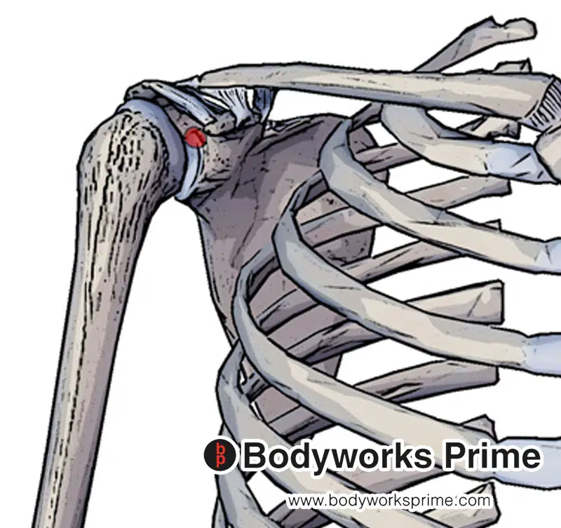
Highlighted in red we can see the origin of the long head of thje biceps brachii at the supraglenoid tubercle.
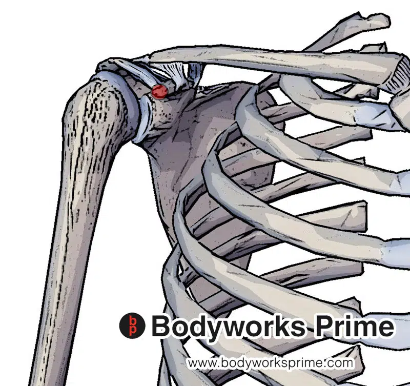
Highlighted in red we can see the origin of the short head of the biceps brachii at the coracoid process of the scapula.
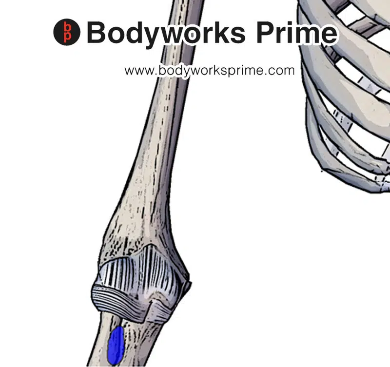
Highlighted in blue we can see the insertion of the biceps brachii to the radial tuberosity.
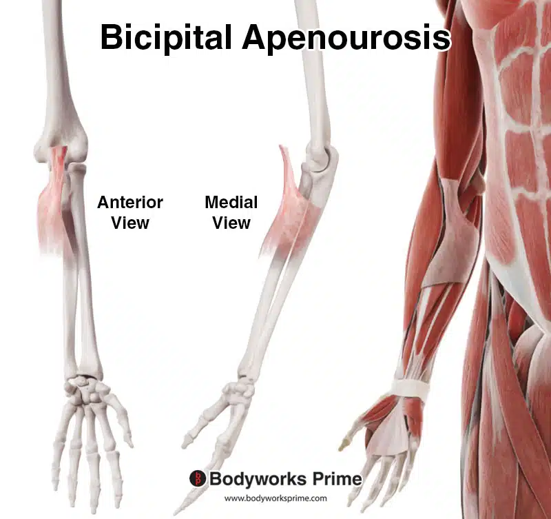
Here we can see the bicipital aponeurosis of the biceps brachii, which connects into the deep fascia of the forearm. The bicipital aponeurosis is considered an insertion of the biceps brachii.
Actions
The biceps brachii serves several important roles in facilitating movement and stability of the arm and shoulder. One of the primary actions of the biceps brachii is to facilitate the flexion of the elbow [18] [19] [20]. Notably, this action is dependent on the angle of the elbow, when the elbow is extended, the muscle acts primarily as an elbow flexor until the elbow reaches 90-degree flexion. Past 90 degrees of flexion, its function is altered based on the orientation of the forearm. If the forearm is pronated, the biceps brachii then becomes the primary muscle involved in forearm supination. If the forearm is supinated, the biceps brachii primarily flexes the elbow [21].
An intriguing aspect of the biceps brachii’s biomechanics is the role played by the long head of the muscle in the dynamic stability of the shoulder joint [22] [23]. Despite ongoing academic debate, evidence from research suggests that this tendon plays a stabilising role at the shoulder joint. Landin, D. et al. (2017) found that beyond 30° of arm elevation, the long head, “even though contracting, does not create a noteworthy elevation moment, which suggests that it cannot serve as a dynamic shoulder stabilizer in higher ranges of elevation” [24]. Some sources often overstate the role of the biceps brachii in arm elevation, claiming the biceps brachii is a strong flexor of the shoulder joint. However, the biceps brachii plays a role as a dynamic stabiliser of the shoulder during elevation of the arm (primarily in the scapular plane) and any shoulder flexion provided is weak comparative to other primary shoulder flexors [25]. Moreover, as previously stated, it seems this stabilisation role is only notable in the first 30 degrees of arm elevation.
The role of the bicipital aponeurosis, a part of the biceps brachii, should also be noted. It not only increases the tension of the main tendon of the biceps brachii but also serves as a protective layer for the median nerve and brachial artery running posteriorly. In addition, it contributes to reinforcing the antebrachial fascia, underscoring its multifaceted function [26] [27] [28].
In summary, the actions of the biceps brachii muscle encompass a range of movements, including flexion of the elbow, supination of the forearm, providing dynamic stability to the shoulder joint, and a weak contribution to shoulder flexion.
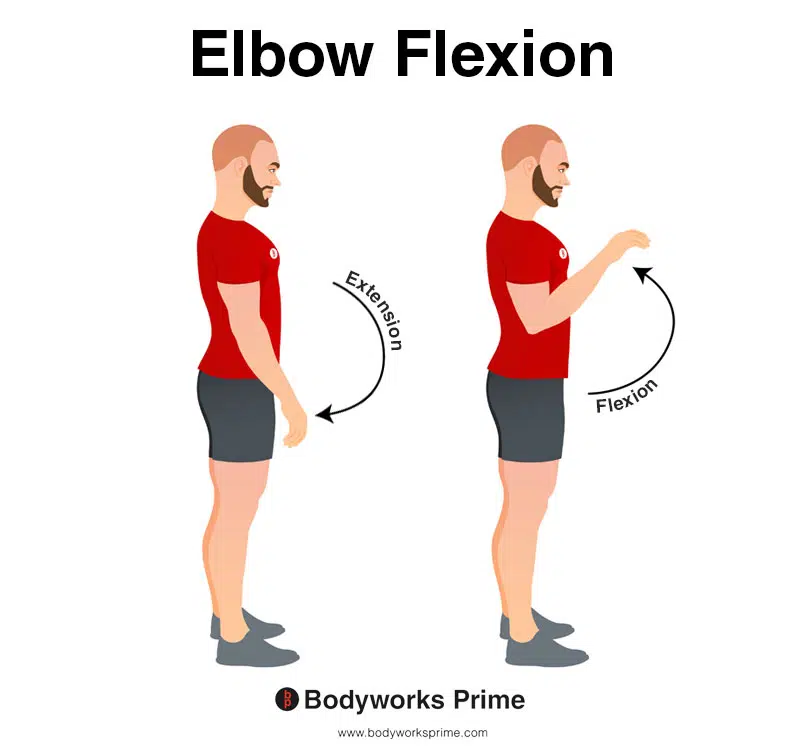
This image demonstrates elbow flexion, which involves bending elbow. The opposite movement of elbow flexion is elbow extension. The biceps brachii flexes the elbow joint. The image depicts flexion with a pronated forearm. However, the biceps brachii more strongly flexes the elbow then the forearm is supinated (palm of hand facing up).
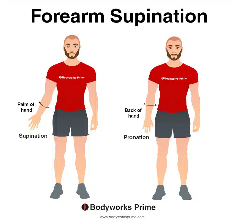
Here we can see the movement of forearm supination, where the forearm rotates laterally so that the palm of the hand is facing up. The oposite of forearm supination is forearm pronation, where the palm faces down. The biceps brachii is able to supinate the forearm.
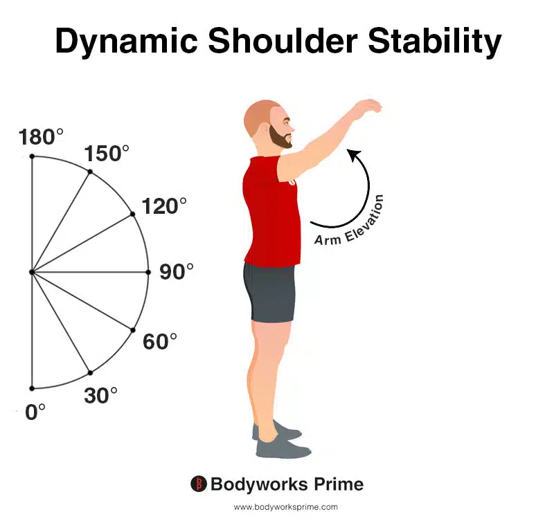
Here we can see a person elevating their arm. The biceps brachii provides shoulder stability during this movement. The stabilisation if provides is primarily in the first 30 degrees of the movement.
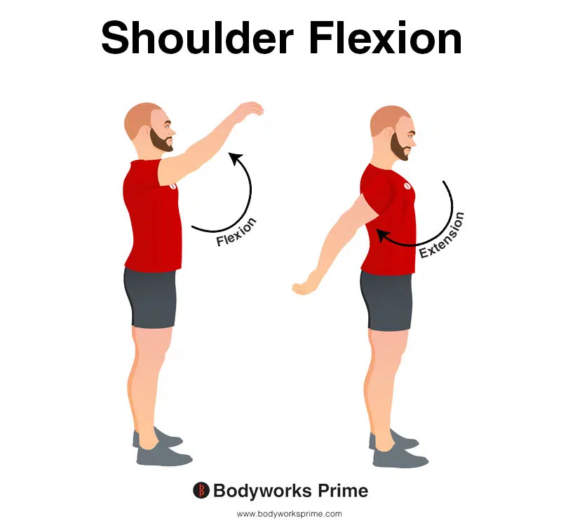
This image shows shoulder flexion. The opposite of this is shoulder extension. The biceps brachii can very weakly assist with flexion of the shoulder.
Innervation
The innervation of the biceps brachii muscle is provided primarily by the musculocutaneous nerve, with roots at the C5 and C6 levels of the spinal cord. The musculocutaneous nerve is a part of the brachial plexus, which is a complex network of nerves which supply innervation to the upper limb [29] [30].
The musculocutaneous nerve branches off from the lateral cord of the brachial plexus, this nerve as a whole, carries fibers from the cervical spinal nerves C5 through C7 (but the biceps brachii is mainly innervated by the part of the nerve associated with C5 and C6). The musculocutaneous nerve typically emerges at the inferior border of the pectoralis major muscle and immediately pierces the coracobrachialis muscle fibers to continue its course through the arm [31].
In the context of the biceps brachii, the musculocutaneous nerve descends between this muscle and the brachialis, which reside in the anterior compartment of the arm, providing innervation as it travels [32]. Variations in the branching pattern to the biceps brachii have been observed, including one single branch serving the entire muscle, or two branches serving either individual heads or the common belly of the muscle [33].
Once it has provided motor fibers to both the biceps brachii and the brachialis, the musculocutaneous nerve continues its course distally, emerging just lateral to the biceps brachii tendon to become the lateral cutaneous nerve of the forearm, also known as the lateral antebrachial cutaneous nerve [34].
Despite being considered one of the more easily identifiable nerves of the brachial plexus, it is essential to note that the musculocutaneous nerve’s course and branching patterns may exhibit variability. For instance, the nerve’s entry point into the coracobrachialis muscle and the distances at which it provides branches to the biceps brachii and brachialis have been found to vary between individuals [35].
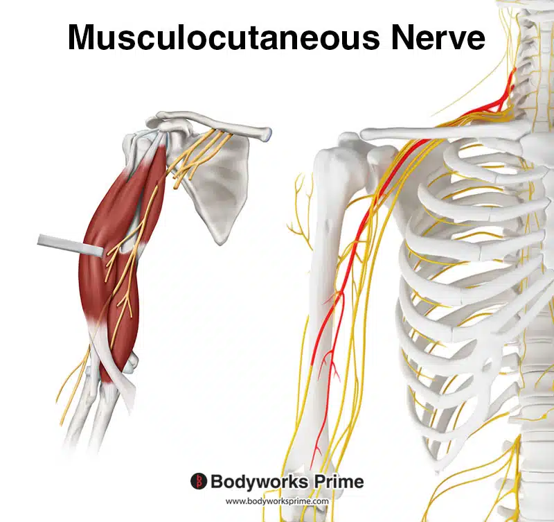
Here we can see the musculocutaneous nerve which innervates the biceps brachii muscle, with roots at the C5 and C6 levels of the spinal cord.
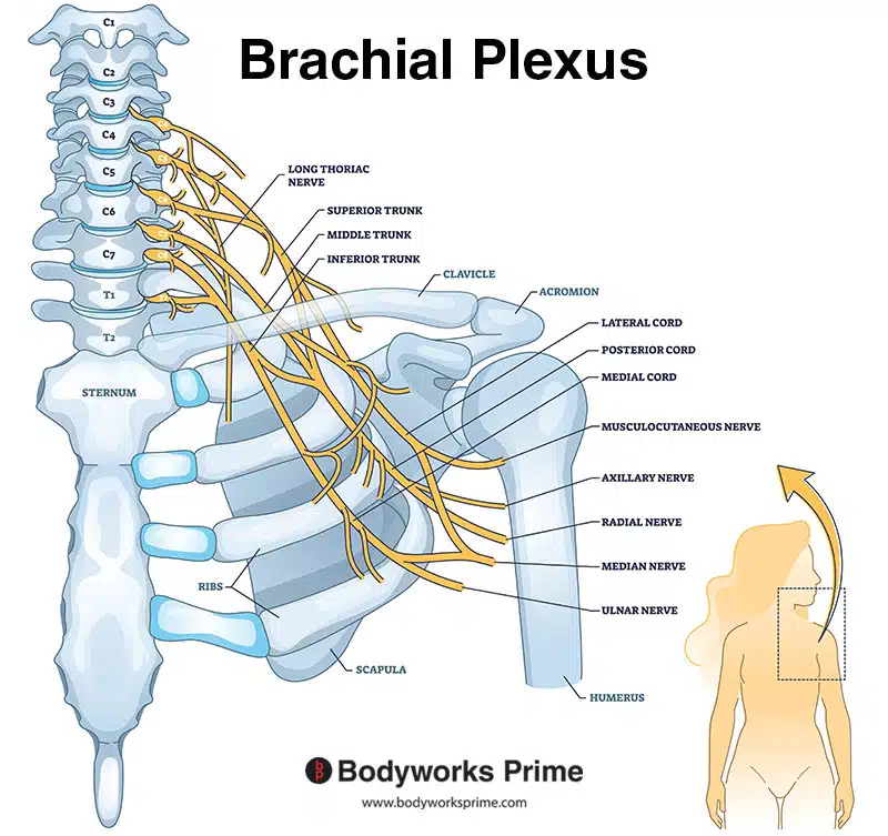
Here we can see an image of the brachial plexus. The musculocutaneous nerve is a part of the brachial plexus.
Blood Supply
The biceps brachii muscle obtains its blood supply mainly via the muscular branches of the brachial artery [36] [37] [38].
The brachial artery, which is a direct extension of the axillary artery, begins at the lower margin of the teres major muscle and it is the primary artery which supplies blood to the upper limb. This artery runs along the ventral surface of the arm, giving rise to several smaller branches before it reaches the cubital fossa [39] [40].
Among these branching arteries are the deep brachial artery, the superior ulnar collateral artery, and the inferior ulnar collateral artery. As the brachial artery arrives at the cubital fossa, it separates into its terminal branches, the radial and ulnar arteries of the forearm [41] [42].
Notably, the brachial artery and its various branches play a significant role in supplying blood to the biceps brachii muscle, among others such as the triceps brachii muscle, and the coracobrachialis muscle. Additionally, the median nerve, a part of the brachial plexus, initially lies lateral to the brachial artery at its proximal segment. However, at its distal segment, the median nerve crosses to the medial side of the brachial artery, lying in the ventral cubital fossa [43] [44].
The inferior ulnar collateral artery is of particular relevance to the biceps brachii muscle. This branch of the brachial artery supplies blood to the biceps brachii muscle, as well as the brachialis muscle and the coracobrachialis muscle [45]. The inferior ulnar collateral artery emerges anterior to the lower segment of the humerus and runs medially along the brachialis muscle before it pierces the medial intermuscular septum. It then forms an anastomosis with the anterior ulnar recurrent artery and the superior ulnar collateral artery [46].
In summary, the primary arterial blood supply to the biceps brachii muscle is a collaborative network involving muscular branches of the brachial artery, and particularly the inferior ulnar collateral artery [47] [48].
Want some flashcards to help you remember this information? Then click the link below:
Biceps Brachii Flashcards
Support Bodyworks Prime
Running a website and YouTube channel can be expensive. Your donation helps support the creation of more content for my website and YouTube channel. All donation proceeds go towards covering expenses only. Every contribution, big or small, makes a difference!
References
| ↑1, ↑5, ↑18, ↑27, ↑37, ↑40 | Moore KL, Agur AMR, Dalley AF. Clinically Oriented Anatomy. 8th ed. Philadelphia: Lippincot Williams & Wilkins; 2017. |
|---|---|
| ↑2, ↑7, ↑19, ↑28, ↑38, ↑42, ↑44 | Standring S. (2015). Gray’s Anatomy: The Anatomical Basis of Clinical Practice, 41st Edn. Amsterdam: Elsevier. |
| ↑3, ↑6, ↑8, ↑12, ↑16, ↑22, ↑29 | Tiwana MS, Charlick M, Varacallo M. Anatomy, Shoulder and Upper Limb, Biceps Muscle. [Updated 2022 Aug 30]. In: StatPearls [Internet]. Treasure Island (FL): StatPearls Publishing; 2023 Jan-. Available from: https://www.ncbi.nlm.nih.gov/books/NBK519538/ |
| ↑4, ↑9, ↑13, ↑17, ↑21, ↑26 | Szewczyk B, Paulsen F, LaPrade RF, Borowski A, Zielinska N, Olewnik Ł. Anatomical variations of the biceps brachii insertion: a proposal for a new classification. Folia Morphol (Warsz). 2023;82(2):359-367. doi: 10.5603/FM.a2022.0022. Epub 2022 Mar 3. PMID: 35239179. |
| ↑10, ↑14, ↑23 | Khan R, Satyapal KS, Naidoo N, Lazarus L. Long head of biceps brachii tendon and transverse humeral ligament morphometry and their associated pathology. Folia Morphol (Warsz). 2020;79(2):359-365. doi: 10.5603/FM.a2019.0075. Epub 2019 Aug 26. PMID: 31448814. |
| ↑11, ↑15, ↑20 | Desai SS, Arbor TC, Varacallo M. Anatomy, Shoulder and Upper Limb, Musculocutaneous Nerve. [Updated 2022 Sep 8]. In: StatPearls [Internet]. Treasure Island (FL): StatPearls Publishing; 2023 Jan-. Available from: https://www.ncbi.nlm.nih.gov/books/NBK534199/ |
| ↑24, ↑25 | Landin D, Thompson M, Jackson MR. Actions of the Biceps Brachii at the Shoulder: A Review. J Clin Med Res. 2017 Aug;9(8):667-670. doi: 10.14740/jocmr2901w. Epub 2017 Jul 1. PMID: 28725314; PMCID: PMC5505302. |
| ↑30, ↑31, ↑32, ↑34, ↑35 | Desai SS, Arbor TC, Varacallo M. Anatomy, Shoulder and Upper Limb, Musculocutaneous Nerve. [Updated 2022 Sep 8]. In: StatPearls [Internet]. Treasure Island (FL): StatPearls Publishing; 2023 Jan-. Available from: https://www.ncbi.nlm.nih.gov/books/NBK534199/. |
| ↑33 | Pacha Vicente D, Forcada Calvet P, Carrera Burgaya A, Llusá Pérez M. Innervation of biceps brachii and brachialis: Anatomical and surgical approach. Clin Anat. 2005 Apr;18(3):186-94. doi: 10.1002/ca.20057. PMID: 15768419. |
| ↑36, ↑47 | Alexander JG, de Fúcio Lizardo JH, da Silva Baptista J. Multiple arterial variations in the upper limb: description and clinical relevance. Anat Sci Int. 2021 Mar;96(2):310-314. |
| ↑39, ↑41, ↑43, ↑45, ↑46, ↑48 | Epperson TN, Varacallo M. Anatomy, Shoulder and Upper Limb, Brachial Artery. [Updated 2022 Jul 25]. In: StatPearls [Internet]. Treasure Island (FL): StatPearls Publishing; 2023 Jan-. Available from: https://www.ncbi.nlm.nih.gov/books/NBK537145/ |


