| Origin | Infraspinous fossa |
| Insertion | Greater tubercle of the humerus (posterior aspect) |
| Action | External rotation of the shoulder joint Stabilisation of the humerus in the glenohumeral joint |
| Nerve | Suprascapular nerve (C5, C6) |
| Artery | Suprascapular and circumflex scapular arteries |
Location & Overview
The infraspinatus muscle is a somewhat thick and triangular shaped muscle of the shoulder and back. It is one of the four rotator cuff muscles of the shoulder and occupies a large part of the infraspinous fossa (which is located on the posterior surface of the scapula). The remaining three rotator cuff muscles are the supraspinatus, teres minor and subscapularis. The rotator cuff is a group of muscles and tendons encapsulating the shoulder joint. The mnemonic ‘SITS’ can be used to more easily memorise the names of these rotator cuff muscles. These muscles facilitate shoulder joint stability and a wide range of movements, making the shoulder one of the most mobile joints in the body [1] [2] [3].
The infraspinatus muscle is located on the posterior surface of the scapula. It’s located inferior to the supraspinatus and superior to the teres minor (situated between these two muscles). The fibres of the infraspinatus muscle run almost parallel to the teres major and minor. Moreover, the infraspinatus muscle is sometimes fused together with the teres minor muscle. The fibres of these muscles are directed towards the shoulder. The supraspinatus and infraspinatus are also closely related anatomically to one another, due to only be separated by the spine of the scapula. The infraspinatus is a deep muscle residing beneath both the trapezius medially and deltoid laterally [4] [5] [6].
Conditions such as tendinitis and muscle strains are common pathologies associated with the infraspinatus muscle. These typically result from overuse, trauma, or lack of sufficient warm-up before engaging in high-intensity physical activities [7] [8] [9].
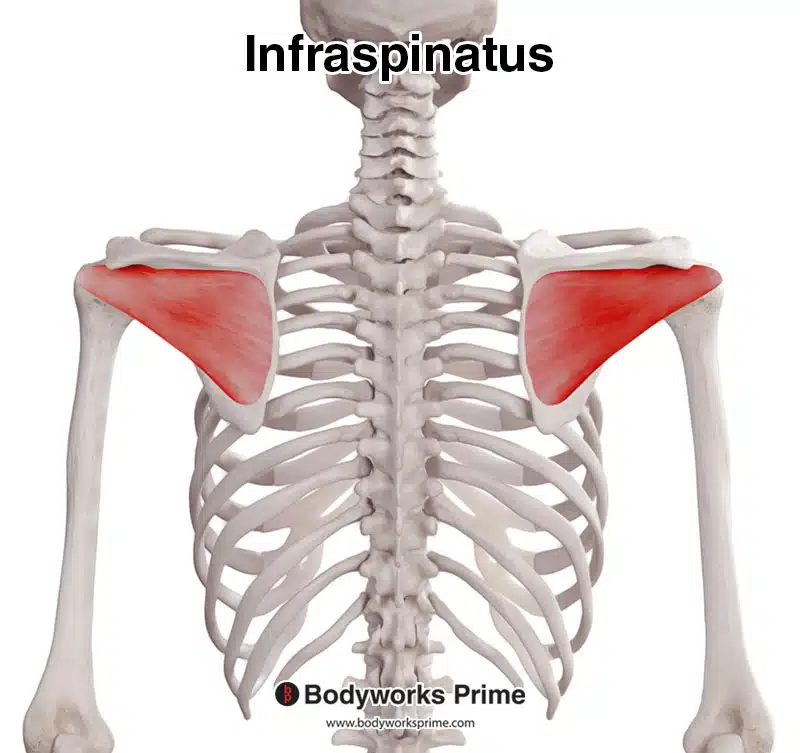
Here we can see the infraspinatus muscle, seen from a posterior view.
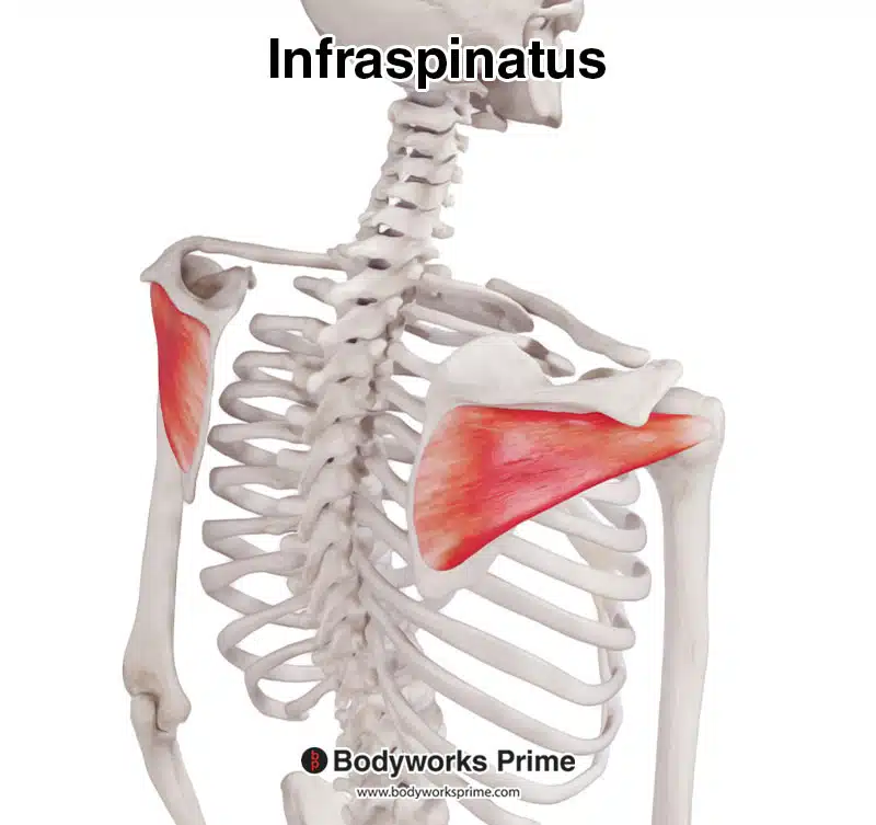
Here we can see the infraspinatus muscle, seen from a posterolateral view.
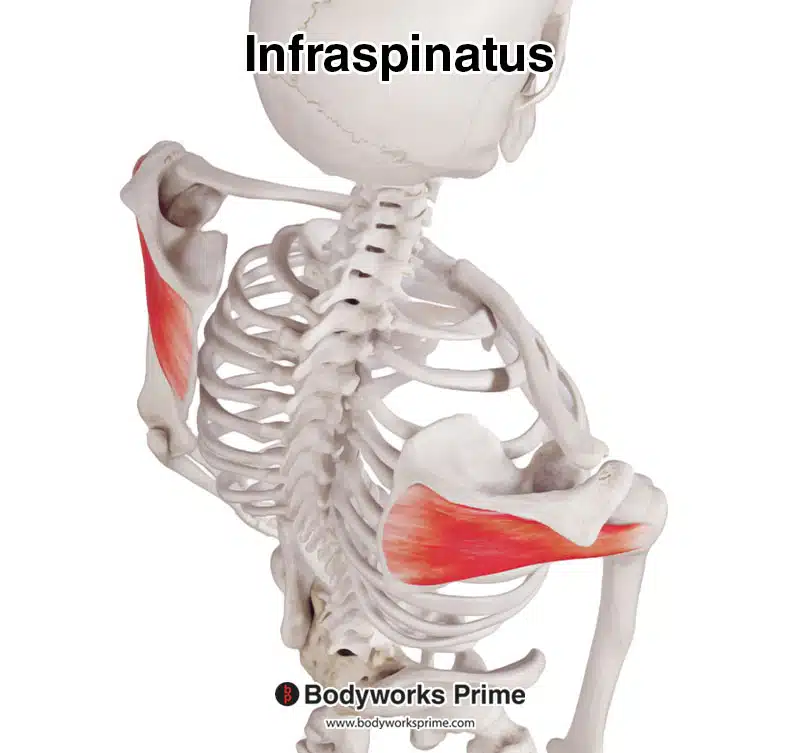
Here we can see the infraspinatus muscle, seen from a superior view.
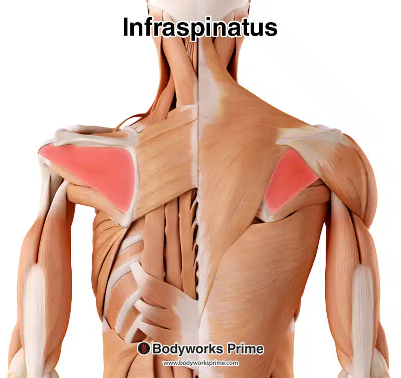
Here we can see the infraspinatus muscle highlighted in red amongst the other muscles of the body. On the left we can see the infraspinatus from a deep view and on the right a superficial view.
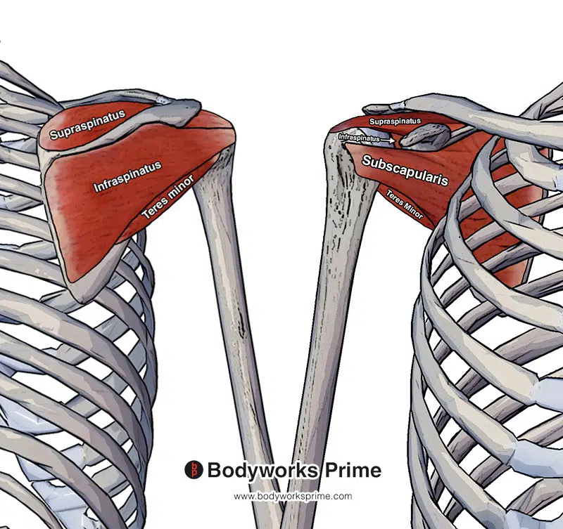
Pictured here we can see all four of the rotator cuff muscles: the supraspinatus, infraspinatus, teres minor and subscapularis. The left side of this picture is from a posterior view (the back) and the right hand side is from an anterior view (the front).
Origin & Insertion
The infraspinatus originates from the infraspinous fossa, located on the posterior scapula and inferior to the spine of the scapula. The infraspinous fossa is divided from the supraspinous fossa by the scapular spine. The spine of the scapula is a bony ridge that runs horizontally across the scapula, towards its superior aspect [10] [11] [12] [13].
The infraspinatus extends laterally from its origin, towards the proximal end and posterior side of the humerus. It then inserts into the middle facet of the greater tuberosity of the humerus. The greater tuberosity of the humerus is a bony prominence on the upper part of the humerus. This location also holds the insertions of the supraspinatus and teres minor muscles, superiorly and inferiorly respectively [14] [15] [16] [17].
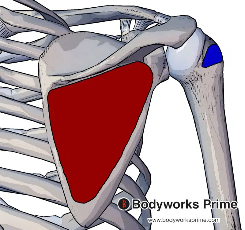
The infraspinatus muscle originates at the infraspinous fossa of the scapula (marked in red) and inserts at the middle facet of the greater tubercle of the humerus (marked in blue).
Actions
As one of the rotator cuff muscles, the infraspinatus muscle has a key role in shoulder joint stability. It exerts a compressive force on the humeral head, stabilising it during movements such as shoulder abduction. The opposing forces generated by the rotator cuff muscles balance the shoulder movements, with the infraspinatus providing the posterior force against the anterior force from the subscapularis muscle. This stabilisation of the glenohumeral joint prevents damage to the surrounding structures. Someone who has an inability to maintain the humeral head in the glenoid fossa may be referred to as having ‘shoulder instability’ [18] [19] [20].
In terms of specific movements, the infraspinatus primarily enables external/lateral rotation of the humerus, due to its insertion at the middle facet of the glenoid tubercle, working in synergy with the teres minor [21] [22] [23].
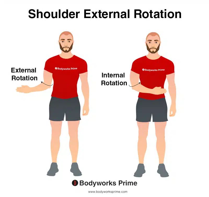
This image demonstrates the movement of external rotation of the shoulder. External rotation involves rotating your arm away from your body, towards the outside. In contrast, internal rotation involves rotating your arm towards the centre of your body, towards the inside. The infraspinatus contributes to external rotation.
Innervation
The infraspinatus muscle is innervated by the suprascapular nerve. This nerve originates from the superior trunk of the brachial plexus, with nerve roots from C5 and C6. From its origin, the suprascapular nerve runs laterally across the lateral cervical region. In addition to the infraspinatus, the suprascapular nerve also innervates the supraspinatus, another rotator cuff muscle. This nerve allows for the transmission of nerve signals between the infraspinatus and the central nervous system [24] [25].
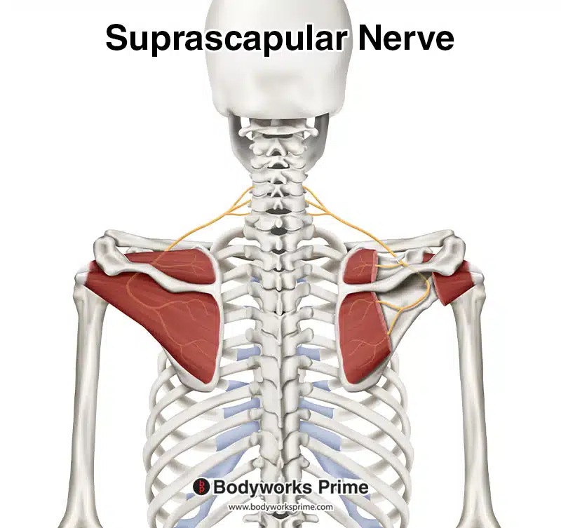
Here we can see the suprascapular nerve which innervates the infraspinatus muscle. This nerve originates from the nerve roots of C5 and C6.
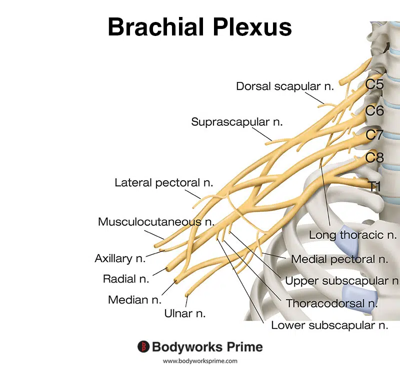
Here we can see the nerves of the brachial plexus. The suprascapular nerve is a part of the brachial plexus.
Blood Supply
The infraspinatus muscle’s blood supply primarily comes from the suprascapular and circumflex scapular arteries. The suprascapular artery, originating from either the thyrocervical trunk or the subclavian artery, courses laterally to the scapular spine to reach the infraspinous fossa on the posterior surface of the scapula. The circumflex scapular artery, a branch of the subscapular artery, courses around the lateral border of the scapula to enter the infraspinous fossa and forms an anastomosis with the suprascapular artery, creating a source of collateral circulation [26].
Want some flashcards to help you remember this information? Then click the link below:
infraspinatus flashcards
Support Bodyworks Prime
Running a website and YouTube channel can be expensive. Your donation helps support the creation of more content for my website and YouTube channel. All donation proceeds go towards covering expenses only. Every contribution, big or small, makes a difference!
References
| ↑1, ↑7, ↑11, ↑15, ↑18, ↑21, ↑24 | Moore KL, Agur AMR, Dalley AF. Clinically Oriented Anatomy. 8th ed. Philadelphia: Lippincot Williams & Wilkins; 2017. |
|---|---|
| ↑2, ↑4, ↑19, ↑22, ↑25 | Standring S. (2015). Gray’s Anatomy: The Anatomical Basis of Clinical Practice, 41st Edn. Amsterdam: Elsevier. |
| ↑3, ↑5, ↑8, ↑12, ↑16, ↑20, ↑23 | Maruvada S, Madrazo-Ibarra A, Varacallo M. Anatomy, Rotator Cuff. [Updated 2023 Mar 27]. In: StatPearls [Internet]. Treasure Island (FL): StatPearls Publishing; 2023 Jan-. Available from: https://www.ncbi.nlm.nih.gov/books/NBK441844/ |
| ↑6, ↑9, ↑13, ↑17, ↑26 | Williams JM, Sinkler MA, Obremskey W. Anatomy, Shoulder and Upper Limb, Infraspinatus Muscle. [Updated 2022 Aug 8]. In: StatPearls [Internet]. Treasure Island (FL): StatPearls Publishing; 2023 Jan-. Available from: https://www.ncbi.nlm.nih.gov/books/NBK513255/ |
| ↑10, ↑14 | Kadi R, Milants A, Shahabpour M. Shoulder Anatomy and Normal Variants. J Belg Soc Radiol. 2017 Dec 16;101(Suppl 2):3. doi: 10.5334/jbr-btr.1467. PMID: 30498801; PMCID: PMC6251069. |


