| Origin | Spinous processes of thoracic T7–T12 Inferior angle of scapula Inferior 3 or 4 ribs Thoracolumbar fascia Iliac crest |
| Insertion | Floor of the intertubercular groove of the humerus |
| Action | Humeral adduction Humeral extension Humeral medial rotation |
| Nerve | Thoracodorsal nerve (C6, C7, C8) |
| Artery | Thoracodorsal artery |
Location & Overview
The latissimus dorsi is a large, flat muscle that occupies a substantial area of the torso’s posterior, spanning from the lower back to the humerus. Its expansive coverage stretches from left to right across the back, with the upper portion partially hidden by the trapezius muscle in the medial dorsal region. The latissimus dorsi plays a crucial role in various upper body movements, such as adduction, extension, and internal rotation of the shoulder joint [1] [2].
As one of the superficial extrinsic muscles of the upper back, the latissimus dorsi is part of a group divided into intrinsic and extrinsic categories based on their movement actions and origins. Extrinsic muscles primarily govern the movement of the humerus and scapula during upper extremity motions. Other superficial extrinsic muscles include the rhomboid major, rhomboid minor, levator scapulae, and the trapezius [3]. Among these, the latissimus dorsi is the largest extrinsic back muscle [4].
The latissimus dorsi’s extensive attachment points provide the muscle with the leverage and force necessary for its various functions. The latissimus dorsi plays an essential role in various physical activities and sports that involve the upper body, such as swimming, rowing, and climbing. It is also an important muscle for the stabilization and control of the shoulder joint during overhead movements and lifting. Because of its size and function, the latissimus dorsi is an important muscle to target during strength training and rehabilitation programs. Some exercises that engage the latissimus dorsi are: pull-ups, lat pulldowns, and bent-over rows, pendlay rows, among others [5] [6].
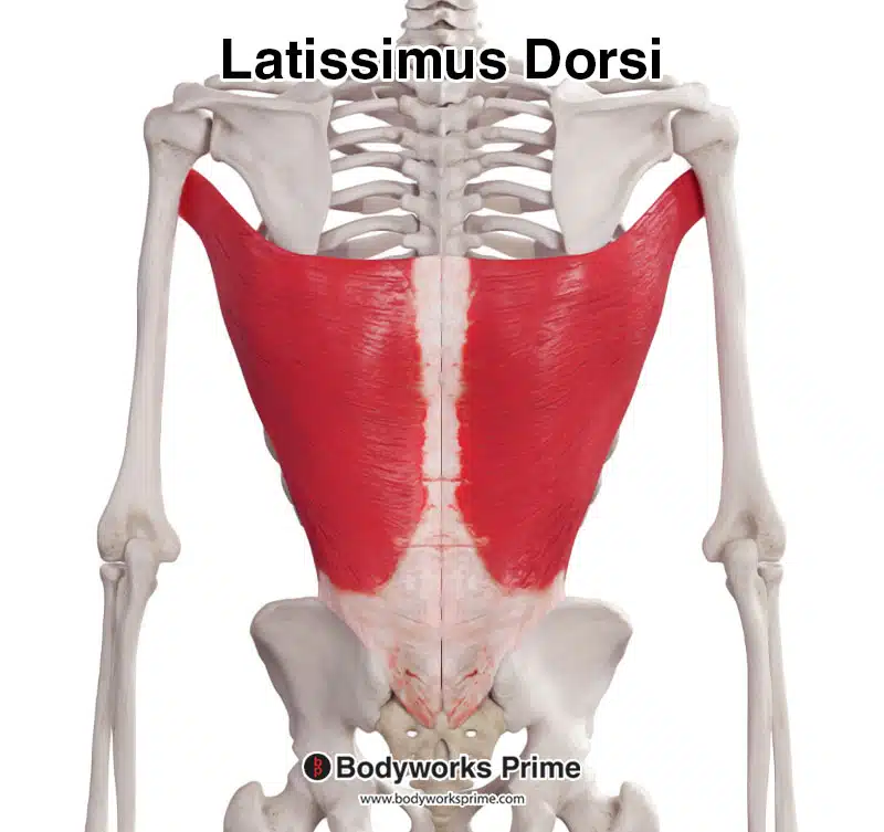
In this image we can see the latissimus dorsi muscle from a posterior view.
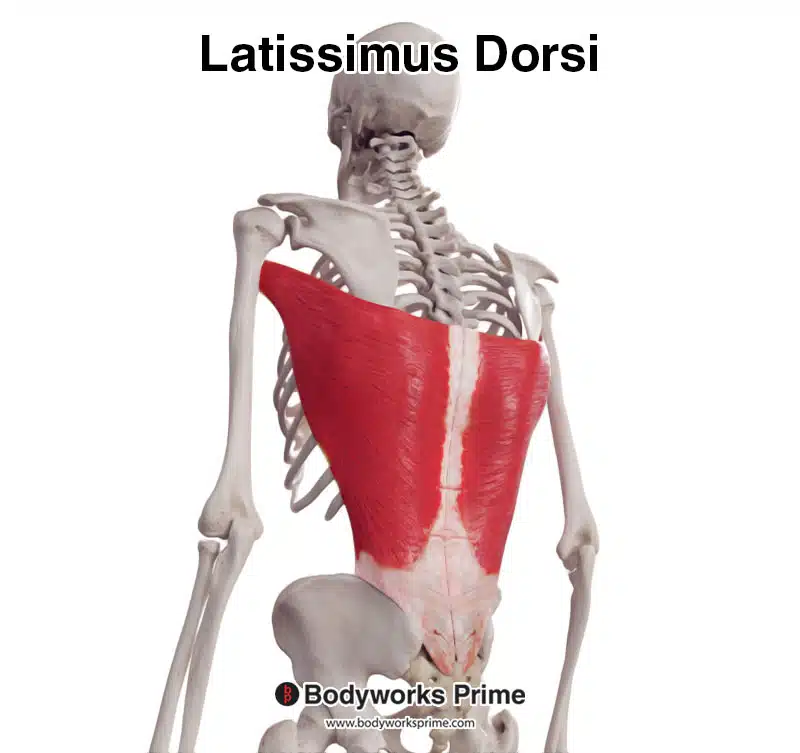
In this image we can see the latissimus dorsi muscle from a posterolateral view.
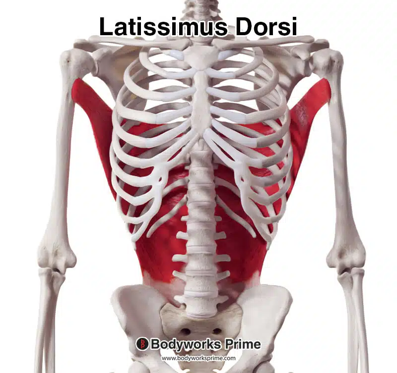
Here we can see the latissimus dorsi muscle from an anterior view.
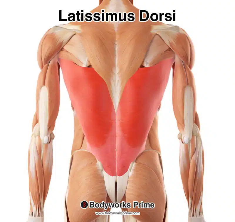
Here we can see the latissimus dorsi muscle highlighted in red amongst the other muscles of the body.
Origin & Insertion
The latissimus dorsi’s origin points include the spinous processes of the thoracic vertebrae T7 to T12, the inferior angle of the scapula, the thoracolumbar fascia (a thick, fibrous sheet connecting the muscle to the lower back), the lower three or four ribs, and the iliac crest of the hip bone [7] [8]
These multiple origin points allow the latissimus dorsi to contribute to a range of upper limb and shoulder movements. The muscle fibers converge as they extend towards the humerus, forming a flat tendon that inserts on the floor of the intertubercular groove, also known as the bicipital groove. This groove is a narrow depression that runs along the anterior surface of the humerus and houses the long head of the biceps tendon. The insertion of the latissimus dorsi on the humerus allows for the muscle to exert force on the upper limb during various actions, providing stability and strength in movements that involve the shoulder joint [9] [10].
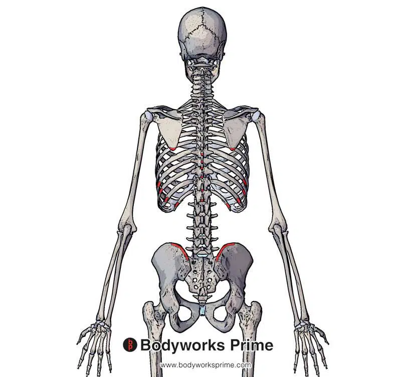
Here we can see the origins of the latissimus dorsi muscle marked in red. The latissimus dorsi origins marked here are: the inferior angle of scapula, the spinous processes of T7–T12, inferior 3 or 4 ribs and the iliac crest.
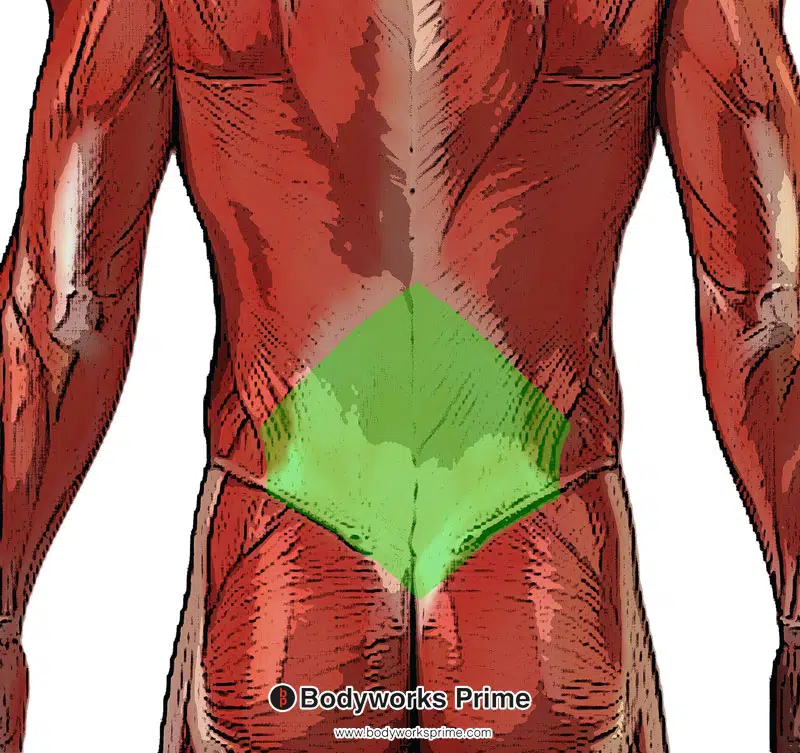
The final origin point of the latissimus dorsi muscle is the thoracolumbar fascia which is marked here in green.
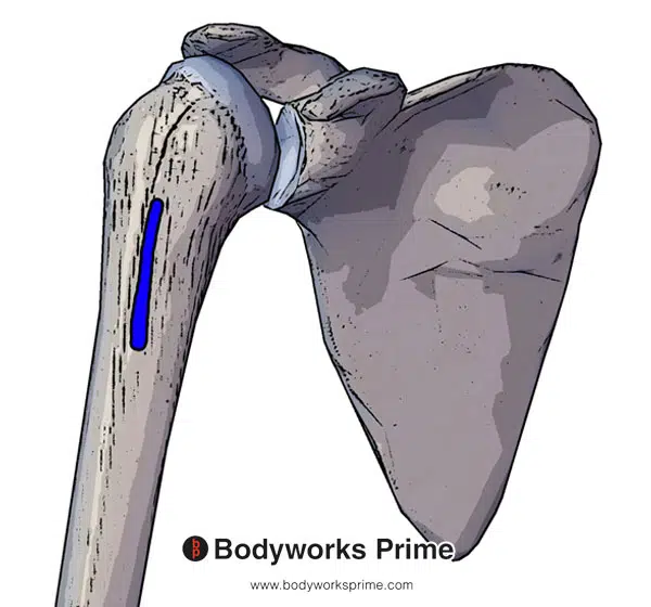
Highlighted here in blue we can see the insertion of the latissimus dorsi on the intertubercular groove of the humerus.
Actions
The latissimus dorsi plays a crucial role in multiple actions involving the humerus and shoulder joint. When this muscle contracts, it generates movement in three primary directions. Firstly, it adducts the humerus, pulling the arm towards the midline of the body. Secondly, it extends the humerus, moving the arm backward in a posterior direction. Lastly, it causes medial rotation of the humerus, turning the arm inward toward the center of the body [11] [12] [13].
In addition to these primary movements, the latissimus dorsi is also involved in lifting the body upwards and forwards when the arms are positioned above the head. This action is evident during exercises such as pull-ups, where the muscle engages to lift up the body by flexing the shoulder joint and pulling the torso towards the hands [14] [15] [16].
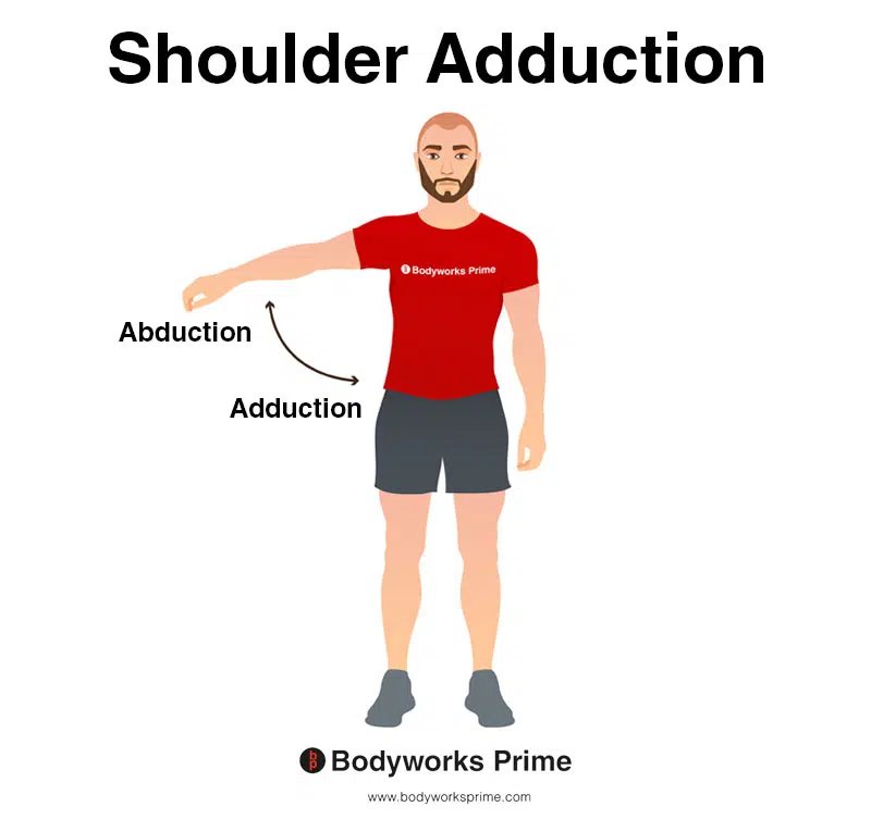
This image shows shoulder adduction. Adduction is where the arm is brought towards the midline of the body, reducing the angle between the arm and the torso. The opposite of adduction is abduction. Abduction occurs as the arm is moved away from the midline, increasing the angle between the arm and the torso. Shoulder adduction is an action of the latissimus dorsi muscle.
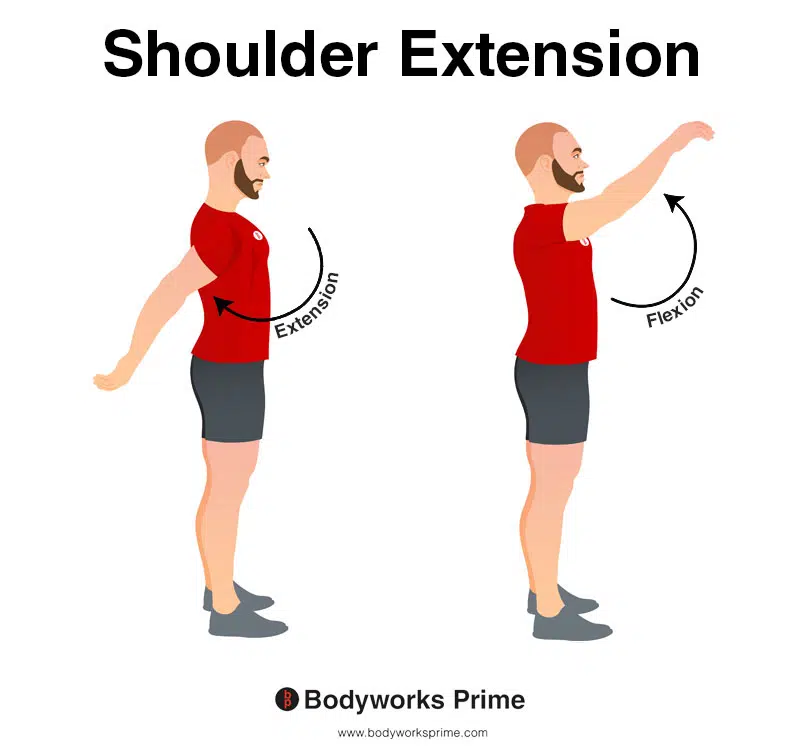
This image shows shoulder extension. Extension is where the arm is moved backward in a posterior direction (towards the back of the body), increasing the angle between the arm and the torso in the sagittal plane. The opposite of this is shoulder flexion which occurs as the arm is moved forward and upward in an anterior direction. Shoulder extension is an action of the latissimus dorsi muscle.

This image shows shoulder internal rotation. Internal rotation is when the arm is rotated inward toward the centre of the body, causing the hand and forearm to move towards the body’s midline. The opposite of this is external rotation, which is when arm is rotated outward away from the centre of the body, moving the hand and forearm away from the body’s midline. Internal rotation is an action of the latissimus dorsi muscle.
Innervation
The latissimus dorsi is innervated by the thoracodorsal nerve (C6, C7, C8), which is sometimes also referred to as the middle subscapular nerve. The thoracodorsal nerve branches from the brachial plexus’ posterior cord [17].
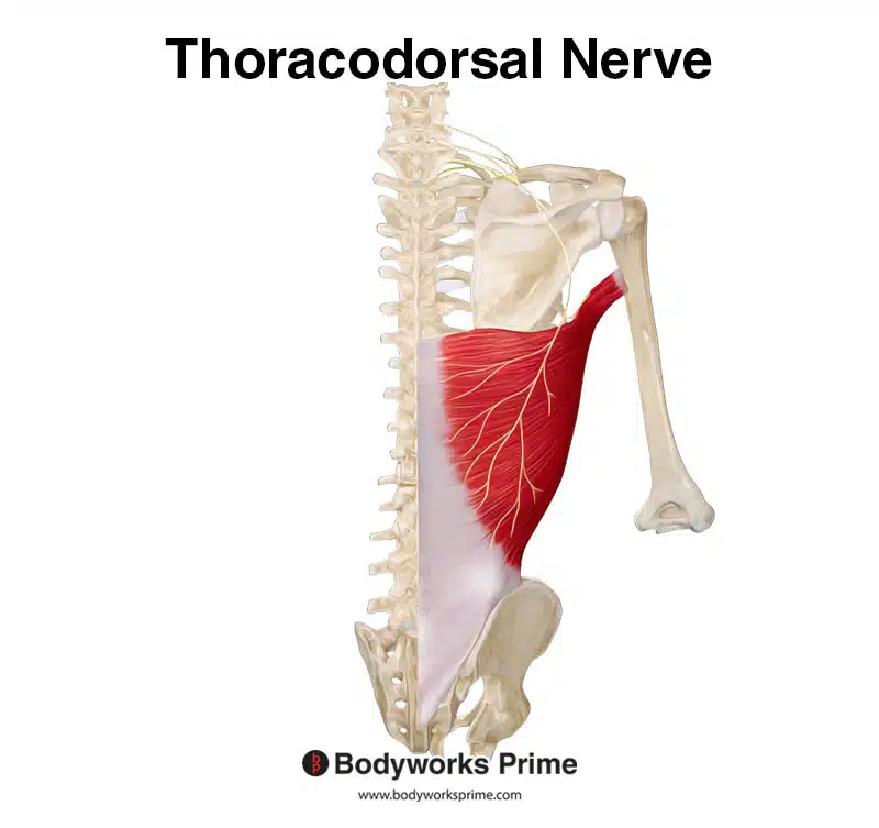
Here we can see the thoracodorsal nerve which innervates the latissimus dorsi, with nerve roots from C6, C7, and C8.
Blood Supply
Blood is supplied to the latissimus dorsi via the thoracodorsal artery. This artery is a continuation of the subscapular artery. The subscapular artery branches off the axillary artery [18].
Want some flashcards to help you remember this information? Then click the link below:
Latissimus Dorsi Flashcards
Support Bodyworks Prime
Running a website and YouTube channel can be expensive. Your donation helps support the creation of more content for my website and YouTube channel. All donation proceeds go towards covering expenses only. Every contribution, big or small, makes a difference!
References
| ↑1, ↑7, ↑9 | Bhatt CR, Prajapati B, Patil DS, Patel VD, Singh BG, Mehta CD. Variation in the insertion of the latissimus dorsi & its clinical importance. J Orthop. 2013;10(1):25-28. Published 2013 Mar 7. doi:10.1016/j.jor.2013.01.002. |
|---|---|
| ↑2, ↑6, ↑8, ↑10, ↑13, ↑16 | Moore KL, Agur AMR, Dalley AF. Clinically Oriented Anatomy. 8th ed. Philadelphia: Lippincot Williams & Wilkins; 2017. |
| ↑3 | Verhaegen F,Debeer P,Moyaert M, The Accessory muscles of the Axilla. Acta orthopaedica Belgica. 2019 Dec; PMID: 32374231 |
| ↑4 | Mitchell B, Imonugo O, Tripp JE. Anatomy, Back, Extrinsic Muscles. [Updated 2021 Aug 4]. In: StatPearls [Internet]. Treasure Island (FL): StatPearls Publishing; 2021 Jan-. Available from: https://www.ncbi.nlm.nih.gov/books/NBK537216/ |
| ↑5, ↑12, ↑15 | Andersen V, Fimland MS, Wiik E, Skoglund A, Saeterbakken AH. Effects of grip width on muscle strength and activation in the lat pull-down. J Strength Cond Res. 2014 Apr;28(4):1135-42. doi: 10.1097/JSC.0000000000000232. PMID: 24662157. |
| ↑11, ↑14 | Donohue BF, Lubitz MG, Kremchek TE. Sports Injuries to the Latissimus Dorsi and Teres Major. Am J Sports Med. 2017 Aug;45(10):2428-2435. doi: 10.1177/0363546516676062. Epub 2016 Dec 20. PMID: 28125914. |
| ↑17 | Anthony DJ, Basnayake BMOD, Ganga NMG, Mathangasinghe Y, Malalasekera AP. An improved technical trick for identification of the thoracodorsal nerve during axillary clearance surgery: a cadaveric dissection study. Patient Saf Surg. 2018 Jun 26;12:18. doi: 10.1186/s13037-018-0164-2. PMID: 29983745; PMCID: PMC6020335. |
| ↑18 | Kumar N,Aithal AP,Nayak SB,Bhaskar R, A rare case of atypical thoracodorsal artery: a challenge for flap reconstruction. Surgical and radiologic anatomy : SRA. 2018 Aug PMID: 29594336. |


