| Origin | Anterior head: Lateral quarter of clavicle & anterior third of lateral acromion Intermediate head: Middle third of lateral aspect of acromion Posterior head: Posterior third of acromion & spine of scapula |
| Insertion | Deltoid tuberosity of humerus |
| Action | Shoulder abduction Shoulder flexion Shoulder extension Horizontal adduction Horizontal abduction Medial rotation Lateral rotation |
| Nerve | Axillary nerve (C5, C6) |
| Artery | Thoracoacromial branch of the axillary artery (primary artery) Posterior circumflex humeral artery Deltoid branches of profunda brachii artery |
Location & Overview
The deltoid muscle, named for its distinctive triangular shape, is a large muscle located in the upper extremity. It covers the shoulder joint, providing it with its rounded contour shape and playing an important role in various movements of the arm [1] [2].
This muscle, positioned in the lateral aspect of the shoulder, can be conceptually divided into three distinct portions based on their fiber orientation – the anterior (clavicular), lateral (acromial), and posterior (spinal). Each of these divisions has specific roles in the movement and stabilization of the shoulder joint [3]. However, a more intricate view of the deltoid’s structure reveals that it consists of seven segments, each separated by their intramuscular tendons. This anatomical configuration suggests a more complex mechanism of action than just the contribution of the broadly defined three segments, hence painting a more precise picture of the deltoid’s role in shoulder dynamics [4]. This will be explored in further detail in the origin and insertions section.
The deltoid muscle is situated superficially, making it the most prominent muscle of the shoulder and upper arm. It essentially envelops and covers the shoulder joint, offering a protective layer to the deeper structures of the shoulder. Some neighbouring muscles include: the trapezius posteriorly, pectoralis major anteriorly, and the latissimus dorsi inferiorly [5] [6].
A unique characteristic of the deltoid muscle is its extensive overlap with the rotator cuff muscles. These muscles work in a coordinated manner to balance the forces acting on the shoulder joint and also provide stability during arm movements. The rotator cuff muscles are the supraspinatus, infraspinatus, teres minor, and the subscapularis. This muscles can be remembered using the mnemonic ‘SITS’ [7] [8].

Pictured here we can see the deltoid muscle from an anterior view.
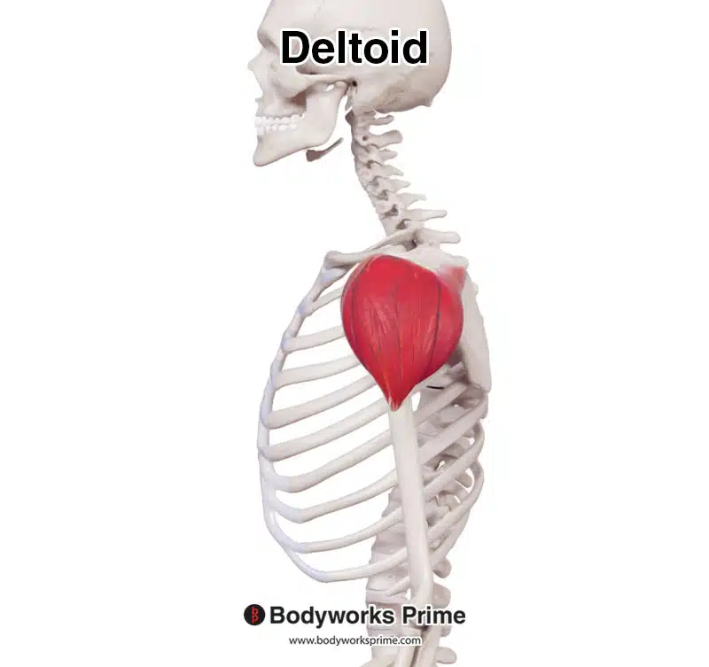
Pictured here we can see the deltoid muscle from a lateral view.
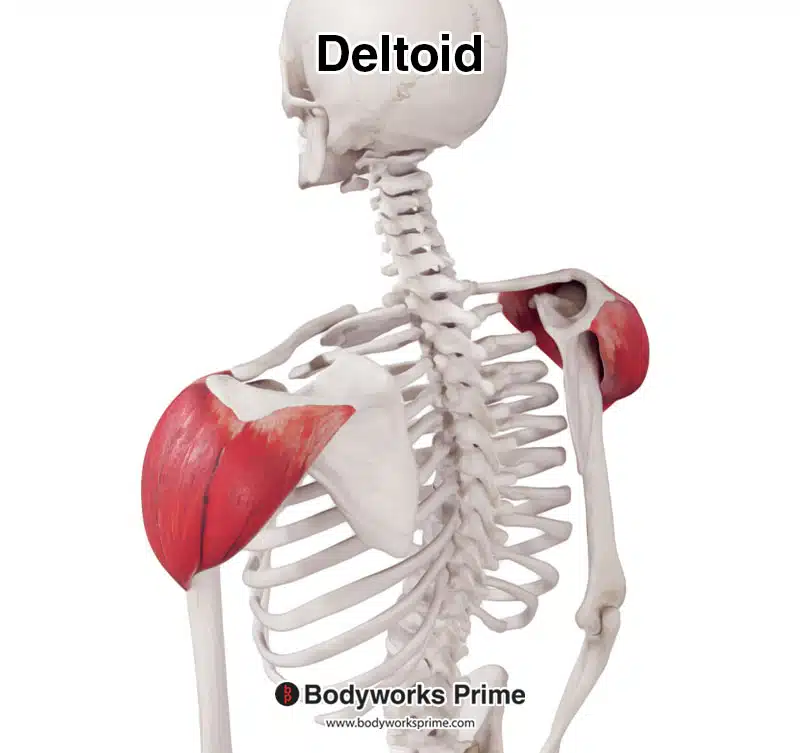
Pictured here we can see the deltoid muscle from a posterior view.
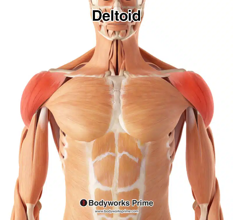
Here we can see the deltoid muscle highlighted in red amongst the other muscles of the body, seen from an anterior view.
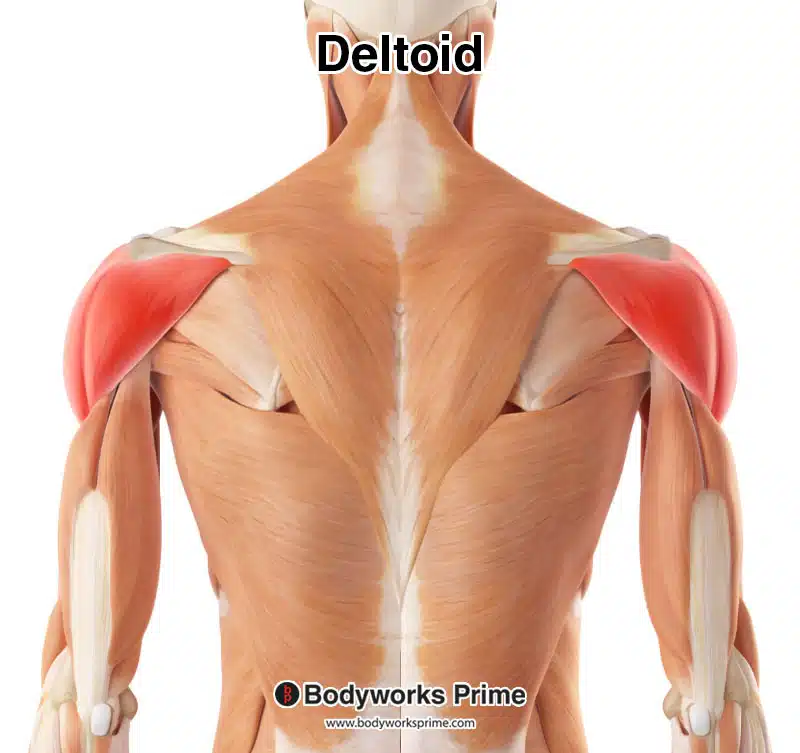
Here we can see the deltoid muscle highlighted in red amongst the other muscles of the body, seen from a posterior view.
Origin & Insertion
The deltoid muscle is known for its origin’s distinctive ‘U’ shape, spanning across the clavicle, acromion, and the spine of the scapula. These points specifically include the lateral third of the clavicle, the lateral acromion, and the spine of the scapula [9] [10].
The clavicle, also known as the collarbone, is a horizontal bone at the base of the neck. The acromion, is a bony process on the scapula and serves as a connection between the clavicle and the shoulder blade, creating the acromioclavicular joint. The spine of the scapula is a noticeable bony ridge that runs across the back of the shoulder blade, providing an attachment point for various muscles, including the deltoid [11] [12].
The deltoid muscle is typically further divided into three segments: the anterior, middle, and posterior segments. The anterior segment originates from the clavicle and the anterior third of the lateral acromion. The middle deltoid originates from the middle third of the lateral aspect of the acromion. The posterior deltoid originates from the posterior third of the acromion and the spine of the scapula. The deltoid muscle extends from these origin points across the shoulder joint to insert onto the deltoid tuberosity of the humerus, which is a rough, raised area on the lateral aspect of the humerus [13].
As previously noted, the deltoid muscle is technically composed of seven segments rather than the traditionally identified three. This segmentation is determined by the presence of three intramuscular tendons, each of which inserts into the deltoid tuberosity on the humerus. From this point of insertion, these tendons extend upwards towards the defined origin points [14].
In the region of the proximal deltoid, the tendons associated with the anterior and posterior segments of the deltoid divide, producing three additional intramuscular tendons. These are then somewhat evenly distributed across the origin points corresponding to their traditional segments. To clarify: the anterior deltoid presents three intramuscular tendons at the proximal end of the muscle, the middle deltoid typically displays one, and the posterior deltoid offers three. This culminates in a total of seven intramuscular tendons. However, at the distal end of the muscle, the six intramuscular tendons of the anterior and posterior deltoid segments consolidate into two intramuscular tendons and join the single middle deltoids tendon to insert on the deltoid tuberosity [15].
It is also worth mentioning that anatomical variations can occur, where some individuals may present an additional intramuscular tendon associated with the middle deltoid. However, such instances are relatively uncommon. Sakoma et al. (2011) found this occurrence to be 6.7% in their study of 30 cadavers (15 males and 15 females) [16].
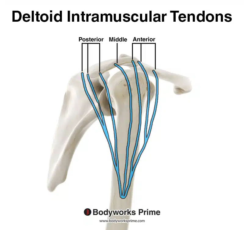
Pictured here we can see the 7 intramuscular tendons of the deltoid muscle. The anatomy of these tendons is my interpretation of the images from the study conducted by Sakoma et al. (2011) [17]. Full reference details are provided in the references section at the bottom of the page, click the numerical citation to go straight to it.
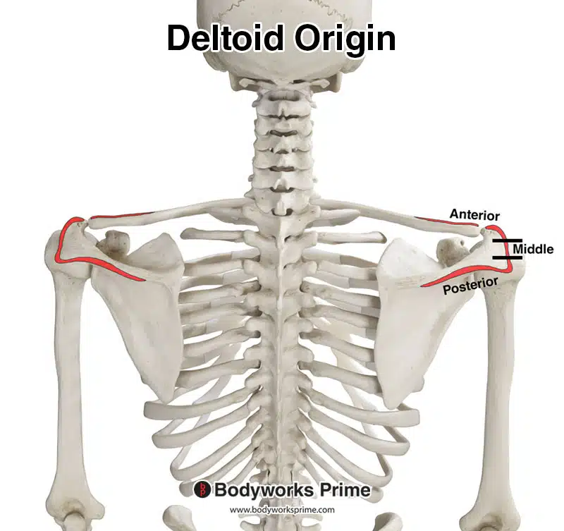
Here we can see the origin points of the deltoid muscle highlighted in red. The two black lines divide the origins of the three heads of the deltoid to demonstrate their respective locations.
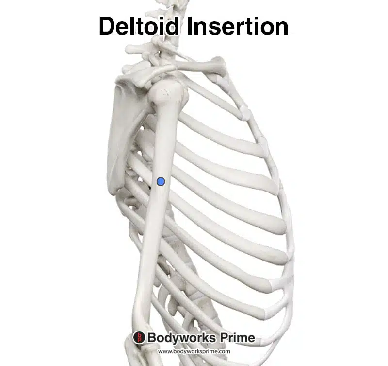
Here we can see the insertion of the deltoid muscle highlighted in blue, on the deltoid tuberosity of the humerus.
Actions
The deltoid’s muscle actions can be divided into its three sections: anterior, middle, and posterior. The deltoid plays a crucial role in facilitating a wide range of shoulder movements. The deltoid can perform shoulder abduction, taking over from the rotator cuff muscles after the initial 15-degree movement to raise the arm from 15 to 100 degrees [18]. Shoulder abduction is the primary caused by the middle and anterior deltoid sections. Though the posterior deltoid does contribute to this also, but to a lesser extent [19]. The anterior deltoid is slightly more active than the middle deltoid during abduction within the scapular plane, though both contribute a very similar amount to the movement [20].
Additionally, the anterior deltoid contributes to shoulder flexion and horizontal adduction. Furthermore, it also assists with medial rotation (internal rotation). Conversely, the posterior deltoid contributes to shoulder extension and horizontal abduction. Additionally, it assists with lateral rotation (external rotation) [21] [22] [23].
The deltoid’s comprehensive role at the shoulder joint extends to its function as a stabiliser. Moreover, all of the deltoid’s sections work together to prevent inferior displacement of the glenohumeral joint when they contract together [24]. An example of this would be during a deadlift, when the humerus is pulled towards the bar by the heavy load. The deltoid assists in keeping the humerus in place in the glenoid cavity, by pulling the humerus superiorly.
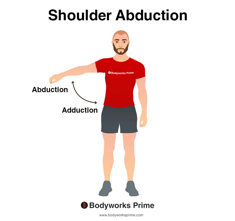
This image demonstrates shoulder abduction. Shoulder abduction involves lifting your arm out to the side of your body. The opposite of abduction is adduction, which involves bringing your arm back towards your body. The deltoid muscle can abduct the shoulder.
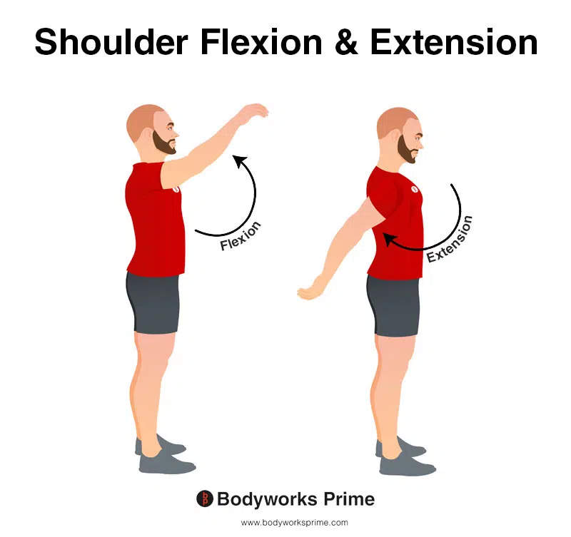
This image shows both shoulder flexion and extension. Flexion is where the arm is moved forward and upward in an anterior direction. The opposite of this is shoulder extension, which occurs as the arm is moved backwards in a posterior direction. The anterior potion of the deltoid contributes to shoulder flexion whereas the posterior portion contributes to shoulder extension.
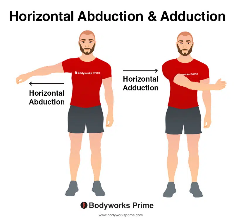
This image demonstrates the action of horizontal abduction and adduction. Horizontal abduction involves moving your arm away from the midline of your body in the transverse plane, such as extending your arms out to the sides from a starting position in front of your body. Conversely, horizontal adduction involves moving your arm towards the midline of your body in the same transverse plane, like bringing your arms back in towards each other. The anterior section of the deltoid is involved in horizontal adduction, whereas the posterior section is involved in horizontal abduction.
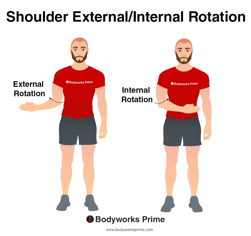
This image demonstrates the movements of shoulder internal and external rotation. Internal rotation involves rotating your arm towards the centre of your body. In contrast, external rotation involves rotating your arm away from your body, towards the outside. The deltoid muscle is involved in both internal and exteranl rotation of the shoulder. With the anterior section usually being responsible for internal rotation and the posterior section external rotation.
Innervation
The deltoid muscle’s primary innervation stems from the axillary nerve, an essential peripheral nerve of the upper limb. The axillary nerve is primarily composed of nerve fibers originating from the C5 and C6 nerve roots of the cervical spine. These segments form part of the upper region of the spinal cord, colloquially known as the neck region. The neurons that emerge from these roots ultimately give rise to the axillary nerve, which branches off from the posterior cord of the brachial plexus, a complex network of nerves located in the shoulder area [25] [26] [27].
The axillary nerve subsequently travels laterally, passing through the quadrangular space alongside the posterior circumflex humeral artery. Upon reaching the deltoid muscle, the axillary nerve divides into anterior and posterior branches, penetrating the muscle from its deep surface. This division allows the nerve fibers to spread throughout the muscle, contributing to its wide-ranging control over shoulder movement [28] [29] [30].
The anterior branch of the axillary nerve primarily provides innervation to the anterior and middle segments of the deltoid muscle. In contrast, the posterior branch mainly serves the posterior segment. Moreover, the nerve’s extensive branching within the muscle ensures an efficient distribution of nerve fibers, facilitating the deltoid’s various actions [31] [32] [33].
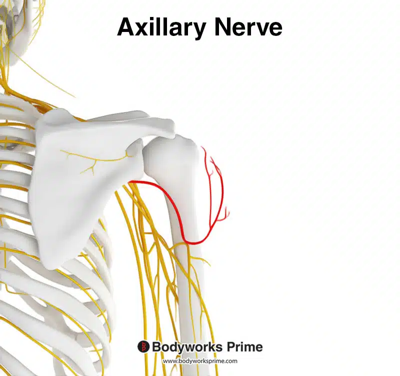
Pictured here you can see the axillary nerve highlighted in red. The deltoid muscle is innervated by the axillary nerve.
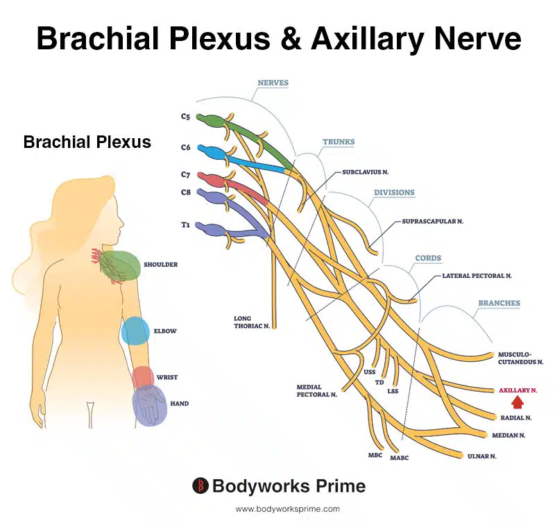
Pictured here you can see the axillary nerve alongside the brachial plexus. The deltoid muscle is innervated by the axillary nerve.
Blood Supply
Blood to the deltoid muscle is primarily provided by the thoracoacromial artery, a branch that stems from the axillary artery. This vessel, notably the second part of the axillary artery, resides behind the pectoralis minor muscle before branching off to supply multiple regions, including the deltoid muscle [34] [35].
In its course, the thoracoacromial artery produces multiple offshoots specifically known as the deltoid branches. These branches navigate in close proximity to the cephalic vein, located within the deltopectoral groove, a noticeable depression between the deltoid and pectoralis major muscles. This vascular network ensures the deltoid muscle receives a steady and reliable blood supply [36] [37].
While the thoracoacromial artery is the chief blood supplier to the deltoid muscle, it is not the only contributor. Additional blood supply comes from the posterior circumflex humeral artery and the profunda brachii artery. However, these vessels play a much lesser role than the thoracoacromial artery. The posterior circumflex humeral artery, although not the predominant provider, dispatches terminal branches that bridge the gap between the deltoid muscle and the proximal humerus, enriching the muscle with the necessary nutrients and oxygen for its functions [38] [39].
Want some flashcards to help you remember this information? Then click the link below:
Deltoid Flashcards
Support Bodyworks Prime
Running a website and YouTube channel can be expensive. Your donation helps support the creation of more content for my website and YouTube channel. All donation proceeds go towards covering expenses only. Every contribution, big or small, makes a difference!
References
| ↑1, ↑10, ↑12, ↑37, ↑39 | Standring S. (2015). Gray’s Anatomy: The Anatomical Basis of Clinical Practice, 41st Edn. Amsterdam: Elsevier. |
|---|---|
| ↑2, ↑6, ↑8 | Peterson SL, Rayan GM. Shoulder and upper arm muscle architecture. J Hand Surg Am. 2011 May;36(5):881-9. doi: 10.1016/j.jhsa.2011.01.008. PMID: 21527142. |
| ↑3, ↑24, ↑26, ↑29, ↑32, ↑35 | Elzanie A, Varacallo M. Anatomy, Shoulder and Upper Limb, Deltoid Muscle. [Updated 2022 May 15]. In: StatPearls [Internet]. Treasure Island (FL): StatPearls Publishing; 2023 Jan-. Available from: https://www.ncbi.nlm.nih.gov/books/NBK537056/ |
| ↑4, ↑13, ↑14, ↑15, ↑16, ↑17 | Sakoma Y, Sano H, Shinozaki N, Itoigawa Y, Yamamoto N, Ozaki T, Itoi E. Anatomical and functional segments of the deltoid muscle. J Anat. 2011 Feb;218(2):185-90. doi: 10.1111/j.1469-7580.2010.01325.x. Epub 2010 Nov 30. PMID: 21118198; PMCID: PMC3042752. |
| ↑5, ↑7, ↑9, ↑11, ↑27, ↑30, ↑33 | Moore KL, Agur AMR, Dalley AF. Clinically Oriented Anatomy. 8th ed. Philadelphia: Lippincot Williams & Wilkins; 2017. |
| ↑18 | Elzanie A, Varacallo M. Anatomy, Shoulder and Upper Limb, Deltoid Muscle. [Updated 2022 May 15]. In: StatPearls [Internet]. Treasure Island (FL): StatPearls Publishing; 2023 Jan-. Available from: https://www.ncbi.nlm.nih.gov/books/NBK537056/ |
| ↑19, ↑20 | Ackland DC, Pak P, Richardson M, Pandy MG. Moment arms of the muscles crossing the anatomical shoulder. J Anat. 2008 Oct;213(4):383-90. doi: 10.1111/j.1469-7580.2008.00965.x. Epub 2008 Aug 6. PMID: 18691376; PMCID: PMC2644775. |
| ↑21 | Miniato MA, Anand P, Varacallo M. Anatomy, Shoulder and Upper Limb, Shoulder. [Updated 2022 Jul 25]. In: StatPearls [Internet]. Treasure Island (FL): StatPearls Publishing; 2023 Jan-. Available from: https://www.ncbi.nlm.nih.gov/books/NBK536933/ |
| ↑22 | Yasojima T, Kizuka T, Noguchi H, Shiraki H, Mukai N, Miyanaga Y. Differences in EMG activity in scapular plane abduction under variable arm positions and loading conditions. Med Sci Sports Exerc. 2008 Apr;40(4):716-21. doi: 10.1249/MSS.0b013e31816073fb. PMID: 18317372. |
| ↑23 | Kuechle DK, Newman SR, Itoi E, Morrey BF, An KN. Shoulder muscle moment arms during horizontal flexion and elevation. J Shoulder Elbow Surg. 1997 Sep-Oct;6(5):429-39. doi: 10.1016/s1058-2746(97)70049-1. PMID: 9356931. |
| ↑25, ↑28, ↑31 | Gurushantappa PK, Kuppasad S. Anatomy of axillary nerve and its clinical importance: a cadaveric study. J Clin Diagn Res. 2015 Mar;9(3):AC13-7. doi: 10.7860/JCDR/2015/12349.5680. Epub 2015 Mar 1. PMID: 25954611; PMCID: PMC4413059. |
| ↑34, ↑36, ↑38 | Smith CD, Booker SJ, Uppal HS, Kitson J, Bunker TD. Anatomy of the terminal branch of the posterior circumflex humeral artery: relevance to the deltopectoral approach to the shoulder. Bone Joint J. 2016 Oct;98-B(10):1395-1398. |


