| Supraspinatus | Origin: Supraspinous fossa of scapula Insertion: Greater tubercle of the humerus (superior aspect) Action: Abduction of humerus, contributes to external rotation (weakly), & stabilisation of the humerus in glenohumeral joint Innervation: Suprascapular nerve (C5, C6) Blood supply: Suprascapular artery |
| Infraspinatus | Origin: Infraspinous fossa Insertion: Greater tubercle of the humerus (posterior aspect) Action: External rotation of the humerus & stabilisation of the humerus in glenohumeral joint Innervation: Suprascapular nerve (C5, C6) Blood supply: Suprascapular and circumflex scapular arteries |
| Teres minor | Origin: Posterior surface of the lateral border of the scapula Insertion: Greater tubercle of the humerus (inferior aspect) Action: External rotation of the humerus, stabilisation of the humerus in glenohumeral joint & can very weakly assist in shoulder adduction Innervation: Axillary nerve (C5, C6) Blood supply: Subscapular artery, circumflex scapular artery & posterior circumflex humeral artery |
| Subscapularis | Origin: Subscapular fossa Insertion: Lesser tubercle of humerus Action: Internal rotation of the humerus, stabilisation of glenohumeral joint & can very weakly assist in shoulder adduction Innervation: Upper and lower branches of the subscapular neve (C5, C6, C7) Blood supply: Subscapular artery |
Location & Overview
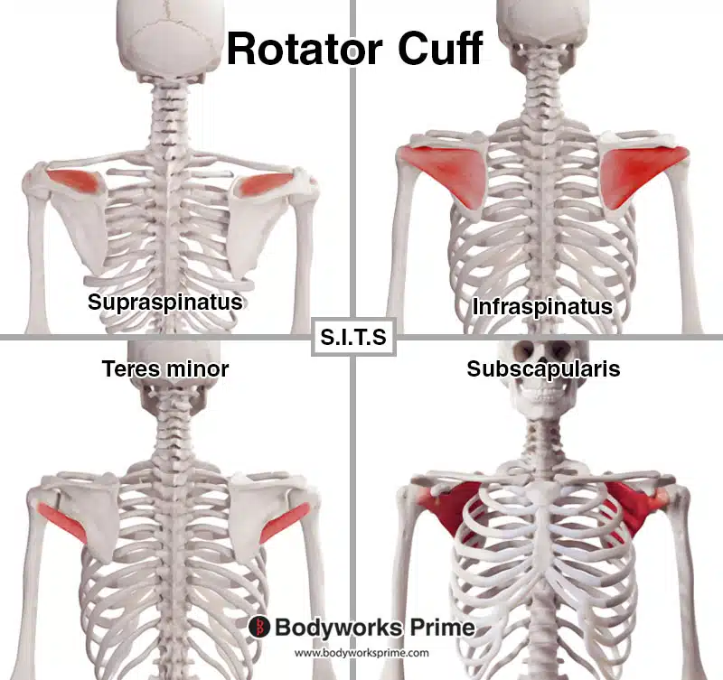
The rotator cuff refers to a collection of muscles in the shoulder which primarily provide stability to the glenohumeral joint. The head of the humerus is a large spherical head which fits inside a cavity called in the glenoid cavity. The head of the humerus and the glenoid cavity form the glenohumeral joint. The rotator cuff is comprised of the following four muscles: the supraspinatus, infraspinatus, teres minor, and the subscapularis. A useful mnemonic for memorising these muscles is ‘SITS’ [1] [2].
All four of the rotator cuff muscles connect the scapula to the humerus. The glenoid cavity is a part of the scapula and the head of the humerus is located at the top of the humerus. The rotator cuff muscle’s tendons blend with the joint capsule. These tendons form a musculotendinous collar which surrounds the superior, posterior, and anterior portions of the joint. The inferior part of the joint does not have a musculotendinous collar and due to this, the inferior part is unprotected. This unfortunately is a cause of most shoulder dislocations, as the humerus can pass inferiorly through this unprotected part [3] [4].
Some fibers of the teres minor muscle interlock and connect with those of the infraspinatus. The supraspinatus and subscapularis tendons also interlock and connect as well and they also envelop part of the biceps tendon [5].
Here we can see each of the rotator cuff muscles separately.
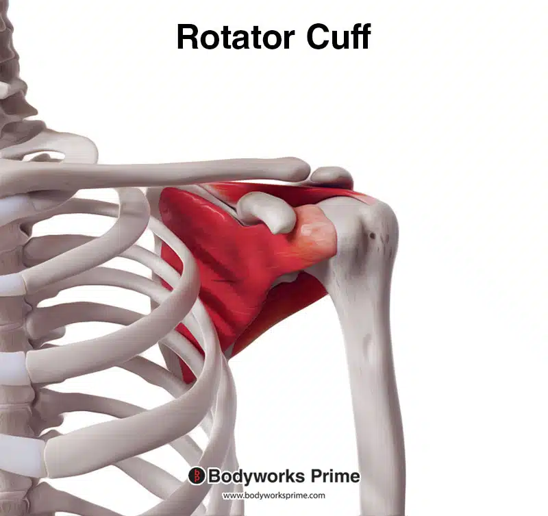
Here we can see the rotator cuff muscles from an anterior view.
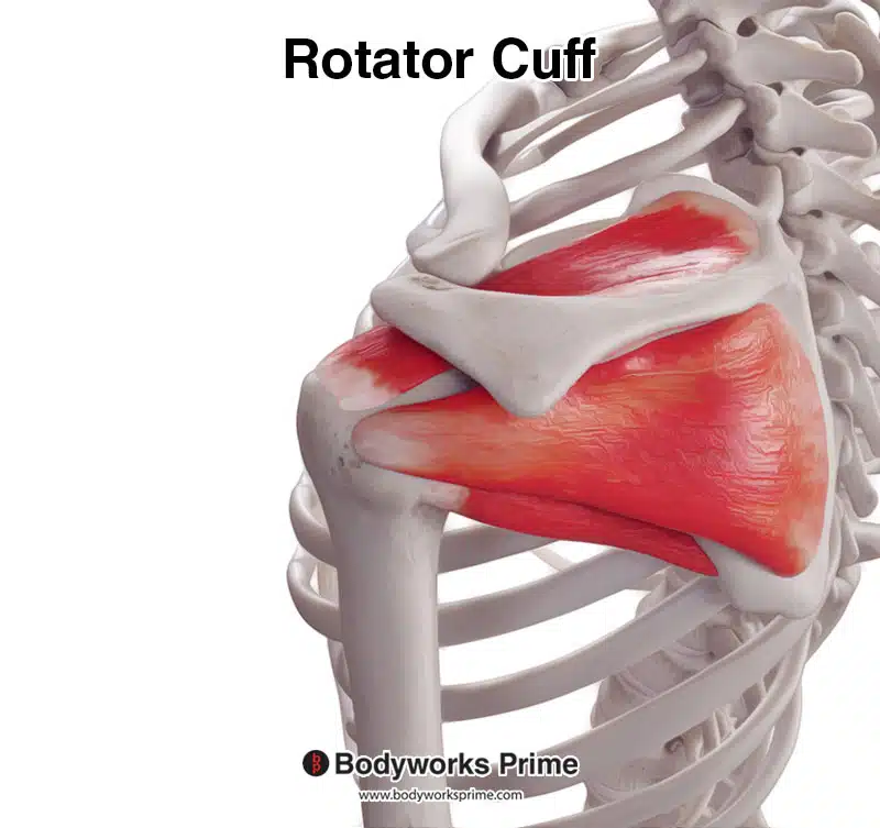
Here we can see the rotator cuff muscles from a posterior view.
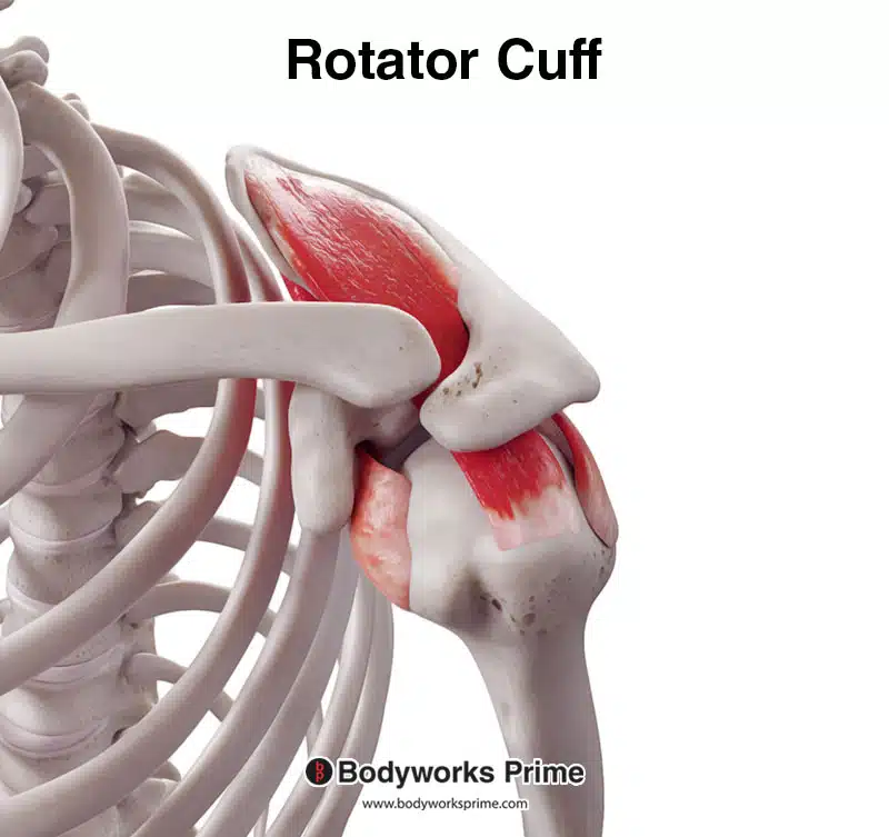
Here we can see the rotator cuff muscles from a superior view.
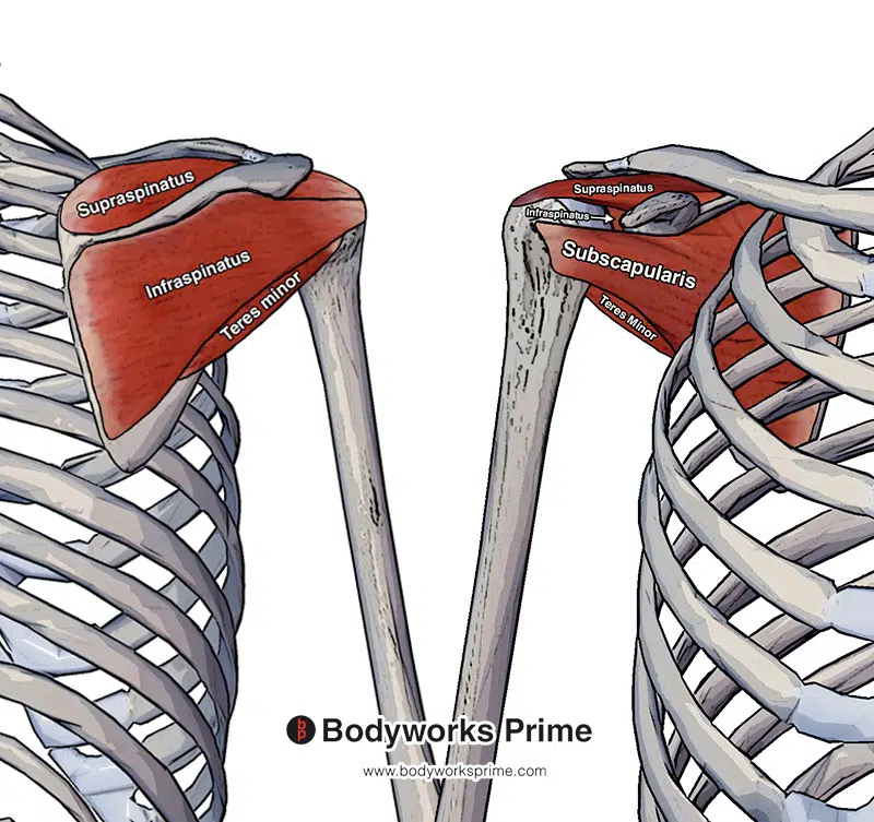
Pictured here we can see all four of the rotator cuff muscles: the supraspinatus, infraspinatus, teres minor and subscapularis. The left side of this picture is from a posterior view (the back) and the right hand side is from an anterior view (the front).
Origin & Insertion
The supraspinatus originates at the supraspinous fossa of the scapula. It then passes under the acromion and above the glenohumeral joint to insert on the greater tuberosity of the humerus (superior aspect) [6] [7] [8].
The infraspinatus originates at the infraspinous fossa of the scapula. It then inserts on the greater tuberosity of the humerus (posterior aspect) just inferior to the insertion of the supraspinatus [9] [10] [11].
The teres minor originates on the lateral border of the scapula and then inserts on the greater tuberosity of the humerus (inferior aspect) just inferior to the insertion of the infraspinatus [12] [13] [14].
The subscapularis is the only rotator cuff muscle originating on the anterior side of the scapula. It originates at the subscapular fossa and then inserts on the lesser tubercle of the humerus [15] [16] [17].
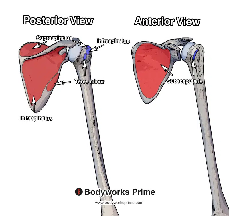
Pictured here we can see the origin and insertion points of the rotator cuff muscles. The origins are highlighted in red and the insertions are highlighted in blue. The supraspinatus originates at the supraspinous fossa of the scapula and inserts on the greater tuberosity of the humerus (superior aspect). The infraspinatus originates at the infraspinous fossa and inserts on the greater tuberosity of the humerus (posterior aspect) just inferior to the insertion of the supraspinatus. The teres minor originates on the lateral border of the scapula and then inserts on the greater tuberosity of the humerus (inferior aspect) just inferior to the insertion of the infraspinatus. The subscapularis originates at the subscapular fossa and then inserts on the lesser tubercle of the humerus.
Actions
The primary role of all the rotator cuff muscles is to stabilise the head of the humerus in the glenoid cavity. This stabilisation is achieved by compressing the head of the humerus against the glenoid [18] [19].
The supraspinatus is responsible for shoulder abduction. The supraspinatus is the primary mover in this abduction during the first 0 to 15 degrees of shoulder abduction. Past this first 0 to 15 degrees, the deltoid muscle is then mainly responsible for shoulder abduction. It also contributes weakly to the lateral rotation of the humerus in certain positions [20] [21] [22].
The infraspinatus is responsible for lateral rotation of the shoulder (also known as external rotation) [23] [24] [25].
The teres minor is also responsible for lateral rotation of the shoulder (also known as external rotation) [26] [27] [28].
The subscapularis muscle is responsible for medial rotation of the shoulder (also known as internal rotation) [29] [30] [31].
In addition to the primary actions of the rotator cuff muscles mentioned above, the teres minor and subscapularis have also been demonstrated to be able to provide very weak assistance during shoulder adduction [32] [33].
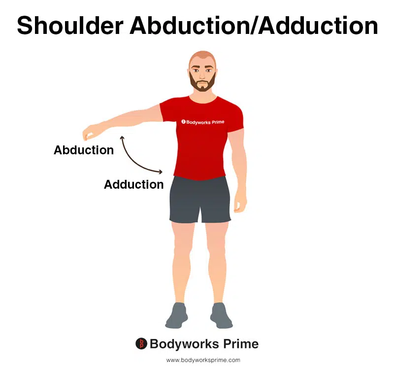
This image demonstrates shoulder abduction and adduction. Shoulder abduction involves lifting your arm out to the side of your body. On the other hand, shoulder adduction involves bringing your arm back towards your body. Shoulder abduction is a primary action of the supraspinatus. Conversely, the teres minor and subscapularis are able to weakly assist other muscles during shoulder adduction.
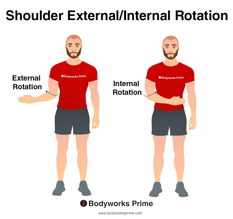
This image demonstrates the movements of shoulder internal and external rotation (also known as medial and lateral rotation respectively). Internal rotation involves rotating your arm towards the centre of your body. In contrast, external rotation involves rotating your arm away from your body, towards the outside. The infraspinatus‘ and teres minor‘s primary actions are external rotation of the shoulder. The supraspinatus also weakly contibutes to external rotation in certain positions. The subscapularis‘ primary action is internal rotation of the shoulder.
Innervation
The innervation of the infraspinatus and supraspinatus is the suprascapular nerve (C5, C6, C7). The suprascapular nerve originates from the superior trunk of the brachial plexus and passes through the suprascapular foramen [34] [35] [36].
The innervation of the teres minor is the axillary nerve (C5, C6). The axillary nerve originates from the posterior cord of the brachial plexus and passes through the quadrangular space into the posterior scapula region [37] [38].
The upper and lower branches of the subscapular neve (C5, C6, C7) innervate the subscapularis muscle. This nerve originates from the posterior cord of the brachial plexus [39] [40].
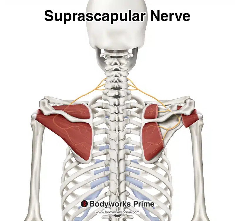
Pictured here we can see the suprascapular nerve which innervates the infraspinatus and supraspinatus.
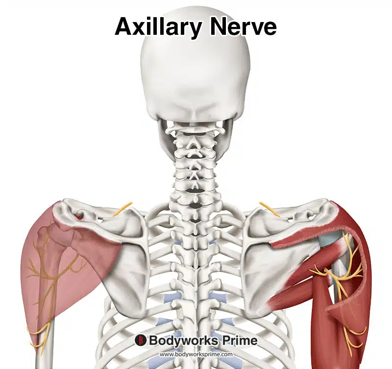
Pictured here we can see the axillary nerve which innervates the teres minor.
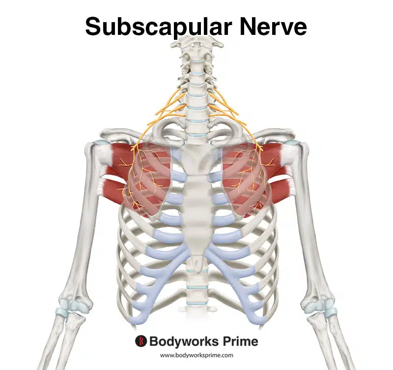
Here we can see the subscapular nerve, which innervates the subscapularis from the nerve roots of C5, C6, and C7.
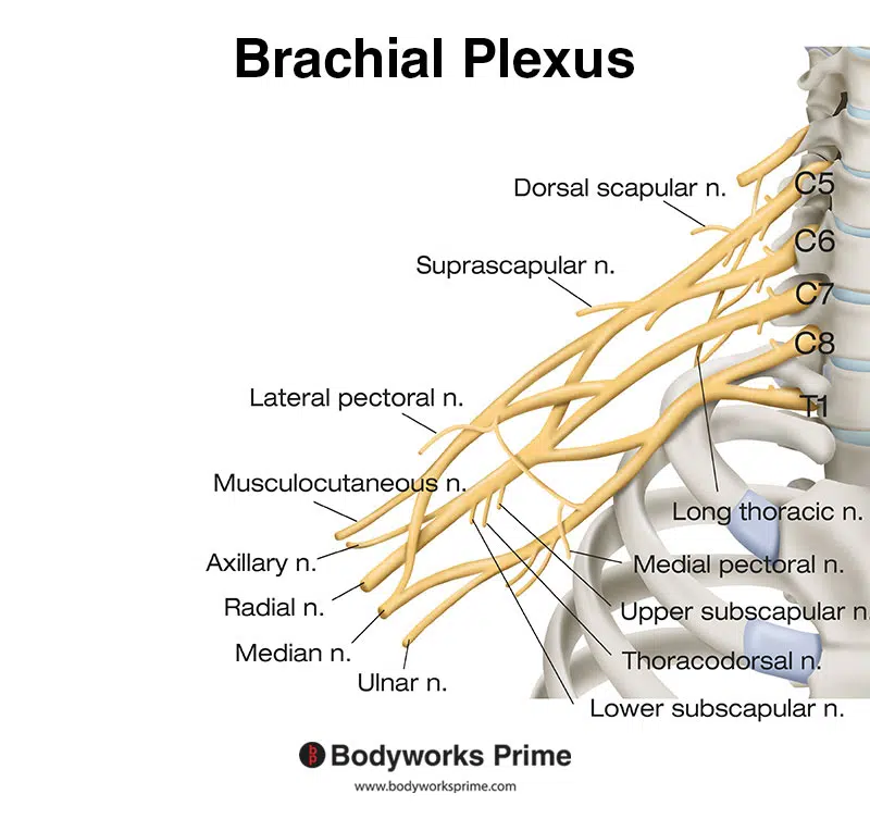
Pictured here we can see the brachial plexus. The suprascapular nerve, subscapular nerve, and axillary nerve are all nerves of the brachial plexus.
Blood Supply
Blood is supplied to the supraspinatus and infraspinatus muscles via the suprascapular artery. This is a branch of the thyrocervical trunk which is a major branch of the subclavian artery. The thyrocervical trunk originates at the base of the neck and enters the region of the posterior scapula just superiorly to the suprascapular foramen [41]. The infraspinatus also receives blood from the circumflex scapular artery. The circumflex scapular artery comes around the lateral border of the scapula and enters into the infraspinous fossa [42].
The teres minor gets its blood supply via the subscapular artery, the circumflex scapular artery and the posterior circumflex humeral artery. The circumflex scapular artery is a branch of the subscapular artery. The subscapular artery and the posterior circumflex humeral artery both arise from the third and most distal part of the axillary artery. The axillary artery travels along the inferior margin of the subscapularis muscle [43].
The subscapularis muscle gets is blood supply via the subscapular artery. The subscapular artery is the largest branch of the axillary artery. The subscapular artery originates from the third part of the axillary artery. The axillary artery travels along the inferior margin of the subscapularis muscle and divides into the circumflex scapular artery and the thoracodorsal artery [44].
Want some flashcards to help you remember this information? Then click the link below:
Supraspinous Flashcards
Infraspinatus Flashcards
Teres Minor Flashcards
Subscapularis Flashcards
Support Bodyworks Prime
Running a website and YouTube channel can be expensive. Your donation helps support the creation of more content for my website and YouTube channel. All donation proceeds go towards covering expenses only. Every contribution, big or small, makes a difference!
References
| ↑1, ↑3, ↑6, ↑9, ↑12, ↑15, ↑18, ↑20, ↑23, ↑26, ↑29, ↑34, ↑37, ↑39, ↑41, ↑44 | Maruvada S, Madrazo-Ibarra A, Varacallo M. Anatomy, Rotator Cuff. [Updated 2022 Mar 31]. In: StatPearls [Internet]. Treasure Island (FL): StatPearls Publishing; 2022 Jan-. Available from: https://www.ncbi.nlm.nih.gov/books/NBK441844/ |
|---|---|
| ↑2, ↑4, ↑8, ↑11, ↑14, ↑17, ↑19, ↑22, ↑25, ↑28, ↑31, ↑33 | Moore KL, Agur AMR, Dalley AF. Clinically Oriented Anatomy. 8th ed. Philadelphia: Lippincot Williams & Wilkins; 2017 |
| ↑5 | Kadi R, Milants A, Shahabpour M. Shoulder Anatomy and Normal Variants. J Belg Soc Radiol. 2017 Dec 16;101(Suppl 2):3. doi: 10.5334/jbr-btr.1467. PMID: 30498801; PMCID: PMC6251069. |
| ↑7, ↑21, ↑24, ↑35 | Jeno SH, Munjal A, Schindler GS. Anatomy, Shoulder and Upper Limb, Arm Supraspinatus Muscle. [Updated 2021 Aug 7]. In: StatPearls [Internet]. Treasure Island (FL): StatPearls Publishing; 2022 Jan-. Available from: https://www.ncbi.nlm.nih.gov/books/NBK537202/ |
| ↑10, ↑36, ↑42 | Williams JM, Sinkler MA, Obremskey W. Anatomy, Shoulder and Upper Limb, Infraspinatus Muscle. [Updated 2021 Aug 13]. In: StatPearls [Internet]. Treasure Island (FL): StatPearls Publishing; 2022 Jan-. Available from: https://www.ncbi.nlm.nih.gov/books/NBK513255/ |
| ↑13, ↑27, ↑38, ↑43 | Juneja P, Hubbard JB. Anatomy, Shoulder and Upper Limb, Arm Teres Minor Muscle. [Updated 2022 May 15]. In: StatPearls [Internet]. Treasure Island (FL): StatPearls Publishing; 2022 Jan-. Available from: https://www.ncbi.nlm.nih.gov/books/NBK513324/ |
| ↑16, ↑30, ↑32, ↑40 | Aguirre K, Mudreac A, Kiel J. Anatomy, Shoulder and Upper Limb, Subscapularis Muscle. [Updated 2021 Aug 7]. In: StatPearls [Internet]. Treasure Island (FL): StatPearls Publishing; 2022 Jan-. Available from: https://www.ncbi.nlm.nih.gov/books/NBK513344/ |


