| Origin | Posterior surface of the lateral border of the scapula |
| Insertion | Greater tubercle of the humerus (inferior aspect) |
| Action | Lateral rotation of the humerus Stabilisation of the humerus in the glenohumeral joint Can weakly assist in shoulder adduction |
| Nerve | Axillary nerve (C5, C6) |
| Artery | Subscapular artery Circumflex scapular artery Posterior circumflex artery |
Location & Overview
The teres minor is a somewhat slender and narrow muscle located in the shoulder region. It is positioned on the lateral border of the scapula and extends to the humerus. The teres minor is inferior to the infraspinatus muscle and superior to the teres major muscle. Sometimes muscle fibres from the infraspinatus can be fused with fibres of the teres minor. It is also deep to the deltoid and trapezius muscles which cover most of the surface of the teres minor [1] [2].
The teres minor forms part of the rotator cuff, along with the supraspinatus, infraspinatus, and subscapularis muscles. A useful mnemonic to remember the name of the rotator cuff muscles is ‘SITS’. Collectively, the rotator cuff muscles are able to provide stabilisation to the glenohumeral joint; which is the ball and socket joint where the head of the humerus sits within the glenoid cavity [3] [4].
Common pathologies associated with the teres minor include rotator cuff tears and tendinopathy, which typically present as shoulder pain and restricted range of motion. These conditions can result from overuse, acute injury, or age-related degeneration. Treatment strategies can range from conservative approaches such as physiotherapy and pain management to surgical interventions in severe cases [5] [6].
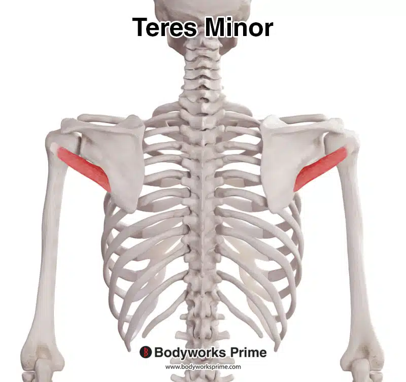
Here we can see the teres minor muscle from a posterior view.
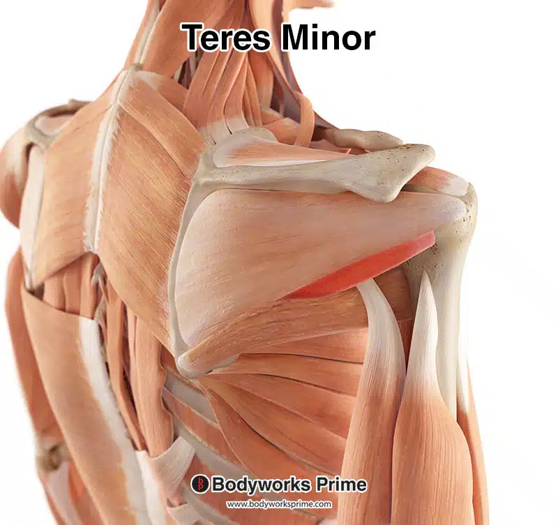
Here we can see the teres minor muscle highlighted in red amongst the other muscles of the body. Superficial muscles such as the trapezius and deltoid have been removed, to better see the teres minor.
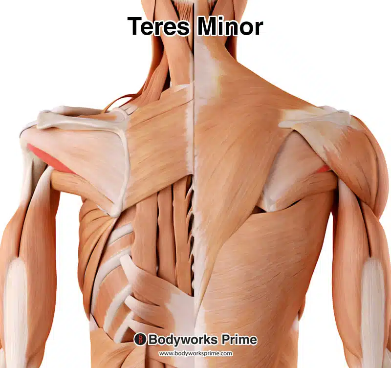
Highlighted in red, the teres minor muscle stands out among the other muscles of the back. On the left, you can see an unobstructed view with the superficial muscles removed, while on the right, they remain intact, providing context to its location. On the right, you can still see a small section of the teres minor.
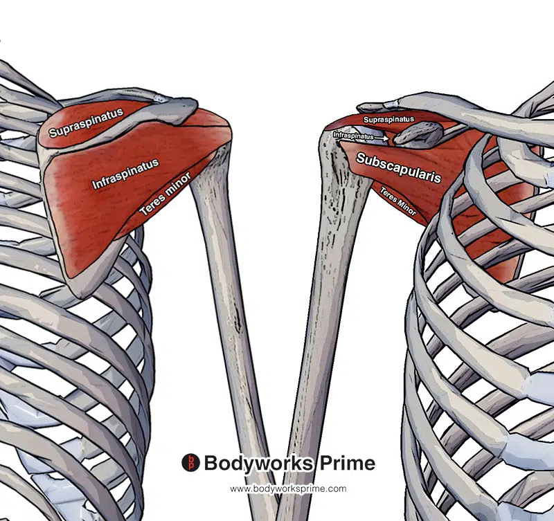
Pictured here we can see all four of the rotator cuff muscles: the supraspinatus, infraspinatus, teres minor and subscapularis. The left side of this picture is from a posterior view (the back) and the right hand side is from an anterior view (the front).
Origin & Insertion
The teres minor originates on the posterior surface of the scapula, specifically on the lateral border. From its origin, the teres minor extends laterally towards the top of the humerus. The insertion is then located on the inferior facet of the greater tubercle of the humerus. The inferior facet is a smooth, flat surface on the lower part of the greater tubercle. The greater tubercle itself is a large, rounded protrusion found on the upper side of the humerus. The upper fibres of the teres minor’s tendon blend into the capsule of the shoulder joint, whereas the lower fibers insert directly onto the humerus [7] [8].
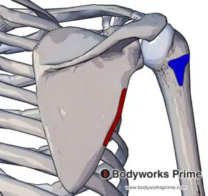
The teres minor originates at the lateral border of the scapula (displayed in red) and inserts at the inferior facet of greater tubercle of the humerus (displayed in blue).
Actions
As a member of the rotator cuff, the teres minor contributes to the stability of the glenohumeral joint by helping secure the head of the humerus within the shallow glenoid cavity of the scapula. Its primary action is the lateral (external) rotation of the arm at the shoulder joint. Additionally, it can provide very weak assistance during adduction of the shoulder [9] [10] [11].
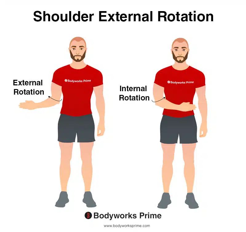
This image demonstrates the movement of external rotation of the shoulder. External rotation involves rotating your arm away from your body, towards the outside. In contrast, internal rotation involves rotating your arm towards the centre of your body, towards the inside. The teres minor contributes to external rotation.
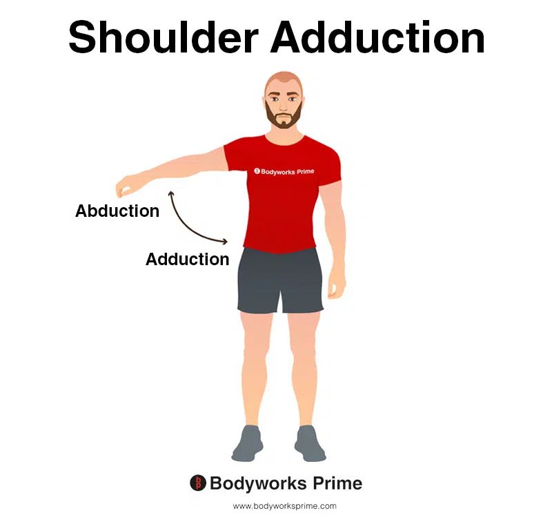
This image shows shoulder adduction. Adduction is where the arm is brought towards the midline of the body, reducing the angle between the arm and the torso. The opposite of adduction is abduction. Abduction occurs as the arm is moved away from the midline, increasing the angle between the arm and the torso. The teres minor contributes to shoulder adduction.
Innervation
The teres minor is innervated by the axillary nerve. This nerve originates from the nerve roots of C5 and C6 and is a part of the the brachial plexus. After originating from C5 and C6 the axillary nerve travels inferiorly and passes through the quadrangular space into the posterior scapula region. The quadrangular space, also known as the quadrilateral space, is a gap situated in the axilla, or armpit region. It is bordered by four structures: the teres minor muscle superiorly, the teres major muscle inferiorly, the long head of the triceps brachii muscle medially, and the humeral shaft laterally. This space allows for the passage of the axillary nerve and the posterior circumflex humeral artery [12] [13] [14].
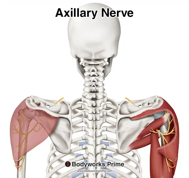
Pictured here we can see the axillary nerve which innervates the teres minor muscle.
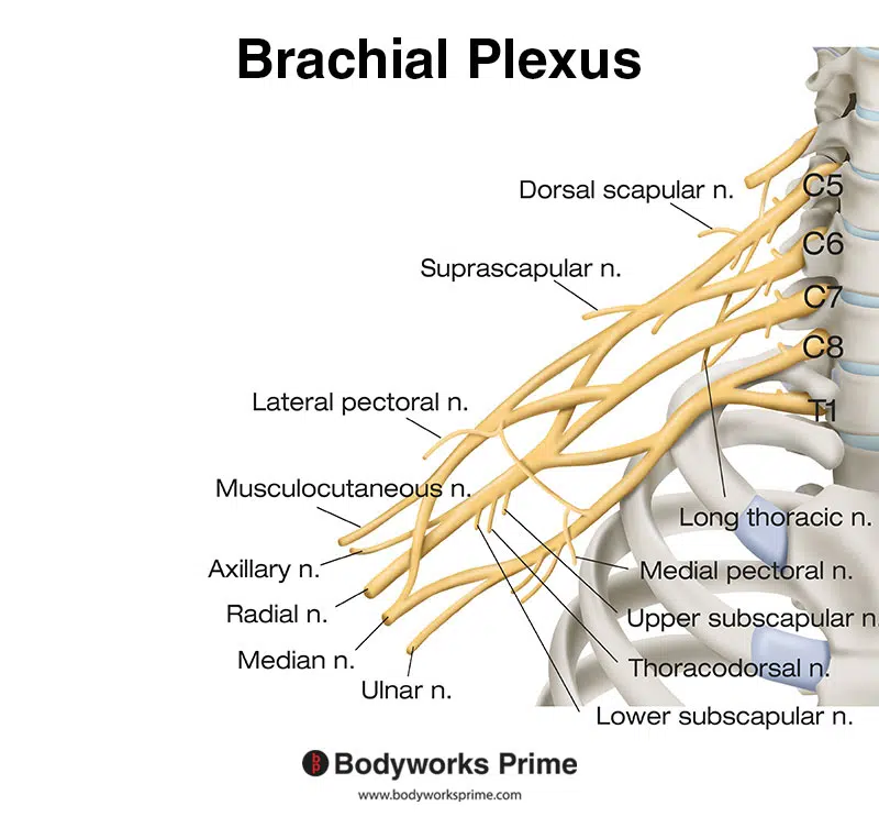
Here we can see the nerves of the brachial plexus. The axillary nerve is a part of the brachial plexus.
Blood Supply
The blood supply to the teres minor is provided primarily by the subscapular artery and its branch, the circumflex scapular artery. It also receives blood supply from the posterior circumflex humeral artery. These arteries branch from the axillary artery [15] [16] [17].
Want some flashcards to help you remember this information? Then click the link below:
Teres Minor Flashcards
Support Bodyworks Prime
Running a website and YouTube channel can be expensive. Your donation helps support the creation of more content for my website and YouTube channel. All donation proceeds go towards covering expenses only. Every contribution, big or small, makes a difference!
References
| ↑1, ↑12, ↑15 | Standring S. (2015). Gray’s Anatomy: The Anatomical Basis of Clinical Practice, 41st Edn. Amsterdam: Elsevier. |
|---|---|
| ↑2, ↑5, ↑7, ↑10, ↑13, ↑16 | Juneja P, Hubbard JB. Anatomy, Shoulder and Upper Limb, Arm Teres Minor Muscle. [Updated 2023 May 8]. In: StatPearls [Internet]. Treasure Island (FL): StatPearls Publishing; 2023 Jan-. Available from: https://www.ncbi.nlm.nih.gov/books/NBK513324/ |
| ↑3, ↑8, ↑11, ↑14, ↑17 | Maruvada S, Madrazo-Ibarra A, Varacallo M. Anatomy, Rotator Cuff. [Updated 2023 Mar 27]. In: StatPearls [Internet]. Treasure Island (FL): StatPearls Publishing; 2023 Jan-. Available from: https://www.ncbi.nlm.nih.gov/books/NBK441844/ |
| ↑4, ↑6, ↑9 | Moore KL, Agur AMR, Dalley AF. Clinically Oriented Anatomy. 8th ed. Philadelphia: Lippincot Williams & Wilkins; 2017. |


