| Origin | Superior two thirds of iliac fossa Inner lip of the iliac crest Lateral aspect of the sacrum |
| Insertion | Lesser trochanter of femur |
| Action | Flexion of the hip joint |
| Nerve | Femoral nerve (L2-L4) |
| Artery | Iliolumbar artery Deep circumflex iliac artery Obturator artery Femoral artery |
Location & Overview
The iliacus muscle connects the hip to the femur and has a fan-shaped structure. It is part of the iliopsoas muscle group, which also includes the psoas major and psoas minor muscles. The term ‘iliopsoas’ is a shortened form of ‘iliopsoas musculotendinous unit’ and is sometimes abbreviated as ‘IPMU’. The iliacus muscle fibers merge with those of the psoas major as they approach the femur, forming a common tendon that inserts onto the lesser trochanter of the femur. Both the iliacus and psoas major muscles pass beneath the inguinal ligament. The psoas minor does not pass under the inguinal ligament because it is connecting onto the hip, rather than the femur [1] [2].
The inguinal ligament is a band of fibrous, tough connective tissue that connects between the anterior superior iliac spine (ASIS) of the hip to the hip’s pubic tubercle. The inguinal ligament is located in the lower part of the abdomen, where the pelvis meets the thigh. If you put your hands on your hips, the inguinal ligament is located roughly where your fingers would be if you traced a line from the bony protrusion at the front of your hip down to the front of your thigh. A picture of the inguinal ligament can be seen in the below images [3] [4] [5].
The iliacus muscle is part of the group of muscles located in the inner hip area. These muscles work together to provide stability and strength to the hip joint, allowing for a range of movements such as walking, running, and jumping. Along with the psoas major, psoas minor, obturator externus, obturator internus, superior gemellus, inferior gemellus, piriformis, and quadratus femoris muscles, the iliacus muscle plays an important role in maintaining proper function of the hip. The iliacus is the primary hip flexor of the hip joint [6].
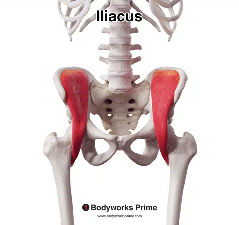
Here we can see the iliacus muscle from an anterior view.
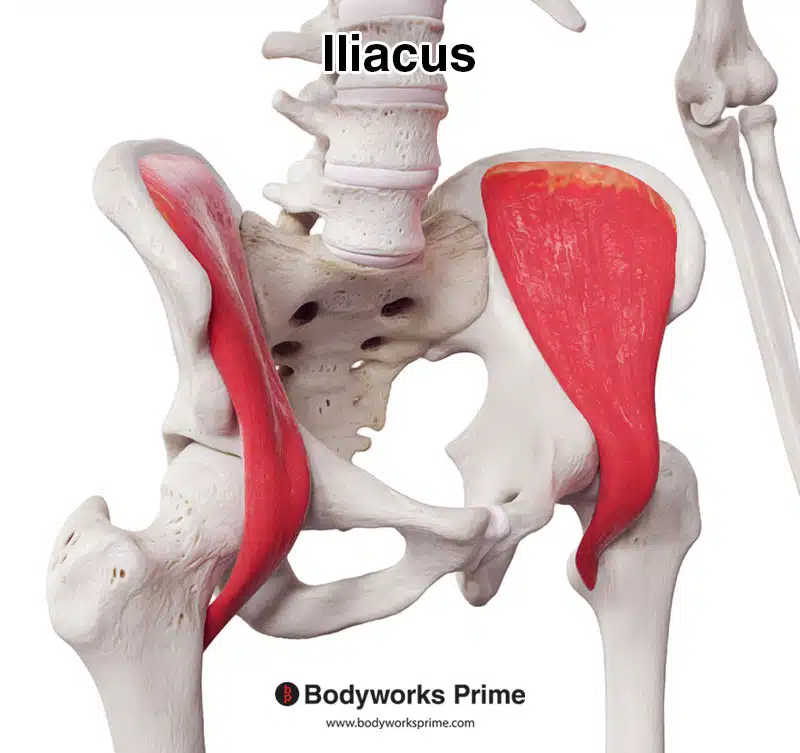
Here we can see the iliacus muscle from a slightly more lateral view, to give a better view of the insertion onto the femur.
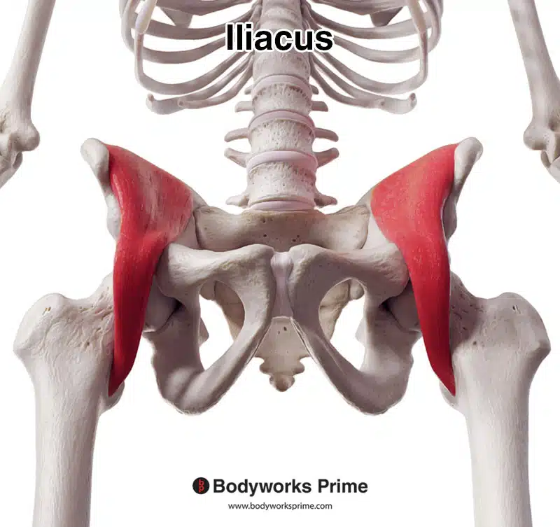
Here we can see the iliacus seen from an inferior view.
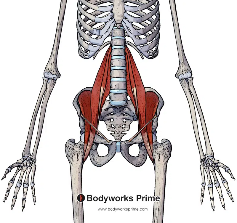
Here we can see pictured the iliopsoas muscle group and the inguinal ligament.
Origin & Insertion
The iliacus originates at the superior two-thirds of the iliac fossa. The superior portion of this origin also connects onto the inner lip of the iliac crest. The origin point also spans onto the lateral aspect of the sacrum. The iliacus muscle then works its way downward in an inferior direction (along with the psoas major) passing deep to the inguinal ligament. Bundles of iliacus muscle’s fibers then merge with fibers of the psoas major muscle as they approach their insertion point and attach onto the lesser trochanter of the femur [7] [8].
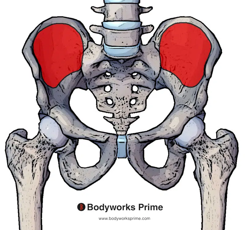
Here we can see the origin of the iliacus marked out in red on the superior two thirds of the iliac fossa, the inner lip of the iliac crest and the lateral aspect of the sacrum.
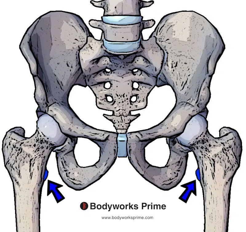
Here we can see the insertion of the iliacus marked out in blue on the lesser trochanter of the femur.
Actions
The main and primary action of the iliacus muscle is to cause flexion of the hip joint. This action can occur by lifting the leg forwards and upwards. If both legs are fixed, then the iliacus can assist in bringing the torso forward towards the legs (i.e bending over). The iliacus works alongside its partner muscle, the psoas major, which forms the iliopsoas muscle group, to perform hip flexion [9] [10] [11] [12].
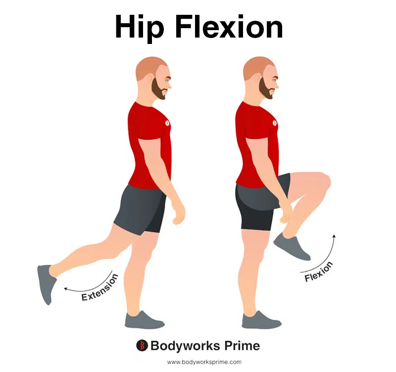
In this image, you can see an example of hip flexion, which is the action of raising your leg up in front of you and causing your hip to bend. If both legs are fixed, the torso will lean forward towards the legs. The opposite movement of hip flexion is hip extension. The primary role of the iliacus muscle is to cause flexion of the hip joint.
Innervation
The iliacus muscle is innervated by the femoral nerve (L2-L4). The femoral nerve is the largest nerve of the lumbar plexus. The lumbar plexus is an a collection of nerves that originate from mostly the lumbar vertebrae.[13].
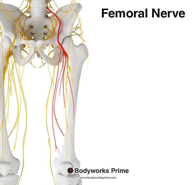
Pictured here is the femoral nerve highlighted in red. The iliacus is innervated by the femoral nerve, from the spinal roots of L2 to L4.
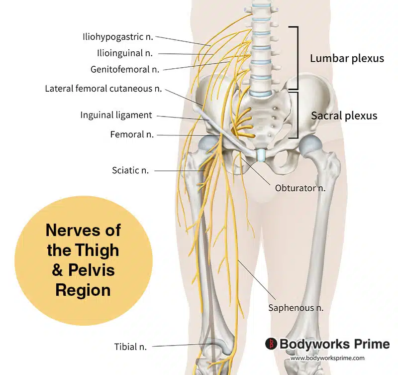
Pictured here we can see the nerves of the thigh and pelvis region. The picture also shows the lumbar plexus, which the femoral nerve is a part of.
Blood Supply
The arteries which supply blood to the iliacus muscle are the iliolumbar artery, deep circumflex iliac artery[14], obturator artery, and femoral artery.
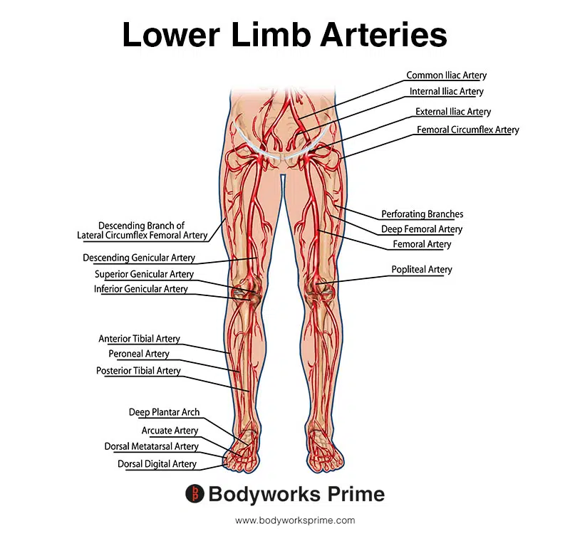
This image shows the arteries of the lower limb.
Want some flashcards to help you remember this information? Then click the link below:
Iliacus Flashcards
Support Bodyworks Prime
Running a website and YouTube channel can be expensive. Your donation helps support the creation of more content for my website and YouTube channel. All donation proceeds go towards covering expenses only. Every contribution, big or small, makes a difference!
References
| ↑1, ↑4, ↑7, ↑9 | Bordoni B, Varacallo M. Anatomy, Bony Pelvis and Lower Limb, Iliopsoas Muscle. [Updated 2022 Apr 29]. In: StatPearls [Internet]. Treasure Island (FL): StatPearls Publishing; 2022 Jan-. Available from: https://www.ncbi.nlm.nih.gov/books/NBK531508/ |
|---|---|
| ↑2, ↑5, ↑10 | Lifshitz L, Bar Sela S, Gal N, Martin R, Fleitman Klar M. Iliopsoas the Hidden Muscle: Anatomy, Diagnosis, and Treatment. Curr Sports Med Rep. 2020 Jun;19(6):235-243. doi: 10.1249/JSR.0000000000000723. PMID: 32516195. |
| ↑3, ↑6, ↑11, ↑13, ↑14 | Moore KL, Agur AMR, Dalley AF. Clinically Oriented Anatomy. 8th ed. Philadelphia: Lippincot Williams & Wilkins; 2017. |
| ↑8 | Siccardi MA, Tariq MA, Valle C. Anatomy, Bony Pelvis and Lower Limb, Psoas Major. [Updated 2021 Aug 11]. In: StatPearls [Internet]. Treasure Island (FL): StatPearls Publishing; 2022 Jan-. Available from: https://www.ncbi.nlm.nih.gov/books/NBK535418/ |
| ↑12 | Anderson CN. Iliopsoas: Pathology, Diagnosis, and Treatment. Clin Sports Med. 2016 Jul;35(3):419-433. doi: 10.1016/j.csm.2016.02.009. Epub 2016 Mar 28. PMID: 27343394. |


