| Origin | Anterior surface of the sacrum (around the level of S2 to S4) Gluteal surface of the ilium Sacrotuberous ligament |
| Insertion | Greater trochanter of the femur (medial side of the superior aspect) |
| Action | Laterally rotates the thigh at hip joint Abducts thigh at hip joint (when hip is flexed past 60⁰) Stabilises head of femur in acetabulum |
| Nerve | Superior gluteal nerve Ventral rami of S1 and S2 |
| Artery | Superior and inferior gluteal arteries Internal pudendal artery |
Location & Overview
The piriformis muscle is situated in the gluteal region and proximal thigh, with a flattened, pear-shaped appearance. It is positioned parallel to the posterior edge of the gluteus medius and lies deep to the gluteus maximus. The name “piriformis” is derived from the Latin words “pirum” and “forma,” which respectively mean “pear” and “shape.” Piriformis syndrome is a common condition associated with this muscle, and typical symptoms include hip pain, buttock pain, and pain that radiates down the back of the leg. These symptoms may include shooting, burning, or aching sensations, as well as numbness and tingling [1] [2].
The piriformis muscle is part of a group of six short muscles that externally rotate the hip joint in the gluteal region. The other five muscles in this group are the obturator internus, quadratus femoris, obturator externus, superior gemellus, and inferior gemellus. The piriformis muscle’s tendon merges with the tendons of the inferior gemellus and superior gemellus, along with the obturator internus muscle, to form the conjoint tendon. This tendon then inserts onto the femur bone [3].
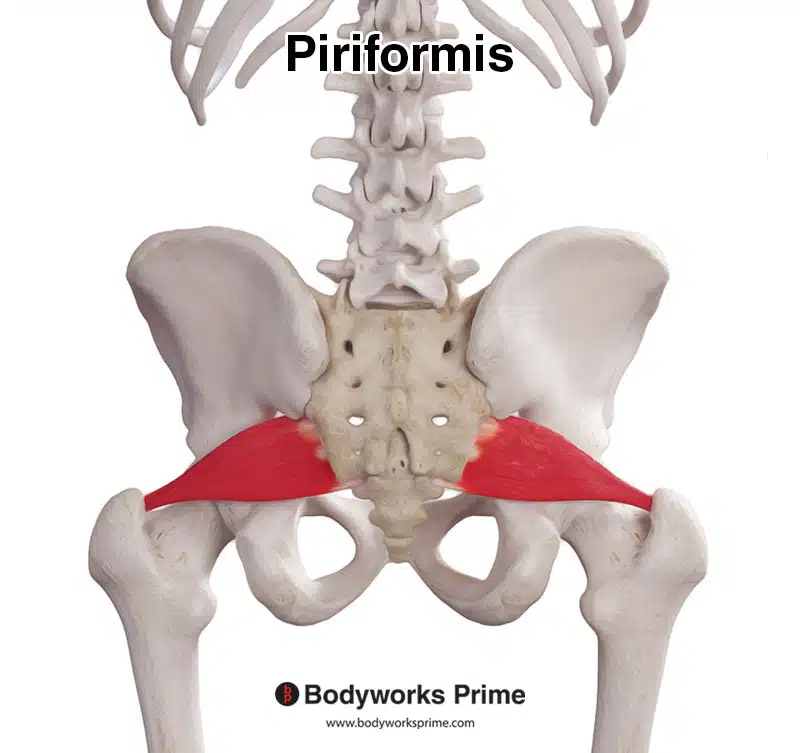
Here we can see the piriformis muscle from a posterior view.
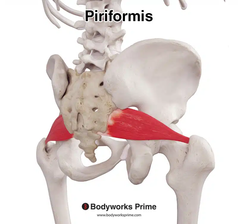
Here we can see the piriformis muscle from a posterolateral view.
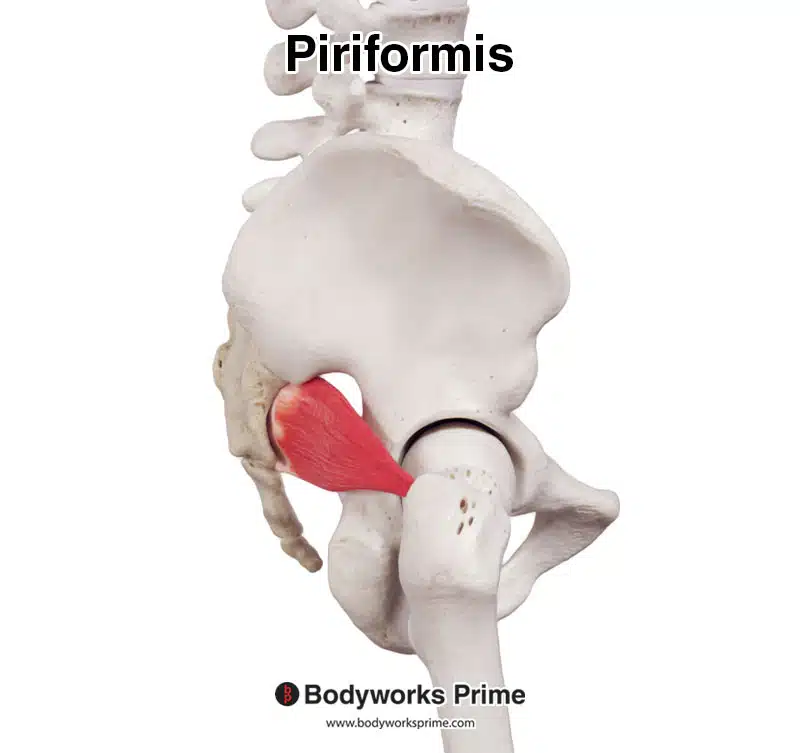
Here we can see the piriformis muscle from a lateral view.
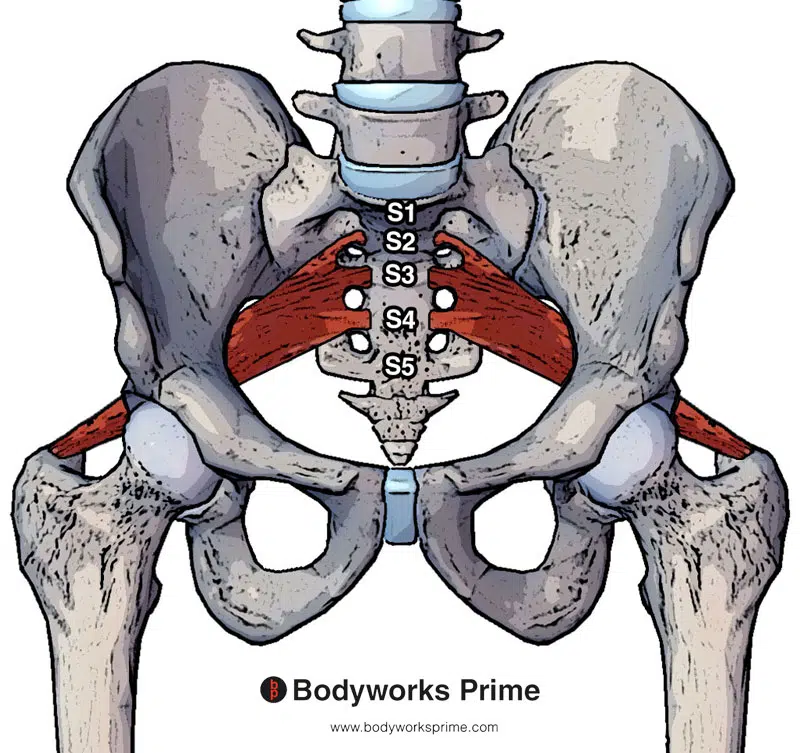
Picture here we can see the piriformis muscle from an anterior view, with the segments of the sacrum labelled. As you can see, it is located between S2 and S4.
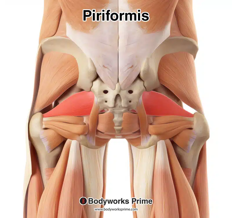
Here we can see the piriformis muscle highlighted in red amongst the other muscles in the area. Superficial muscles such as the gluteus maximus and gluteus medius have been removed so that we can see the piriformis.
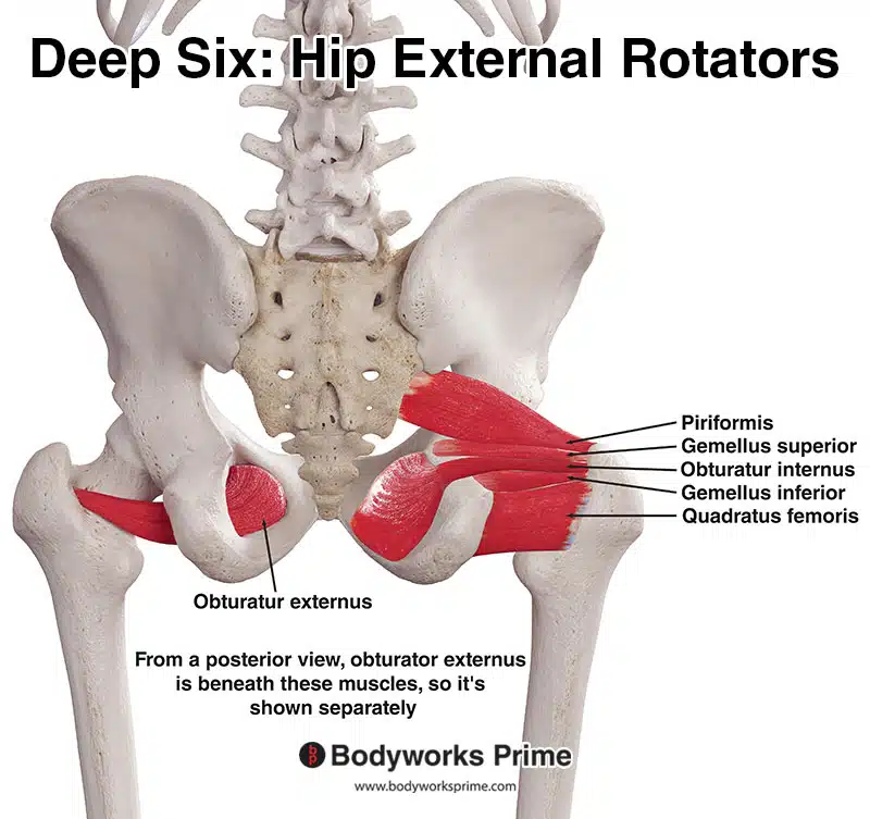
Pictured here are the deep six hip external rotator muscles: the piriformis, gemellus superior, gemellus inferior, obturator internus, obturator externus, and quadratus femoris. These muscles play an important role in stabilising the hip joint and facilitating hip lateral rotation.
Origin & Insertion
The piriformis muscle originates from various regions, including the anterior surface of the sacrum between levels S2 to S4, the spinal area of the gluteal muscles, the capsule of the adjacent sacroiliac joint, the gluteal surface of the ilium near the margin of the greater sciatic notch, and occasionally the sacrotuberous ligament. Its insertion point is located on the medial side of the superior aspect of the greater trochanter of the femur [4].
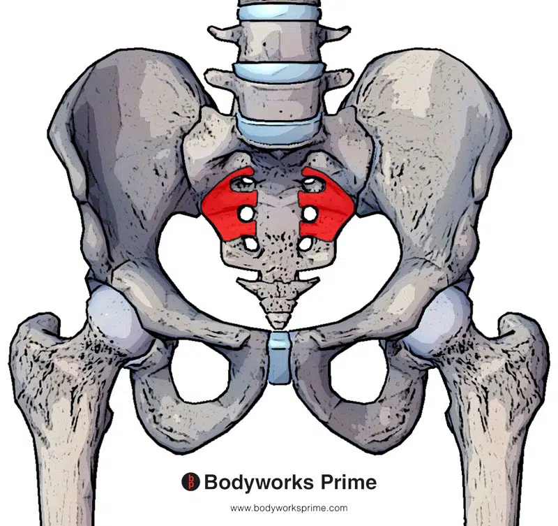
Pictured here is the main origin of the piriformis muscle on the anterior surface of the sacrum (between S2 and S4).
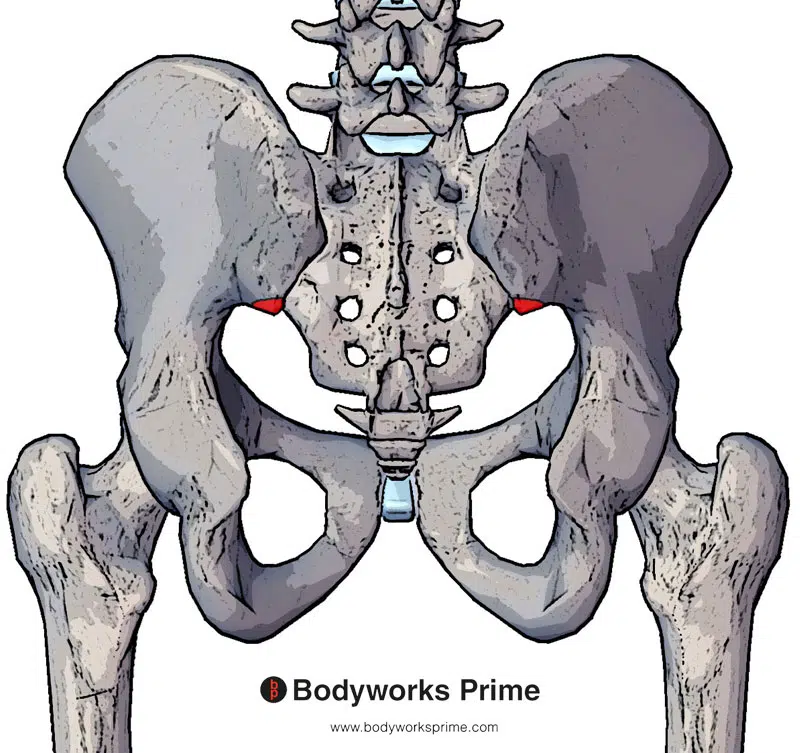
Pictured here is another origin of the piriformis on the gluteal surface of the ilium (near to the posterior inferior iliac spine).
Pictured here is the sacrotuberous ligament which is another origin of the piriformis muscle. The piriformis would be connecting onto the superior portion of this ligament.
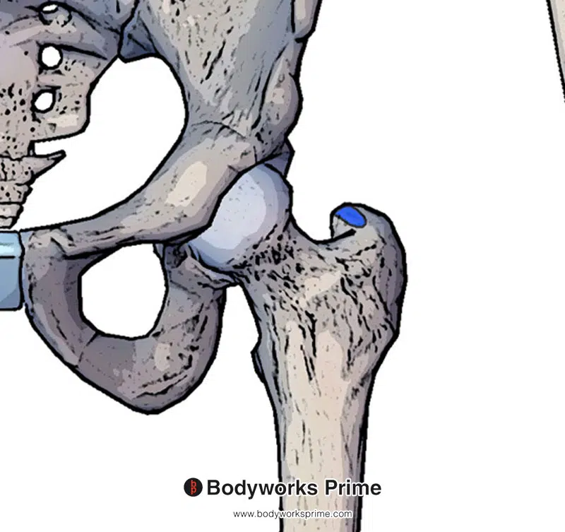
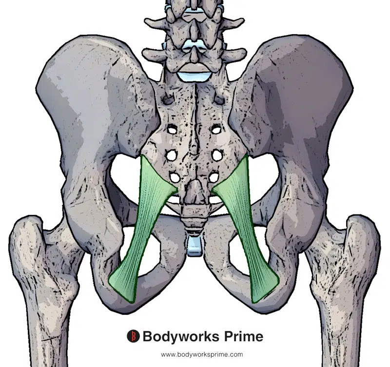
Pictured here is the insertion of the piriformis muscle which is on the greater trochanter of the femur (on the medial side of the superior aspect of it).
Actions
The piriformis muscle is a lateral rotator of the hip (also known as external rotation). The quadratus femoris, inferior gemellus, obturator internus, and obturator externus can also assist in this movement. When the hip is extended, the piriformis is in the most advantageous position to assist in lateral rotation of the femur. When the hip joint is flexed (past around 60⁰ of hip flexion), this is when the piriformis muscle is then in a more advantageous position for hip abduction [5] [6] [7].
Variations in the body, tendon, and tendon insertion of the piriformis muscle can impact its actions. Although it has been suggested that the piriformis muscle functions as an internal rotator of the hip joint, this ability is dependent on an individual’s unique anatomical structure. Therefore controversy exists regarding the precise actions of the piriformis muscle. The truth is that the piriformis muscle’s role as an internal rotator varies depending on the individual’s anatomy and where the muscle inserts. In most cases, the piriformis has a variation that makes it unsuitable for aiding in internal rotation of the hip joint [8] [9] [10].
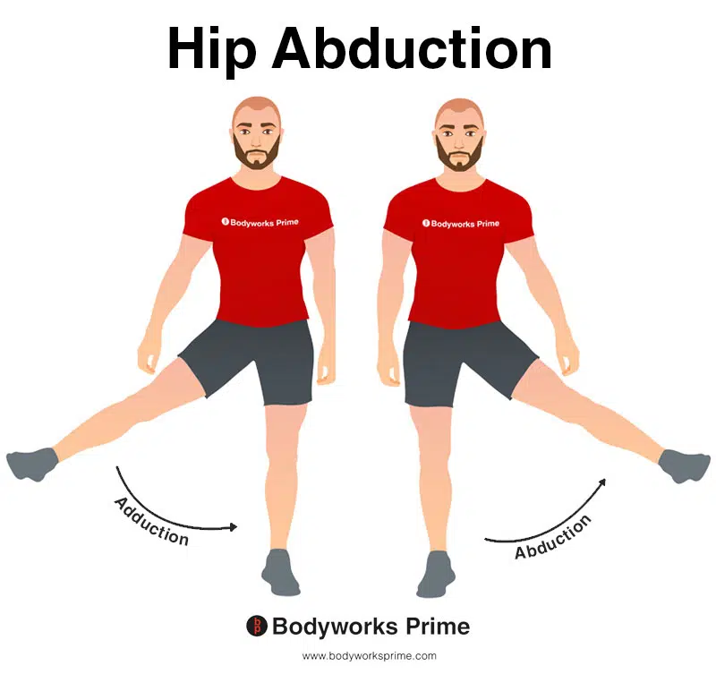
This image shows an example of hip abduction, which involves moving the leg out to the side (laterally). The opposite of hip abduction is hip adduction. The piriformis muscle is an abductor of the hip joint when hip is flexed past 60 degrees. Hip flexion involves lifting the leg up in front of you (towards the anterior of the body). Put simply, the piriformis can assist in hip abduction once the leg is lifted forwards and the hip joint reaches 60 degrees of flexion.
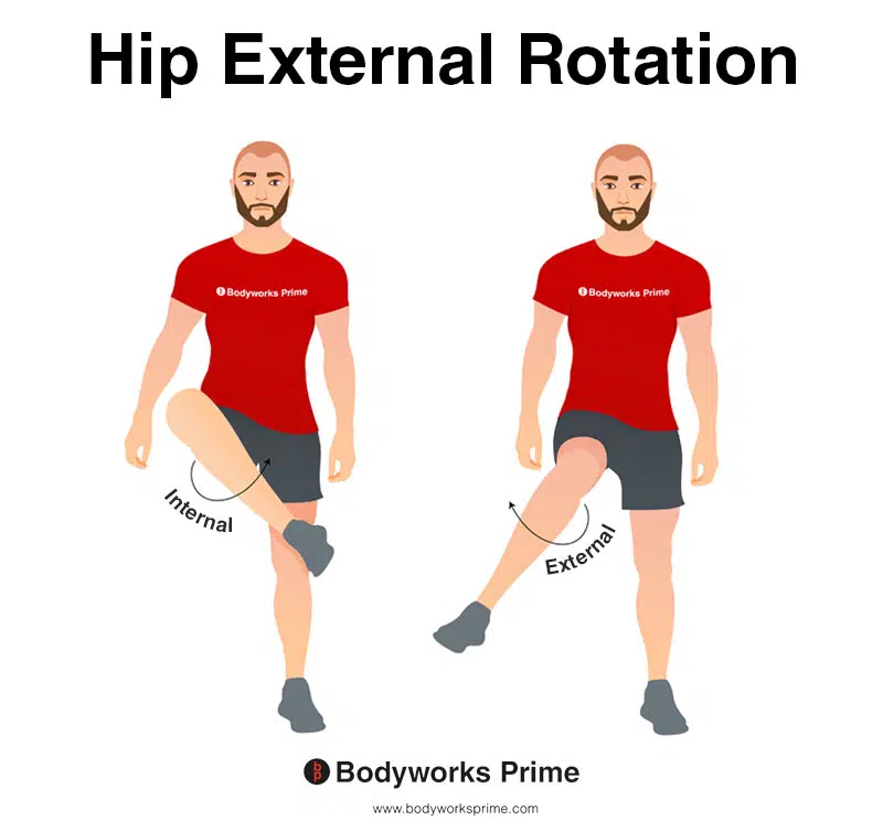
This image depicts an example of hip external rotation, which involves rotating the leg from the hip joint outwards (laterally). External rotation is also referred to as lateral rotation. The opposite of external rotation (lateral rotation) is internal rotation (medial rotation). The piriformis muscle is an external rotator of the hip joint.
Remember that the piriformis also stabilises head of femur in acetabulum (an action not shown in the above pictures)
Innervation
Previously the piriformis muscle was said to be innervated by the ‘nerve to piriformis’. However, recent cadaver research suggests that: “a single ‘nerve to piriformis’ does not exist in the majority of specimens thus this term should be abandoned” [11]. Instead, the most common innervations of the piriformis are the superior gluteal nerve and the ventral rami of S1 and S2. [12] [13] [14]
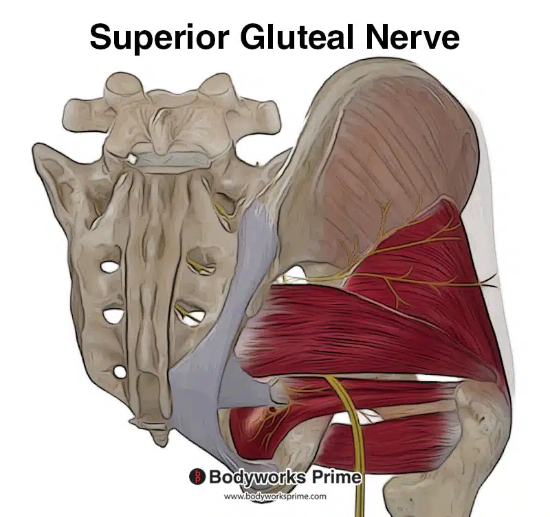
Pictured here you can see the superior gluteal nerve. Branches of the inferior part of this nerve extend to innervate the piriformis muscle. Moreover, the piriformis receives direct innervation from the ventral rami of the vertebra (S1 & S2).
Blood Supply
Blood is supplied to the piriformis via the superior gluteal artery, inferior gluteal artery, and internal pudendal artery which are branches of the internal iliac artery [15] [16].
Want some flashcards to help you remember this information? Then click the link below:
Piriformis Flashcards
Support Bodyworks Prime
Running a website and YouTube channel can be expensive. Your donation helps support the creation of more content for my website and YouTube channel. All donation proceeds go towards covering expenses only. Every contribution, big or small, makes a difference!
References
| ↑1, ↑5, ↑9, ↑12, ↑16 | Chang C, Jeno SH, Varacallo M. Anatomy, Bony Pelvis and Lower Limb, Piriformis Muscle. [Updated 2021 Oct 11]. In: StatPearls [Internet]. Treasure Island (FL): StatPearls Publishing; 2022 Jan-. Available from: https://www.ncbi.nlm.nih.gov/books/NBK519497/ |
|---|---|
| ↑2 | Hicks BL, Lam JC, Varacallo M. Piriformis Syndrome. [Updated 2022 Feb 12]. In: StatPearls [Internet]. Treasure Island (FL): StatPearls Publishing; 2022 Jan-. Available from: https://www.ncbi.nlm.nih.gov/books/NBK448172/ |
| ↑3 | Lezak B, Massel DH. Anatomy, Bony Pelvis and Lower Limb, Gemelli Muscles. 2021 Sep 7. In: StatPearls [Internet]. Treasure Island (FL): StatPearls Publishing; 2022 Jan–. PMID: 32491352. |
| ↑4 | Chang C, Jeno SH, Varacallo M. Anatomy, Bony Pelvis and Lower Limb, Piriformis Muscle. [Updated 2021 Oct 11]. In: StatPearls [Internet]. Treasure Island (FL): StatPearls Publishing; 2022 Jan-. Available from: https://www.ncbi.nlm.nih.gov/books/NBK519497/ |
| ↑6 | Demirel A, Baykara M, Koca TT, Berk E. Ultrasound elastography findings in piriformis muscle syndrome. Indian J Radiol Imaging. 2018 Oct-Dec;28(4):412-418. doi: 10.4103/ijri.IJRI_133_18. PMID: 30662201; PMCID: PMC6319104. |
| ↑7 | Lung K, Lui F. Anatomy, Abdomen and Pelvis, Superior Gluteal Nerve. 2021 Aug 11. In: StatPearls [Internet]. Treasure Island (FL): StatPearls Publishing; 2022 Jan–. PMID: 30571029. |
| ↑8 | Windisch G, Braun EM, Anderhuber F. Piriformis muscle: clinical anatomy and consideration of the piriformis syndrome. Surg Radiol Anat. 2007 Feb;29(1):37-45. doi: 10.1007/s00276-006-0169-x. Epub 2007 Jan 10. PMID: 17216293. |
| ↑10 | Benzon HT, Katz JA, Benzon HA, Iqbal MS. Piriformis syndrome: anatomic considerations, a new injection technique, and a review of the literature. Anesthesiology. 2003 Jun;98(6):1442-8. doi: 10.1097/00000542-200306000-00022. PMID: 12766656. |
| ↑11, ↑13 | Iwanaga J, Eid S, Simonds E, Schumacher M, Loukas M, Tubbs RS. The majority of piriformis muscles are innervated by the superior gluteal nerve. Clin Anat. 2019 Mar;32(2):282-286. doi: 10.1002/ca.23311. Epub 2018 Dec 21. PMID: 30408241. |
| ↑14 | Fishman LM, Hosseini M. Piriformis syndrome – a diagnosis comes into its own. Muscle Nerve. 2019 Apr;59(4):395-396. doi: 10.1002/mus.26417. Epub 2019 Feb 4. PMID: 30623985. |
| ↑15 | Goidescu OC, Enyedi M, Tulin AD, et al. Overview of the anatomical basis of the piriformis syndrome-dissection with magnetic resonance correlation. Exp Ther Med. 2022;23(2):113. doi:10.3892/etm.2021.11036 |


