| Origin | Transverse processes of L1-L5 vertebrae Vertebral bodies of T12-L5 vertebrae and the adjacent intervertebral discs |
| Insertion | Lesser trochanter of the femur |
| Action | Flexion of the hip joint (primary action) Weak assistance in lateral flexion of the trunk Weak assistance in external rotation of the femur Provides dynamic stability to the femur and the hip |
| Nerve | Anterior rami of the first, second and third lumbar nerves (L1-L3) |
| Artery | Lumbar branch of iliolumbar artery (primary source) Obturator artery External iliac artery Femoral artery |
Location & Overview
The primary function of the psoas major muscle is to flex the hip joint. This muscle is part of the psoas muscle group, which includes the psoas minor (although the psoas minor muscle may be absent in some individuals). The psoas major muscle is situated in the lumbar region of the trunk, located laterally on both sides of the vertebral column. It sits medially to the quadratus lumborum muscle and posterior to the psoas minor muscle [1] [2] [3] [4].
Both the psoas major and iliacus muscles are located deep to the inguinal ligament and pass through the gap created between the ligament and the pelvis to reach their insertion point on the lesser trochanter of the femur. The inguinal ligament is a tough, fibrous band of connective tissue that connects the anterior superior iliac spine (ASIS) of the hip to the pubic tubercle of the hip. This ligament is located in the lower part of the abdomen where the pelvis meets the thigh. If you place your hands on your hips, you can locate the inguinal ligament approximately where your fingers would be if you traced a line from the bony protrusion at the front of your hip down to the front of your thigh [5] [6] [7] [8]. The second image below this text shows the psoas major and iliacus passing under the inguinal ligament.
At the distal end, the psoas major muscle combines with the iliacus muscle to form a shared tendon that connects to the lesser trochanter of the femur. The combination of the psoas major, minor and the iliacus is referred to as the ‘iliopsoas muscle’, which is also sometimes known as the ‘iliopsoas musculotendinous unit (IPMU)’. The psoas major and iliacus muscles work together to flex the hip joint, lifting the upper leg towards the body. On the other hand, the psoas minor muscle, which does not cross the hip joint, is unable to assist in hip flexion [9] [10] [11] [12].
The name of the psoas major muscle is believed to have originated from the Ancient Greek word ‘ψόᾱ’, meaning ‘psoas’, which translates to ‘muscles of the loins’ in English.
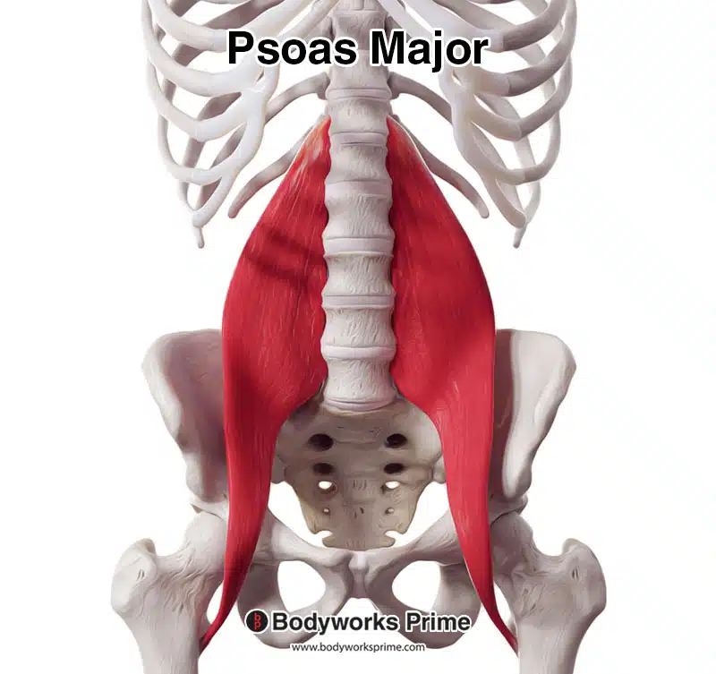
Here we can see the psoas major muscle from an anterior view.
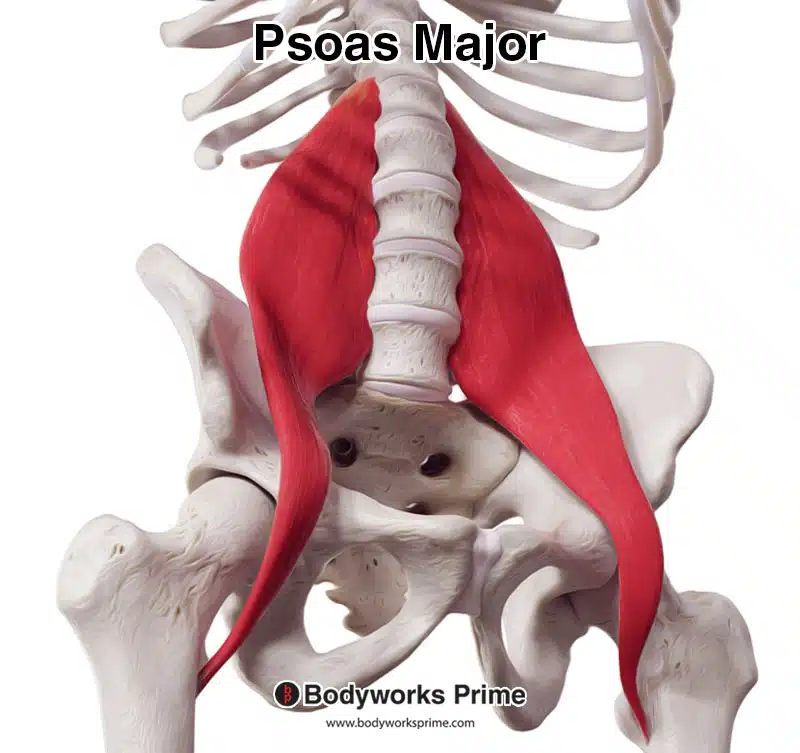
Here we can see the psoas major muscle from a more lateral view.
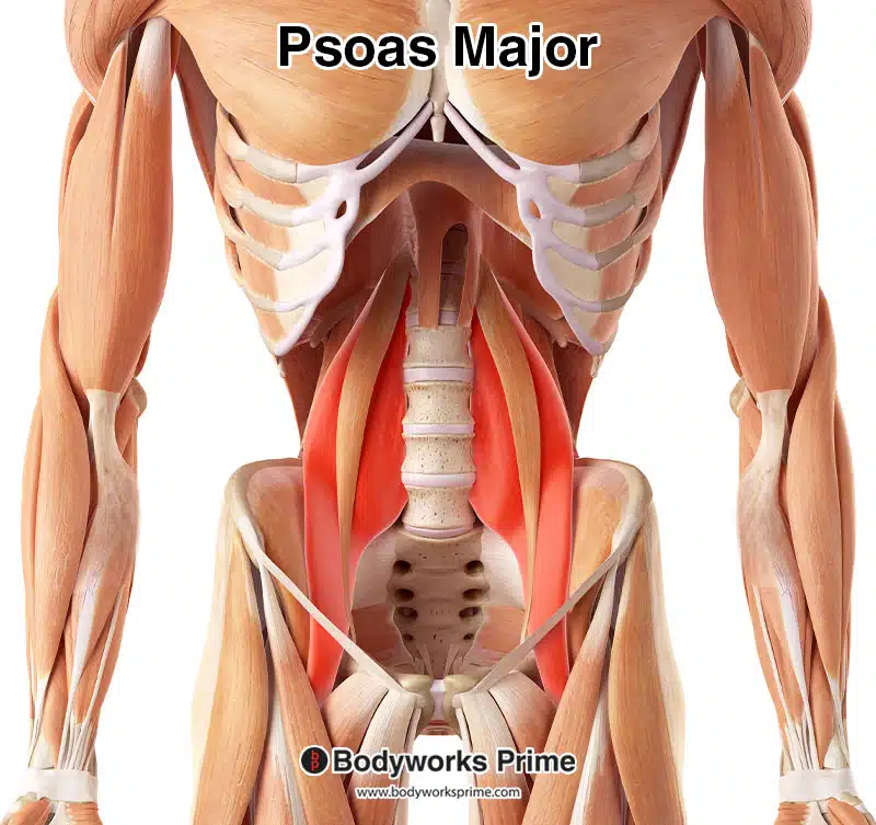
Here we can see the psoas major muscle highlighted in red amongst the other muscles of the body. Because the psoas major is a deep muscle, muscles like the abdominal muscles have been removed to reveal the psoas major. The visible muscle lying over the top of the psoas major is the psoas minor. It is also important to note that there are internal organs in between the psoas muscles and the abdominal muscles. This is why it is not possible to palpate the psoas muscles.
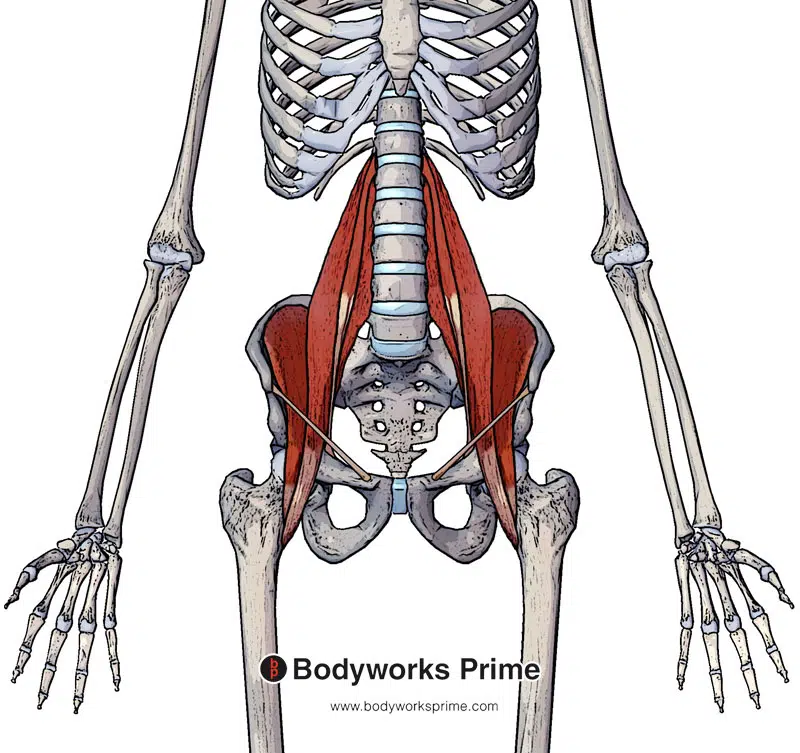
Next, we can see the psoas major, psoas minor and iliacus muscle (collectively known as the iliopsoas muscle). You can also see the inguinal ligament and how the psoas major and iliacus pass behind it through the gap between the inguinal ligament and the pelvis.
Origin & Insertion
The psoas major muscle originates on the transverse processes and the lateral surfaces of the bodies of the lumbar vertebrae L1 to L5. It also connects onto the vertebral bodies of T12 to L5. The vertebral origin also connects onto the intervertebral discs between these vertebrae. The psoas major then works its way down in an inferolateral direction passing underneath the inguinal ligament to insert onto the lesser trochanter of the femur[13] [14] [15].
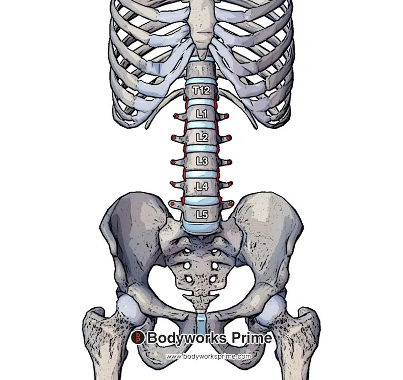
Marked out in red on the vertebra are the origin points of the psoas major. These origin points are on the transverse processes of L1-L5 vertebrae in addition to the vertebral bodies of T12-L5 vertebrae and the adjacent intervertebral discs.
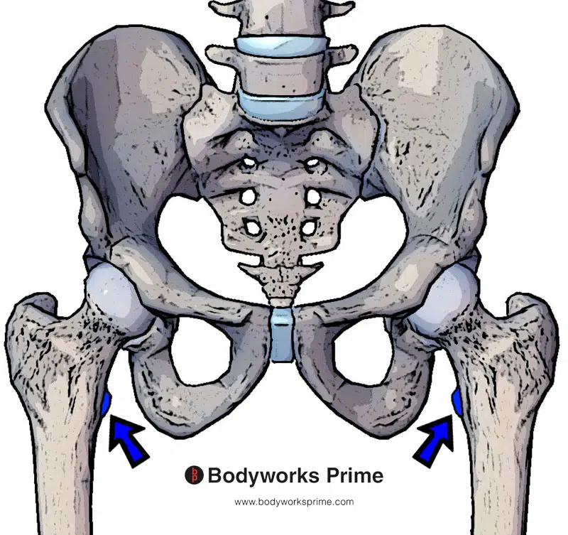
Marked out in blue on both femurs you can see the psoas major’s insertion point which is on the lesser trochanter of the femur.
Actions
The psoas major muscle’s primary function is to flex the hip joint, while it can also provide weak assistance in external rotation (i.e. lateral rotation) of the femur in the hip joint. It can also perform lateral flexion of the torso, but its role in this movement is weaker compared to other muscles such as the internal oblique and external oblique muscles. Additionally, the psoas major achieves this lateral flexion by only flexing one side at a time through unilateral contraction. Some studies suggest that the psoas major also plays a dynamic stability role, helping to maintain the femur in place in the acetabulum during movement [16] [17] [18] [19] [20] [21] [22] [23].
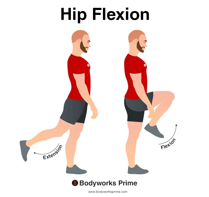
In this image, you can see an example of hip flexion, which is the action of raising your leg up in front of you and causing your hip to bend. If both legs are fixed, the torso will lean forward towards the legs. The opposite movement of hip flexion is hip extension. Hip flexion is the psoas major’s primary action.
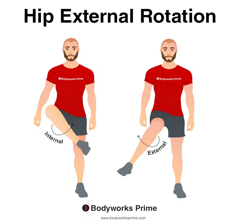
This image depicts an example of hip external rotation, which involves rotating the leg from the hip joint outwards (laterally). External rotation is also referred to as lateral rotation. The opposite of external rotation (lateral rotation) is internal rotation (medial rotation). The psoas major can provide weak assistance in external rotation of the femur in the hip joint.
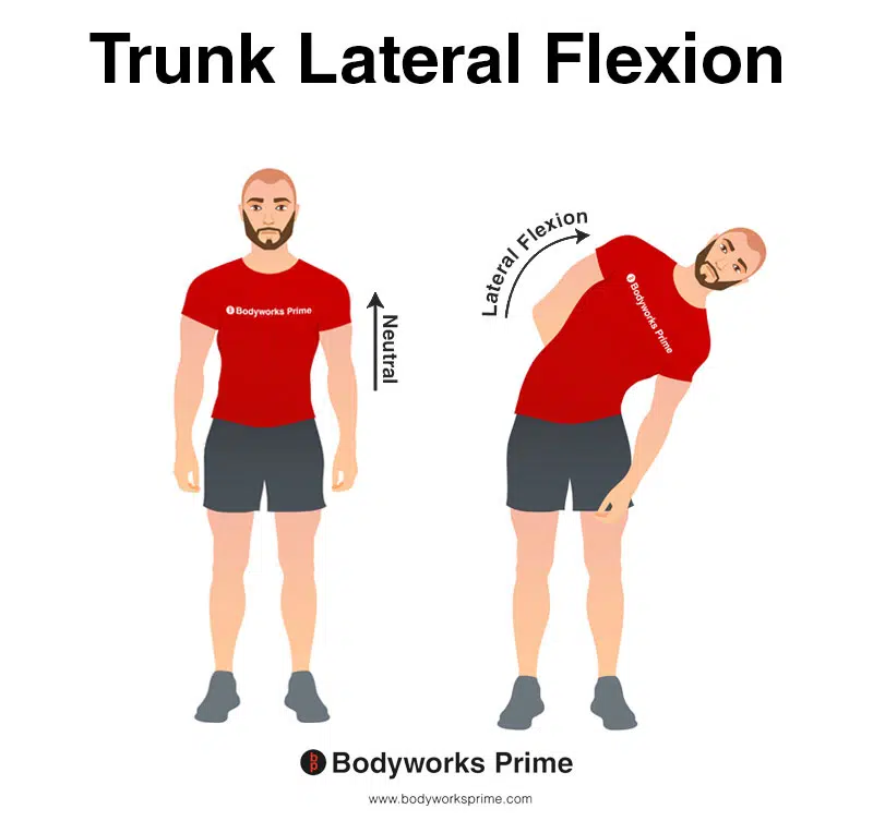
This image demonstrates trunk lateral flexion, which involves bending the torso to the side. The psoas major can provide weak assistance to other muscles during lateral flexion of the trunk.
Innervation
The psoas major muscle is innervated by the anterior rami of the first, second and third lumbar nerves (L1-L3) [24] [25].
Blood Supply
Blood is supplied to the psoas major mainly via the lumbar branch of iliolumbar artery. It also receives a blood supply from the obturator, external iliac, and femoral arteries [26] [27].
Want some flashcards to help you remember this information? Then click the link below:
Psoas Major Flashcards
Support Bodyworks Prime
Running a website and YouTube channel can be expensive. Your donation helps support the creation of more content for my website and YouTube channel. All donation proceeds go towards covering expenses only. Every contribution, big or small, makes a difference!
References
| ↑1, ↑5, ↑9, ↑16, ↑24, ↑26 | Siccardi MA, Tariq MA, Valle C. Anatomy, Bony Pelvis and Lower Limb, Psoas Major. [Updated 2021 Aug 11]. In: StatPearls [Internet]. Treasure Island (FL): StatPearls Publishing; 2022 Jan-. Available from: https://www.ncbi.nlm.nih.gov/books/NBK535418/ |
|---|---|
| ↑2, ↑6, ↑10, ↑14, ↑20, ↑25, ↑27 | Bordoni B, Varacallo M. Anatomy, Bony Pelvis and Lower Limb, Iliopsoas Muscle. [Updated 2022 Apr 29]. In: StatPearls [Internet]. Treasure Island (FL): StatPearls Publishing; 2022 Jan-. Available from: https://www.ncbi.nlm.nih.gov/books/NBK531508/ |
| ↑3, ↑7, ↑11 | Sajko S, Stuber K. Psoas Major: a case report and review of its anatomy, biomechanics, and clinical implications. J Can Chiropr Assoc. 2009;53(4):311-318 |
| ↑4, ↑8, ↑12, ↑15, ↑21 | Moore KL, Agur AMR, Dalley AF. Clinically Oriented Anatomy. 8th ed. Philadelphia: Lippincot Williams & Wilkins; 2017 |
| ↑13, ↑19 | Sajko S, Stuber K. Psoas Major: a case report and review of its anatomy, biomechanics, and clinical implications. J Can Chiropr Assoc. 2009;53(4):311-318. |
| ↑17 | Arbanas J, Klasan GS, Nikolic M, Jerkovic R, Miljanovic I, Malnar D. Fibre type composition of the human psoas major muscle with regard to the level of its origin. J Anat. 2009;215(6):636-641. doi:10.1111/j.1469-7580.2009.01155.x |
| ↑18 | Hirase T, Mallett J, Barter LE, Dong D, McCulloch PC, Harris JD. Is the Iliopsoas a Femoral Head Stabilizer? A Systematic Review. Arthrosc Sports Med Rehabil. 2020;2(6):e847-e853. Published 2020 Nov 17. doi:10.1016/j.asmr.2020.06.006 |
| ↑22 | Skyrme AD, Cahill DJ, Marsh HP, Ellis H. Psoas major and its controversial rotational action. Clin Anat. 1999;12(4):264-5. doi: 10.1002/(SICI)1098-2353(1999)12:4<264::AID-CA4>3.0.CO;2-L. PMID: 10398385. |
| ↑23 | Reimann R, Sodia F, Klug F. Die umstrittene Rotationswirkung ausgewählter Muskeln im Hüftgelenk [Controversial rotation function of certain muscles in the hip joint]. Ann Anat. 1996 Aug;178(4):353-9. German. PMID: 8928937. |


