| Origin | Superior nuchal line External occipital protuberance Nuchal ligament Spinous processes of C7 to T12 vertebrae |
| Insertion | Lateral third of clavicle Acromion Spine of scapula |
| Action | Elevates scapula Depresses scapula Scapula adduction Scapula rotation Neck extension and lateral flexion. |
| Nerve | Spinal accessory nerve (cranial nerve XI) (motor fibres) C3, C4 cervical spinal nerves (pain and proprioceptive fibres) |
| Artery | Cervical artery Dorsal scapular artery Posterior intercostal arterial branches |
Location & Overview
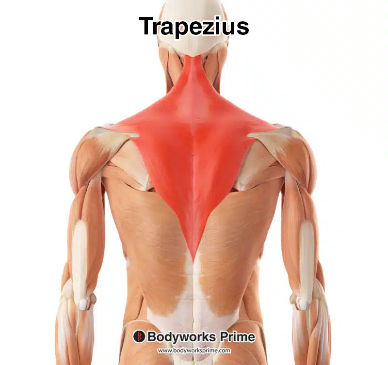
The trapezius is often divided into three functional regions based on the orientation of its fibers: the superior (or descending) portion, the intermediate (or horizontal) portion, and the inferior (or ascending) portion [1]. The superior portion descends from the top of the neck to the clavicle, the intermediate portion extends horizontally from the spine to the medial border of the acromion, and the inferior portion ascends from the lower back to the spine of the scapula [2] [3].
Muscle Fibre Summary:
Superior fibres = descending trapezius
Intermediate fibres = horizontal trapezius
Inferior fibres = ascending trapezius
The primary and most popular exercise for strengthening the trapezius muscle is the shrug. Shrugs can be performed using various types of equipment, such as dumbbells, barbells, cables, or a Smith machine. Adjusting grip positioning (wide, intermediate, or narrow) can offer variability in the stimulus provided. For example, a narrower grip can deliver a greater stretch on the trapezius at the bottom of the lift. A common mistake when performing shrugs is using excessively heavy weights. It is important to select a weight that allows you to shrug as high as possible and pause for at least a second. Performing more repetitions with a compromised range of motion and technique is less effective than full range of motion training. However, heavy partials can provide a novel stimulus when programmed sensibly to complement a well-rounded program. Other excellent trapezius strengthening exercises include Y raises and prone trap raises, upright rows (performed with a high range of motion), face pulls, band pull-aparts, and incline dumbbell shrugs.
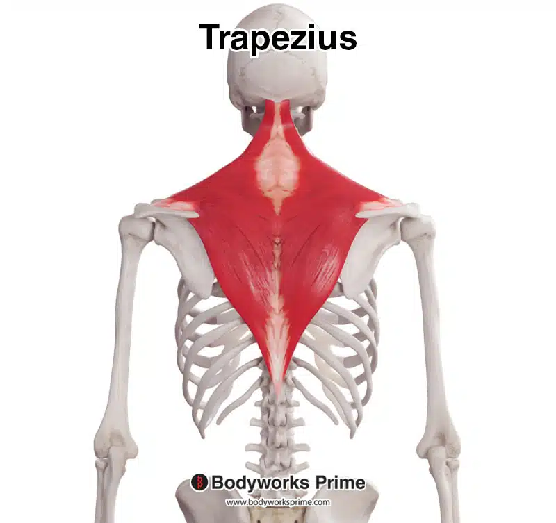
Here we can see the trapezius muscle from a posterior view.
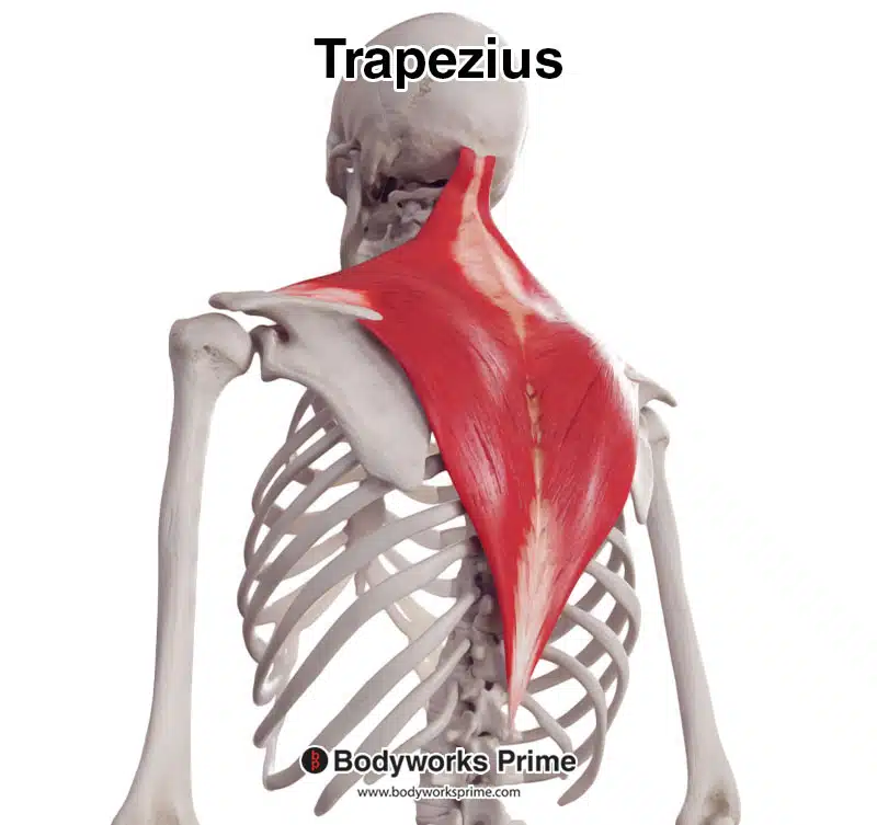
Here we can see the trapezius muscle from a posterolateral view.
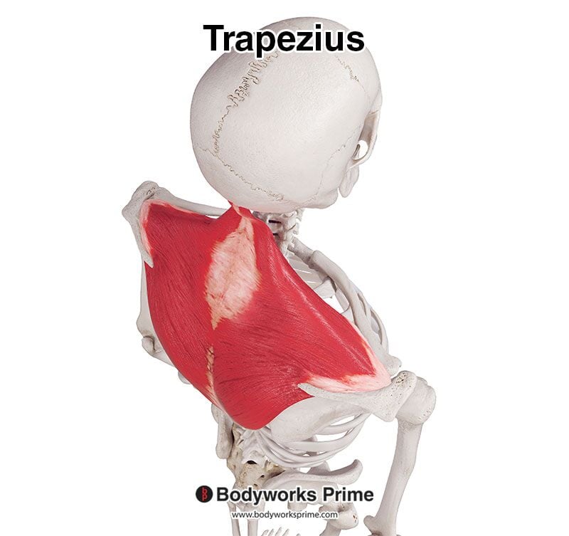
Here we can see the trapezius muscle from a superior view.
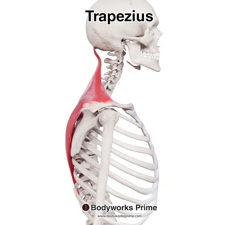
Here we can see the trapezius muscle from lateral view.
Here we can see the trapezius muscle highlighted in red amongst the other muscles of the body.
Origin & Insertion
The trapezius muscle originates from several points along the skull and the vertebra. The most superior point of origin is the external occipital protuberance, which is a prominent bony projection at the base of the skull, on the back of the head. The muscle also originates from the superior nuchal line, which is a ridge that runs laterally from the external occipital protuberance. From here, the origin continues down the nuchal ligament, which is a strong fibrous tissue that runs along the posterior aspect of the neck. The nuchal ligament connects the skull to the cervical and thoracic vertebrae. The trapezius muscle then originates from the spinous processes of the C7 to T12 vertebrae. The spinous processes are the bony projections off the back of the vertebra and they can be felt down the midline of the back [4] [5].
The trapezius muscle fibers extend from these origins to insert onto various points on the shoulder girdle. The superior fibers, also known as the descending trapezius, insert onto the lateral third of the clavicle, which is a long, curved bone that forms the front part of the shoulder girdle. The intermediate fibers, or horizontal trapezius, attach to the medial border of the acromion, which is a bony process on the scapula that forms the highest point of the shoulder. Finally, the inferior fibers, or ascending trapezius, insert onto the spine of the scapula, which is a bony ridge that runs horizontally across the posterior aspect of the scapula [6] [7].
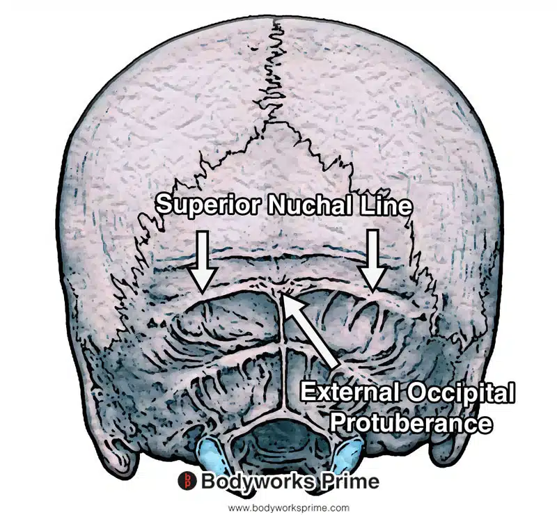
Here we can see the superior nuchal line and external occipital protuberance. They are origin points of the trapezius muscle.
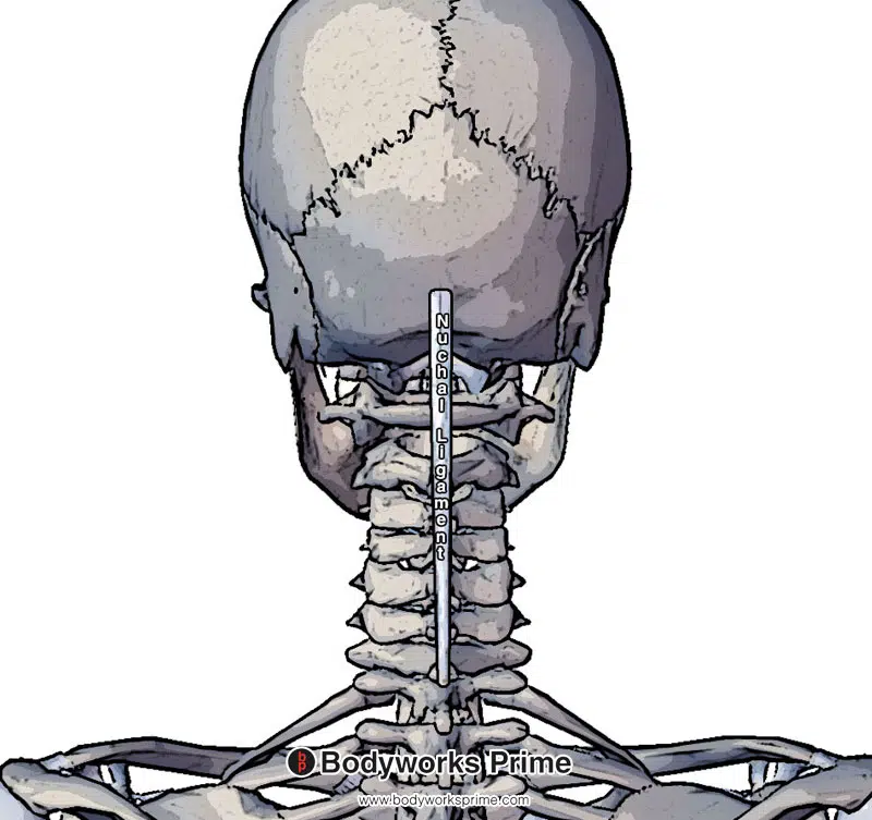
Here we can see the nuchal ligament. It is one of the origin points of the trapezius muscle.
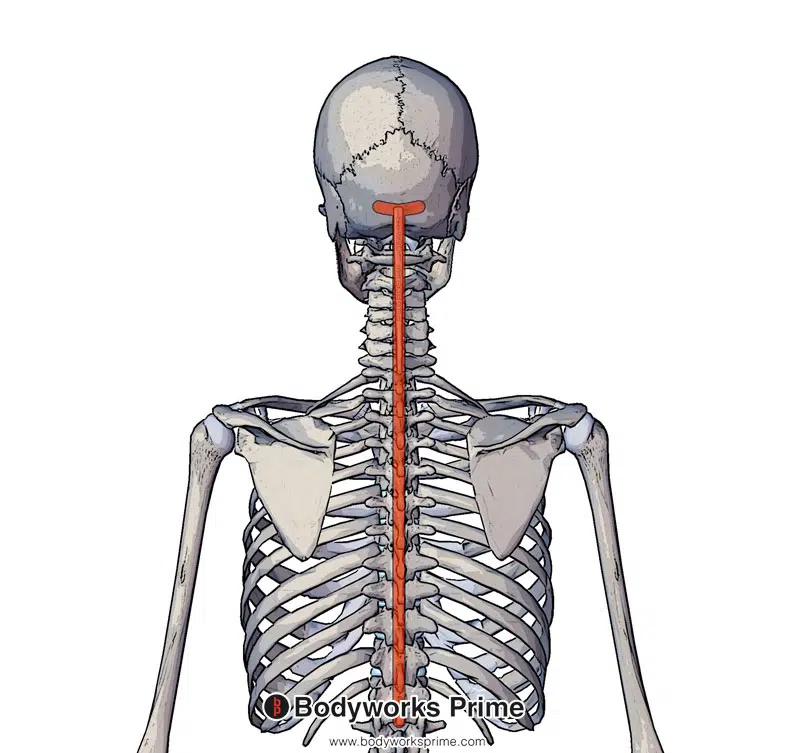
Here we can see the origin of the trapezius muscle marked in red. The trapezius originates at the superior nuchal line, external occipital protuberance, nuchal ligament and spinous processes of C7 – T12 vertebrae.
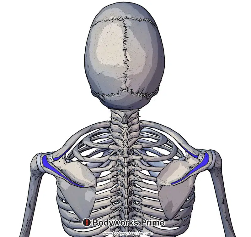
Here we can see the insertion of the trapezius muscle marked in blue. The trapezius inserts on the lateral third of the clavicle, acromion and spine of scapula.
Actions
The trapezius muscle has several actions involving the scapula and neck. Elevation and depression of the scapula are the first two actions, with the superior fibers contributing to scapula elevation and the inferior fibers to scapula depression. Elevation of the scapula can be observed when shrugging the shoulders, while depression of the scapula occurs when lowering the shoulders back down [8] [9].
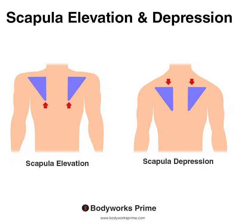
This image shows both scapula elevation and depression. In scapula elevation, the shoulder blade moves superiorly, as in the action of shrugging the shoulders. Conversely, scapula depression occurs when the shoulder blade moves inferiorly, as in the action of lowering the shoulders from a shrugged position. The trapezius muscle can both elevate and depress the scapula.
Another action of the trapezius is scapula adduction or retraction. This is primarily performed by the intermediate fibers. Adduction of the scapula pulls the shoulder blades closer together, towards the spine. This can be seen when pinching the shoulder blades together [10] [11].
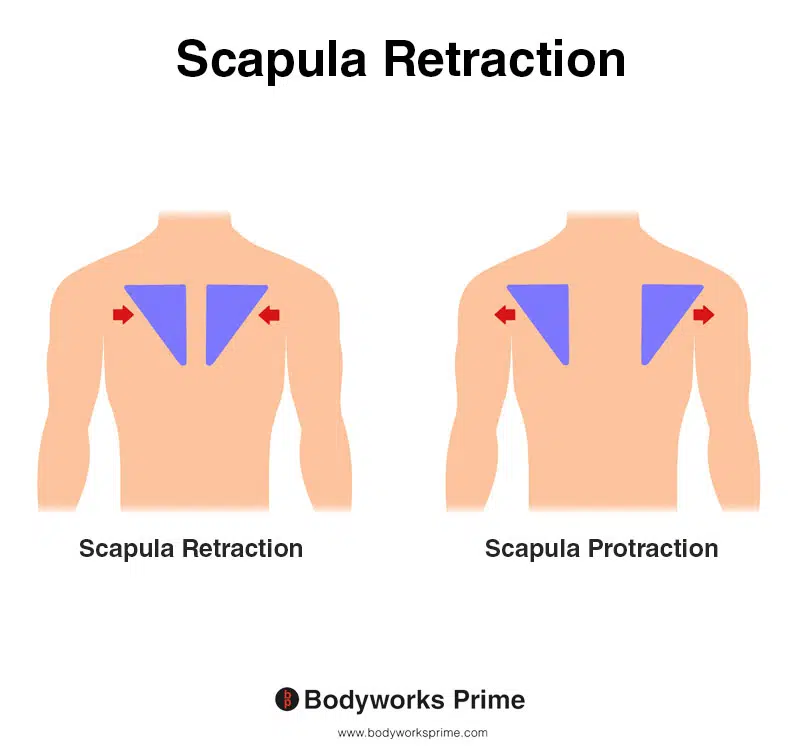
This image shows scapula retraction, also known as scapula adduction, where the shoulder blades move towards the spine, as in the action of pinching the shoulder blades together. The opposite of scapula retraction is scapula protraction, also known as scapula abduction, where the shoulder blades move away from the spine, as in the action of reaching forward. The trapezius muscle is able to perform scapula retraction.
The superior and inferior fibers of the trapezius also work together to assist in scapula rotation, helping to angle the glenohumeral joint of the scapula upwards. The superior fibers can rotate the scapula medially (upwardly), while the inferior fibers can rotate it laterally (downwardly). Scapula rotation is essential for overhead movements, such as reaching up to grab something from a high shelf [12] [13].
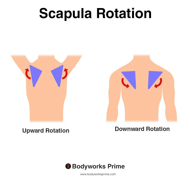
This image shows both scapula upward and downward rotation. In upward rotation, the shoulder blade moves laterally and superiorly, tilting the glenoid cavity upward. This movement enables the arm to be raised overhead, while maintaining joint alignment. On the other hand, scapula downward rotation occurs when the shoulder blade moves medially and inferiorly, tilting the glenoid cavity downward. This movement helps to lower the arm and return it to a resting position, as well as maintain joint stability during various arm movements. The trapezius muscle can both upwardly and downwardly rotate the scapula.
Lastly, the trapezius muscle is also responsible for neck extension and neck lateral flexion. Neck extension involves tilting the head back, while neck lateral flexion involves tilting the head from side to side [14] [15].
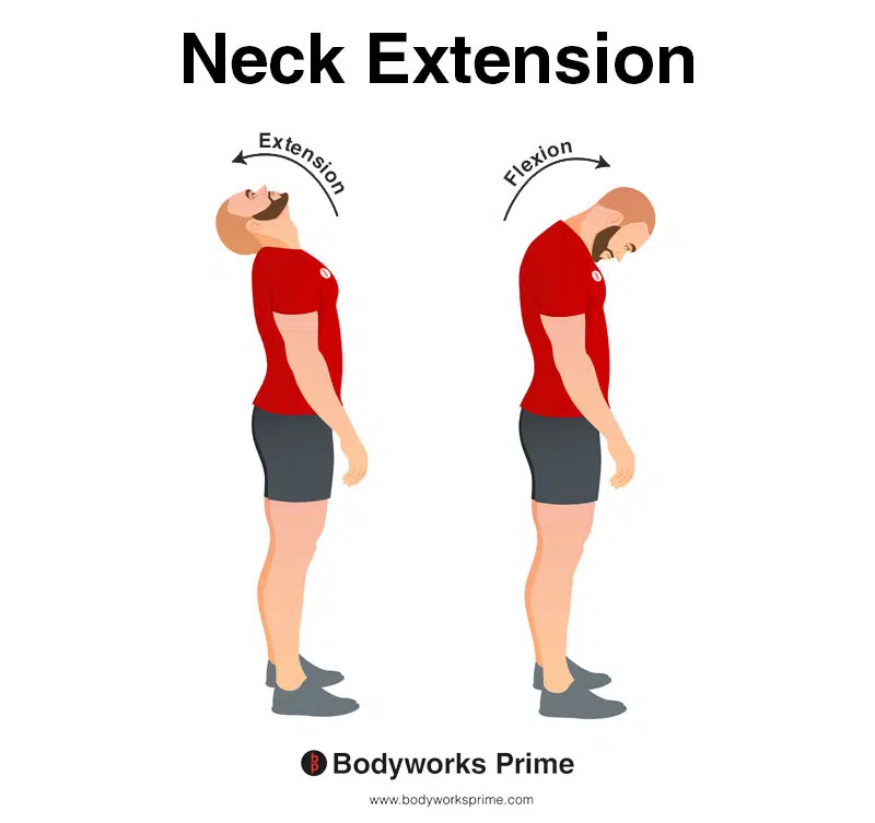
This image demonstrates neck extension, which involves bending the neck backwards. The opposite of neck extension is neck flexion. An action of the trapezius muscle is extension of the neck.
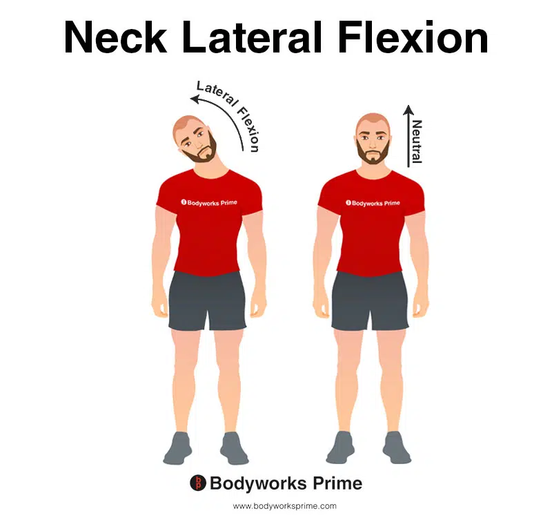
This image demonstrates neck lateral flexion, which involves bending the neck to the side. An action of the trapezius muscle is lateral flexion of the neck.
Innervation
Innervation of the trapezius muscle is provided by both motor and sensory nerves. The primary motor nerve which innervates the trapezius muscle is the spinal accessory nerve. This nerve is also known as cranial nerve XI. The nerve arises from the brainstem and travels through the jugular foramen before entering the posterior cervical triangle. Once in the triangle, the spinal accessory nerve courses through the deep tissues where it innervates the trapezius muscle, providing the required motor function for the trapezius’ various actions [16] [17].
Moreover, the trapezius muscle is not only involved in motor function but also in sensory perception, particularly with regard to pain and proprioception. Sensory innervation of the trapezius is facilitated by cervical spinal nerves C3 and C4, which emerge from the cervical plexus—a complex web of nerves created by the anterior rami of cervical spinal nerves. The input from nerves C3 and C4 provides the trapezius with importnat information regarding the muscle’s position, tension, and movement dynamics [18] [19].
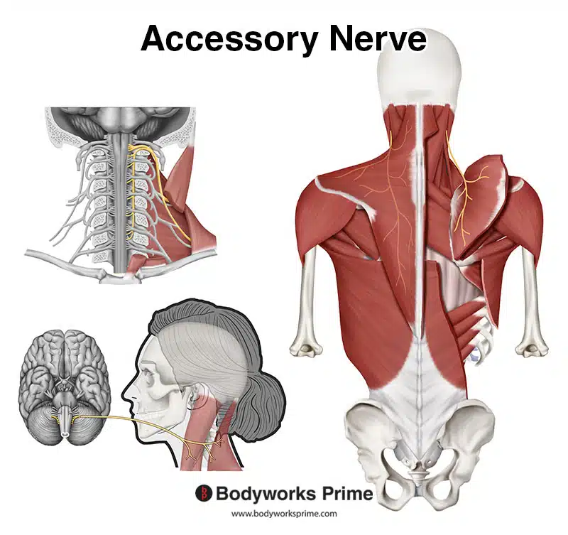
Pictured here we can see the accessory nerve which innervates the trapezius muscle.
Blood Supply
The trapezius muscle primarily receives its blood supply from the transverse cervical artery, a branch of the thyrocervical trunk. The thyrocervical trunk itself originates from the subclavian artery, a significant artery responsible for supplying the upper limb and upper thoracic area. The transverse cervical artery traverses the posterior cervical triangle, ensuring blood flow to the trapezius and adjacent muscles [20] [21].
As well as the transverse cervical artery, blood is also supplied to the superior portion of the trapezius muscle by the dorsal scapular artery. This artery originates from either the subclavian artery or the transverse cervical artery and follows along the medial border of the scapula, supplying blood to the levator scapulae, rhomboids, and the superior trapezius [22] [23].
The deep portions of the trapezius muscle receive their blood from the posterior intercostal arterial branches. These branches arise from the aorta (the main artery that carries blood away from your heart) and the branches then travel between the ribs, which then provides a blood supply to the muscles within the thoracic region [24] [25].
Want some flashcards to help you remember this information? Then click the link below:
Trapezius Muscle Flashcards
Support Bodyworks Prime
Running a website and YouTube channel can be expensive. Your donation helps support the creation of more content for my website and YouTube channel. All donation proceeds go towards covering expenses only. Every contribution, big or small, makes a difference!
References
| ↑1 | Johnson G, Bogduk N, Nowitzke A, House D. Anatomy and actions of the trapezius muscle. Clin Biomech (Bristol, Avon). 1994 Jan;9(1):44-50. doi: 10.1016/0268-0033(94)90057-4. PMID: 23916077. |
|---|---|
| ↑2, ↑8, ↑10, ↑12, ↑14 | Moore KL, Agur AMR, Dalley AF. Clinically Oriented Anatomy. 8th ed. Philadelphia: Lippincot Williams & Wilkins; 2017. |
| ↑3, ↑5, ↑7, ↑17, ↑19, ↑21, ↑23, ↑25 | Standring S. (2015). Gray’s Anatomy: The Anatomical Basis of Clinical Practice, 41st Edn. Amsterdam: Elsevier. |
| ↑4, ↑6, ↑9, ↑11, ↑13, ↑15, ↑16, ↑18 | Ourieff J, Scheckel B, Agarwal A. Anatomy, Back, Trapezius. [Updated 2021 Jul 26]. In: StatPearls [Internet]. Treasure Island (FL): StatPearls Publishing; 2021 Jan-. Available from: https://www.ncbi.nlm.nih.gov/books/NBK518994/ |
| ↑20, ↑22, ↑24 | Netterville JL, Wood DE. The lower trapezius flap. Vascular anatomy and surgical technique. Arch Otolaryngol Head Neck Surg. 1991 Jan;117(1):73-6. doi: 10.1001/archotol.1991.01870130079020. PMID: 1986765. |


