| Origin | Long head: infraglenoid tubercle of scapula Medial head: below the radial groove Lateral head: above the radial groove |
| Insertion | Olecranon process of ulna |
| Action | All heads: extension of elbow Long head: adduction and extension of the arm at the shoulder joint |
| Nerve | Radial nerve (C6, C7, C8) |
| Artery | Deep brachial artery (profunda brachii artery) Superior ulnar collateral artery (primarily to medial head) Long head: posterior circumflex humeral artery |
Location & Overview
The triceps brachii, commonly known simply as the “triceps,” is a prominent elbow extensor situated on the posterior aspect of the upper arm. It derives its name from Latin, with ‘brachii’ signifying ‘of the arm’ and ‘triceps’ indicating its three-headed structure. The triceps brachii has three heads: a long head, a lateral head, and a medial head, illustrated in the images below this text. These heads work in close proximity to each other and give the triceps brachii its distinctive horseshoe like shape [1] [2].
The triceps brachii is located at the posterior of the humerus bone. It spans from the shoulder to the elbow, sharing the upper arm region with muscles such as the biceps brachii (anteriorly placed) and the brachialis (underneath the biceps brachii). At the shoulder, it is proximal to the deltoid muscle, a muscle which forms the rounded contour of the shoulder [3].
This muscle, due to its size, occupies a substantial portion of the arm’s posterior aspect. Its lateral head contributes to the muscle’s external rounded appearance, while the long head, more medially placed, forms part of the muscle’s overall bulk. The medial head, although less visible, still adds to the mass and strength of the triceps brachii [4].
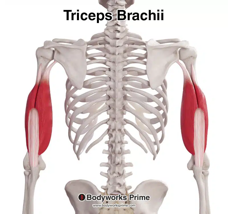
Pictured here we can see the triceps brachii from a posterior view.

Pictured here we can see the triceps brachii from a posterolateral view.
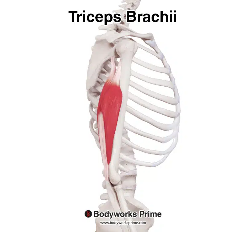
Pictured here we can see the triceps brachii from a lateral view.
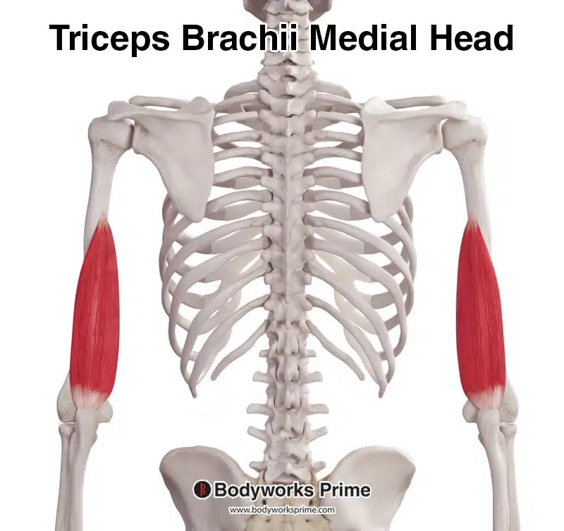
Pictured here we can see just the medial head of the triceps brachii. This head is deep to the long and lateral heads so is covered up in the previous images.
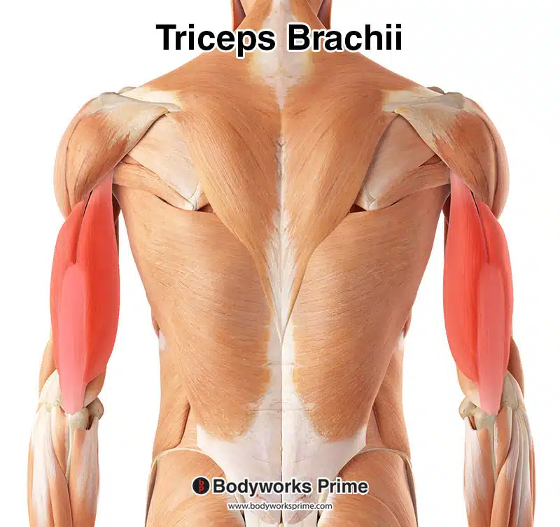
Here we can see the triceps brachii highlighted in red, seen from a posterior view, amongst the other muscles of the body.
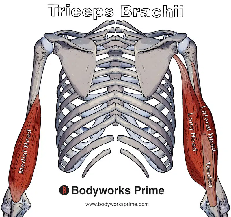
Pictured here we can see all three heads of the triceps brachii muscle as well as their common tendon.
Origin & Insertion
The triceps brachii’s distinctive three-headed structure gives it a trio of origin points, each associated with a different part of the upper arm’s skeletal structure. The muscle then converges to a single common tendon attaching to the olecranon process of the ulna [5] [6] .
The triceps’ long head originates on the infraglenoid tubercle of the scapula. This bony landmark is a small bony protrusion on the lower part of the scapula’s lateral aspect (the side of the shoulder blade facing away from the body). This is the only head of the triceps that crosses the shoulder joint, which in turn allows it to act on the shoulder joint [7] [8]. This is further discussed in the actions section.
The lateral head originates above the radial groove. The radial groove is a shallow depression that runs obliquely down the posterior aspect of the humerus. This groove provides a pathway for the radial nerve and the deep branch of the brachial artery [9] [10].
As for the medial head of the triceps, this originates just below the radial groove. This head is mostly concealed by the overlying lateral and long heads, but it still is able to add significant power to the triceps muscle [11] [12].
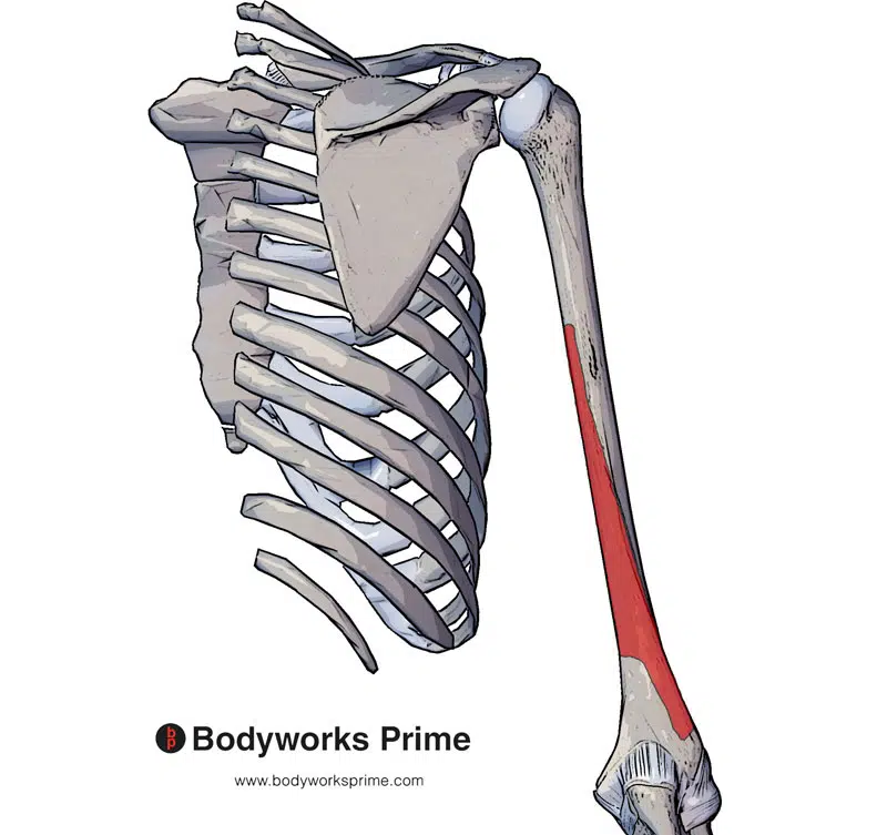
The fibers of these three tricep heads converge to form a single tendon, a tough band of connective tissue. This tendon inserts onto the olecranon process of the ulna. The olecranon process of the ulna is a large, curved bony prominence that can easily be felt on the back of the elbow. The ulna is one of the two bones of the forearm, with the olecranon process being the pointy part of your elbow that sticks out when you bend your arm [13] [14]. Research by Madsen, M. et al. (2006) proposed that “The medial head of the triceps has a tendon that is distinct from, and deep to, the common tendon of the long and lateral heads” suggesting that the medial head has an additional deep attachment [15].
The combination of these three diverse origin points and the shared insertion point on the olecranon of the ulna results in a versatile muscle capable of executing a range of arm movements at both the elbow and shoulder which is discussed further in the next section.
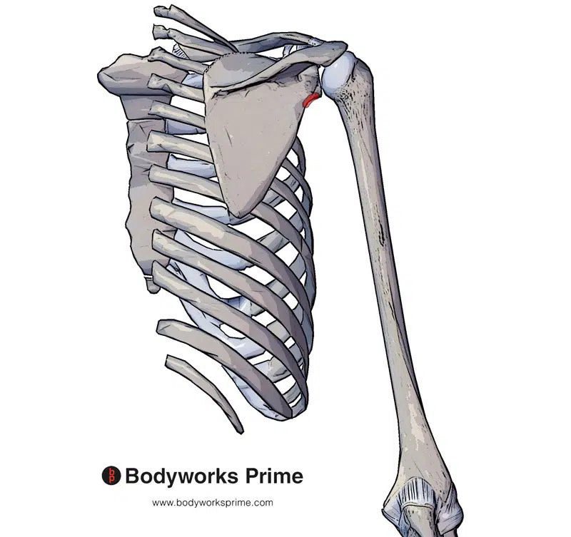
Pictured here we can see the origin of the long head of the triceps brachii at the infraglenoid tubercle of scapula (origin highlighted in red).
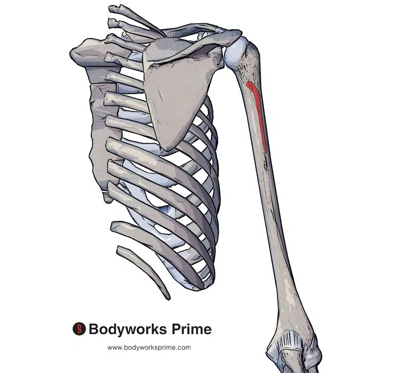
Pictured here we can see the origin of the lateral head which originates above the radial groove (origin highlighted in red).
Pictured here we can see the origin of the medial head of the triceps brachii which is below the radial groove (origin highlighted in red).
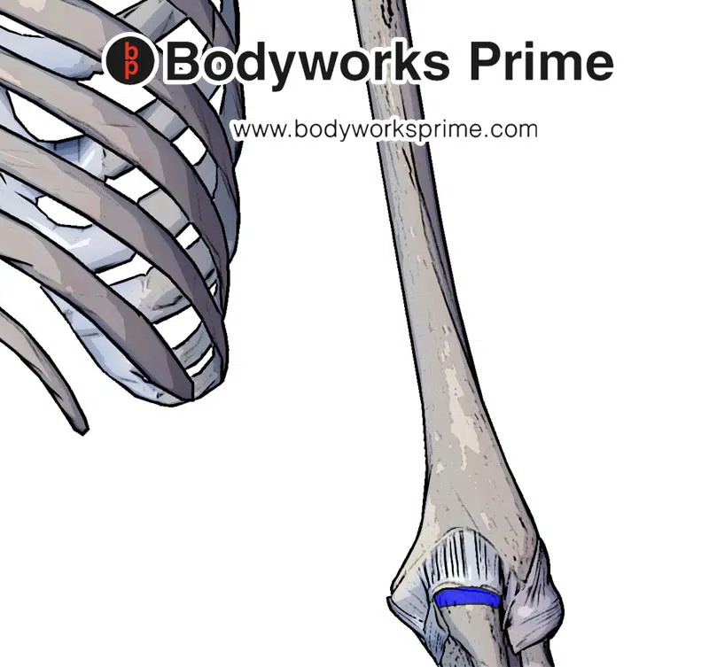
Pictured here we can see the insertion of the triceps brachii at the olecranon process of ulna.
Actions
All the heads of the triceps brachii can assist in extension of elbow. However, because the long head crosses the shoulder joint; the long head can also assist in adduction and extension of the arm at the shoulder joint [16] [17].
The primary action of the triceps brachii as a whole is to extend the elbow joint. When this action occurs, it straightens the arm, counteracting the bicep brachii muscle’s action of bending the arm at the elbow [18] [19]. Research by Kholinne, E. et al. (2018) proposed that “each head of the triceps brachii has a different pattern of force and activity during different shoulder elevations. The long head contributes to elbow extension more at shoulder elevation and the medial head takes over at 90° and above of shoulder elevation” illustrating the complex interaction of the heads of the triceps brachii [20].
The three different heads of the triceps do not always act in the same way. While they all contribute to elbow extension, the long head, due to its unique attachment to the scapula, has additional functions. Since it crosses the shoulder joint, it also assists in adduction and extension of the arm at the shoulder joint. Furthermore, the long head also plays a role in stabilising the shoulder joint too [21] [22].
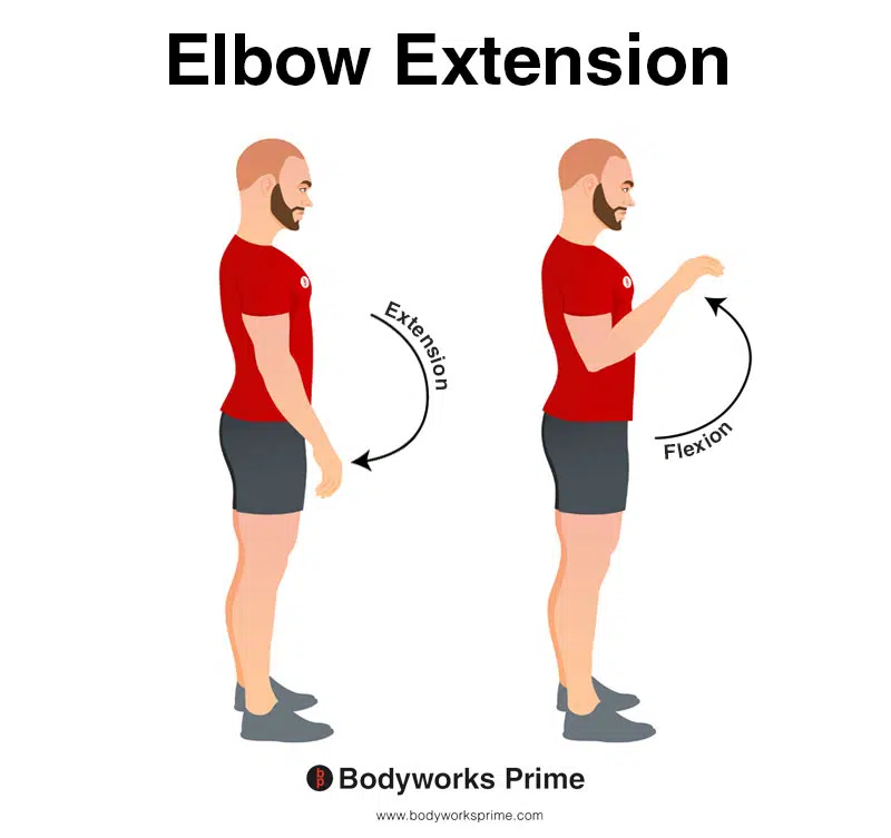
This image demonstrates elbow extension, which involves straightening the elbow. The opposite movement of elbow extension is elbow flexion. This action is the primary function of the triceps brachii muscle.
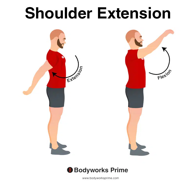
This image shows shoulder extension. Extension is where the arm is moved backward in a posterior direction (towards the back of the body), increasing the angle between the arm and the torso in the sagittal plane. The opposite of this is shoulder flexion which occurs as the arm is moved forward and upward in an anterior direction. Only the long head can assist with shoulder extension.
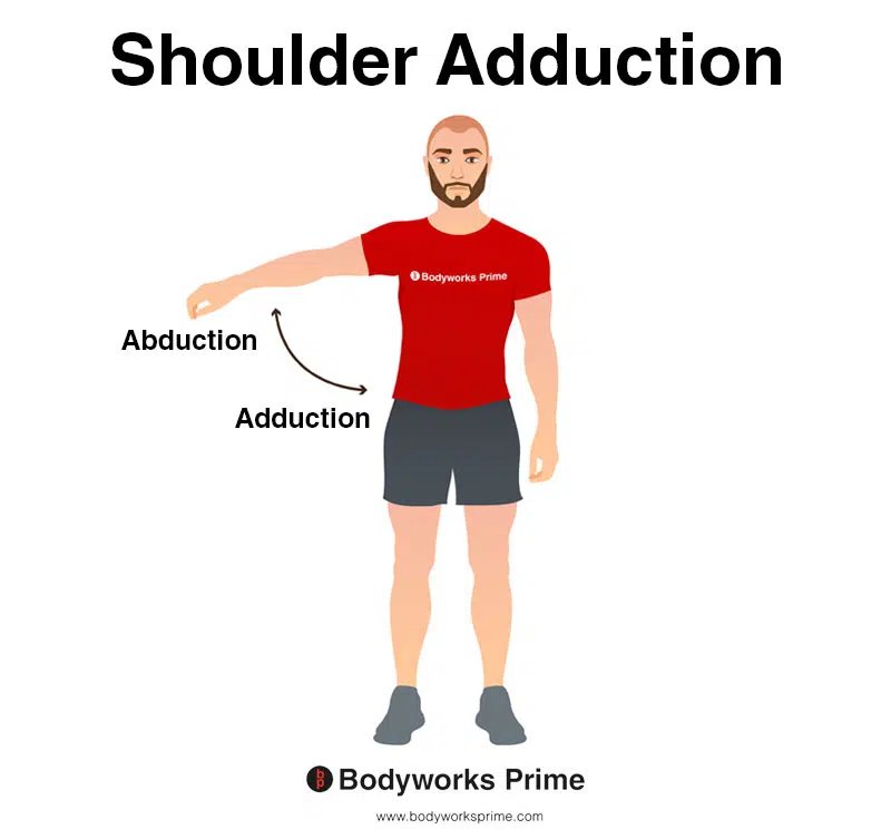
This image shows shoulder adduction. Adduction is where the arm is brought towards the midline of the body, reducing the angle between the arm and the torso. The opposite of adduction is abduction. Abduction occurs as the arm is moved away from the midline, increasing the angle between the arm and the torso. Only the long head can assist with shoulder adduction.
Innervation
The three heads of the triceps brachii muscle is innervated by the radial nerve, specifically the fibers from the sixth, seventh, and eighth cervical nerve roots, commonly represented as C6, C7, and C8 [23] [24].
The radial nerve is one of the five main nerves of the brachial plexus. The brachial plexus is a complex network of nerves that originate from the spinal cord’s cervical region. The C6, C7, and C8 nerve roots emerge from the spinal cord in the neck region, joining together with the first thoracic root (T1) to form the brachial plexus (along with C5). This intricate network travels down the neck, under the collarbone, and into the armpit, branching out to provide innervation to the entire upper limb [25] [26].
The radial nerve arises from the brachial plexus, extending along the posterior aspect of the upper limb. After working its way around the humerus in the ‘spiral groove’, a winding path along the upper arm bone, the radial nerve reaches the posterior compartment of the arm, where it gives off branches to the triceps brachii [27] [28].
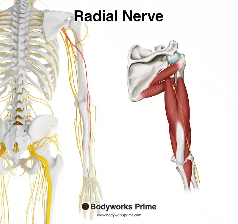
This image shows the radial nerve highlighted in red on the left and a depiction of its location in relation to the triceps brachii on the right.
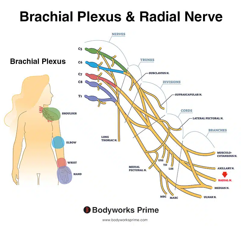
Here we can see the brachial plexus with the radial nerve indicated with the red arrow.
Blood Supply
Blood is supplied to the triceps brachii muscle primarily via two arteries: the deep brachial artery (also known as the profunda brachii artery) and the superior ulnar collateral artery [29] [30].
The deep brachial artery is the primary supplier of blood to the triceps brachii muscle. This artery originates from the brachial artery, which is a continuation of the axillary artery beyond the border of the teres major muscle. Upon its origin, the deep brachial artery courses posteriorly between the long and medial heads of the triceps brachii. It travels along the radial nerve in the radial (spiral) groove of the humerus, supplying blood to all three heads of the triceps brachii muscle[31] [32].
The superior ulnar collateral artery, a branch of the brachial artery, also contributes to the blood supply of the triceps brachii, particularly the medial head. It travels with the ulnar nerve posterior to the medial epicondyle of the humerus, providing additional blood flow to the triceps brachii muscle [33] [34].
In addition to these primary arteries, the long head of the triceps brachii also receives blood from the posterior circumflex humeral artery. This artery arises from the axillary artery, typically beyond the lateral border of the pectoralis minor muscle, and this artery courses posteriorly around the surgical neck of the humerus through the quadrangular space with the axillary nerve. The posterior circumflex humeral artery provides branches that supply the shoulder joint and the surrounding muscles, including the deltoid, the teres major and teres minor, and importantly, the long head of the triceps brachii [35].
Want some flashcards to help you remember this information? Then click the link below:
Triceps Brachii Flashcards
Support Bodyworks Prime
Running a website and YouTube channel can be expensive. Your donation helps support the creation of more content for my website and YouTube channel. All donation proceeds go towards covering expenses only. Every contribution, big or small, makes a difference!
References
| ↑1, ↑3, ↑4, ↑22, ↑23, ↑25, ↑27 | Moore KL, Agur AMR, Dalley AF. Clinically Oriented Anatomy. 8th ed. Philadelphia: Lippincot Williams & Wilkins; 2017. |
|---|---|
| ↑2, ↑15, ↑17 | Madsen M, Marx RG, Millett PJ, Rodeo SA, Sperling JW, Warren RF. Surgical anatomy of the triceps brachii tendon: anatomical study and clinical correlation. Am J Sports Med. 2006 Nov;34(11):1839-43. doi: 10.1177/0363546506288752. Epub 2006 May 30. PMID: 16735585. |
| ↑5, ↑7, ↑9, ↑11, ↑13, ↑18, ↑21, ↑29, ↑31, ↑33 | Standring S. (2015). Gray’s Anatomy: The Anatomical Basis of Clinical Practice, 41st Edn. Amsterdam: Elsevier. |
| ↑6, ↑8, ↑10, ↑12, ↑14, ↑19 | Tiwana MS, Sinkler MA, Bordoni B. Anatomy, Shoulder and Upper Limb, Triceps Muscle. [Updated 2022 Aug 30]. In: StatPearls [Internet]. Treasure Island (FL): StatPearls Publishing; 2023 Jan-. Available from: https://www.ncbi.nlm.nih.gov/books/NBK536996/ |
| ↑16, ↑20 | Kholinne E, Zulkarnain RF, Sun YC, Lim S, Chun JM, Jeon IH. The different role of each head of the triceps brachii muscle in elbow extension. Acta Orthop Traumatol Turc. 2018 May;52(3):201-205. doi: 10.1016/j.aott.2018.02.005. Epub 2018 Mar 2. PMID: 29503079; PMCID: PMC6136322. |
| ↑24, ↑26, ↑28 | Glover NM, Murphy PB. Anatomy, Shoulder and Upper Limb, Radial Nerve. 2022 Aug 29. In: StatPearls [Internet]. Treasure Island (FL): StatPearls Publishing; 2023 Jan–. PMID: 30521261. |
| ↑30, ↑32, ↑34 | Epperson TN, Varacallo M. Anatomy, Shoulder and Upper Limb, Brachial Artery. [Updated 2022 Jul 25]. In: StatPearls [Internet]. Treasure Island (FL): StatPearls Publishing; 2023 Jan-. Available from: https://www.ncbi.nlm.nih.gov/books/NBK537145/ |
| ↑35 | Tang A, Varacallo M. Anatomy, Head and Neck, Posterior Humeral Circumflex Artery. 2022 Aug 8. In: StatPearls [Internet]. Treasure Island (FL): StatPearls Publishing; 2023 Jan–. PMID: 30855867. |


