| Origin | Anterior and lateral surface of the femur & lateral intermuscular septum |
| Insertion | Quadriceps tendon, which inserts into the patellar ligament, which inserts onto the tibial tuberosity |
| Action | Extends the knee |
| Nerve | Femoral nerve (L2, L3, L4) |
| Artery | Femoral artery |
Location & Overview
The vastus intermedius muscle is one of the four quadricep muscles, along with the vastus lateralis, vastus medialis, and rectus femoris. The vastus intermedius is located deep to the rectus femoris, between the vastus medialis and vastus lateralis. It is almost hidden by the other muscles and cannot be palpated [1]. Some studies have also reported a fusion of the vastus intermedius and vastus lateralis muscles [2] [3]. In extreme cases of this fusion, the two muscle present as an almost complete fusion [4].
Pathologies involving the vastus intermedius muscle are less commonly reported compared to other quadricep muscles, likely due to its deep location. However, strains and contusions can still occur, typically as a result of overuse or direct trauma [5]. Weakness of the vastus intermedius muscle may contribute to instability of the knee joint and increased risk of injury [6].
Strengthening exercises targeting the vastus intermedius muscle are essential for maintaining knee joint stability and overall lower limb function. Exercises that engage the vastus intermedius include squats, lunges, leg presses, and leg extensions [7] [8]. Incorporating these exercises into a well-rounded training program can help prevent injuries and enhance athletic performance.
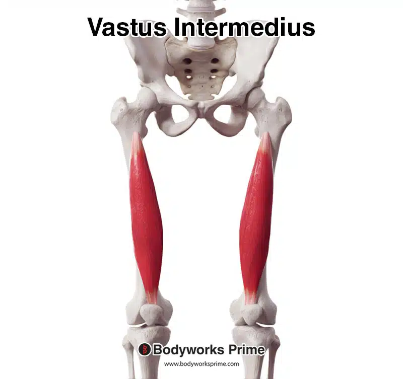
Here we can see the vastus intermedius from an anterior view.
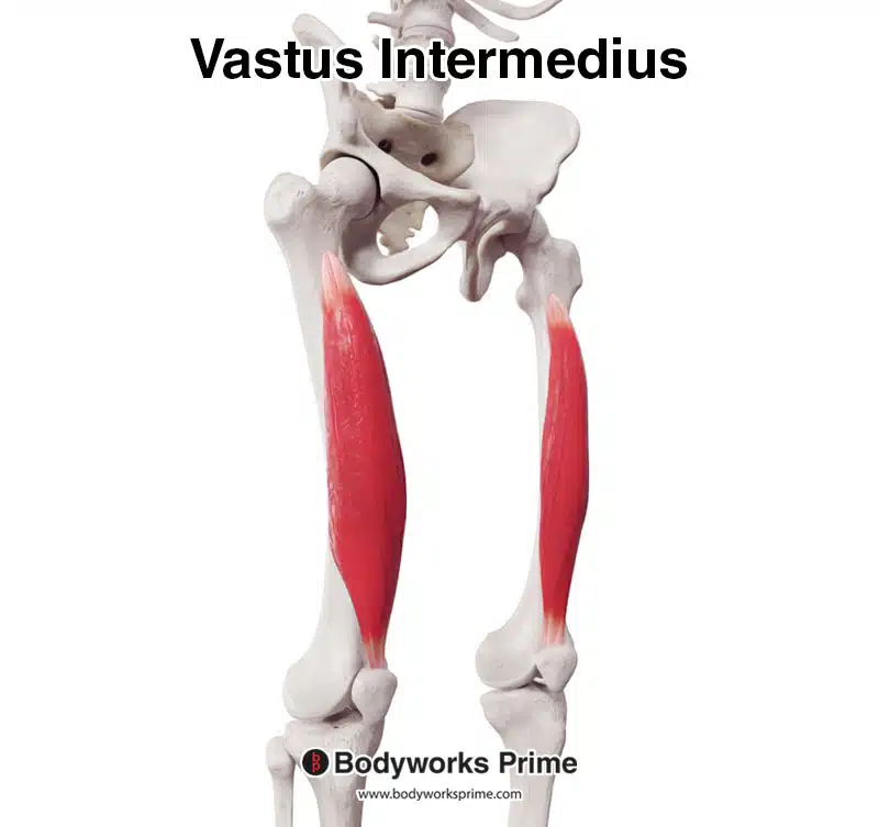
Here we can see the vastus intermedius from an anterolateral view.
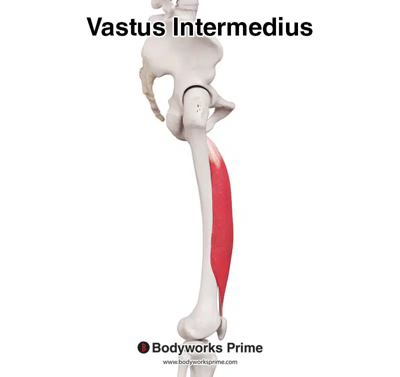
Here we can see the vastus intermedius from a lateral view.
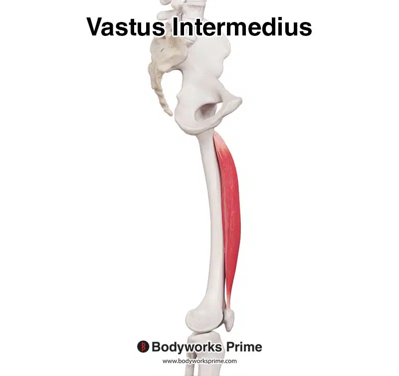
Here we can see the vastus intermedius from a medial view.
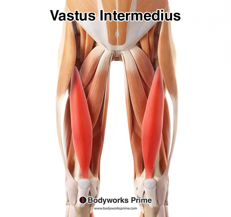
Here we can see the vastus intermedius highlighted in red amongst the other muscles of the thigh. Because the vastus intermedius is a deep muscle, the other quadriceps muscles have been removed from this image to reveal the vastus intermedius.
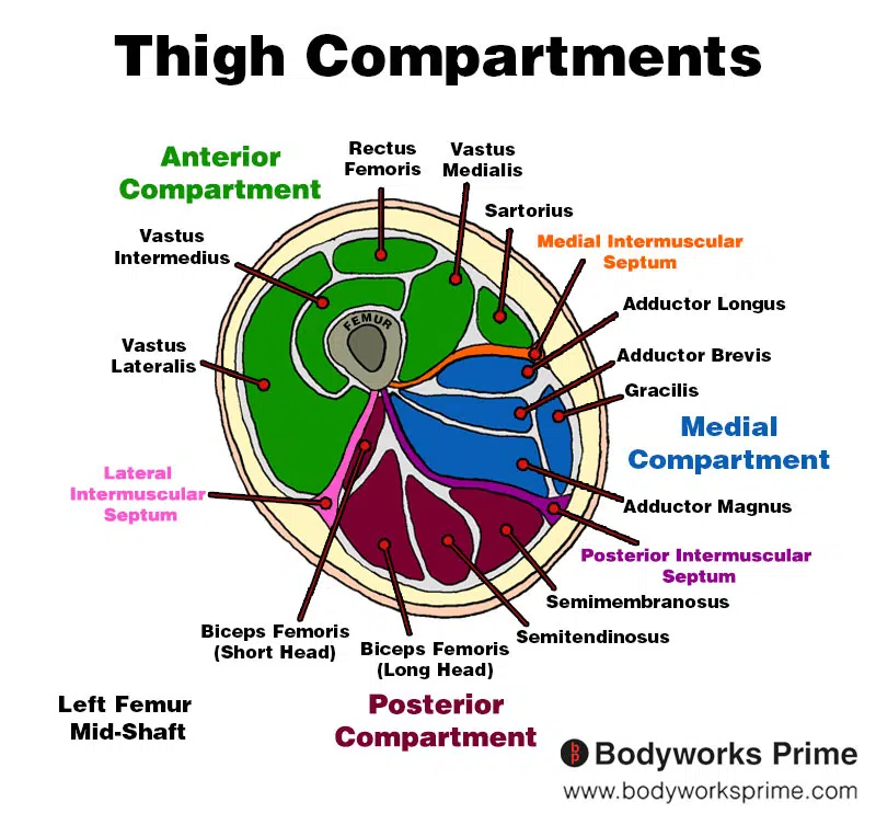
Here we can see an image of the compartments of the thigh. We can see the vastus intermedius in the anterior compartment, the section coloured in green.
Origin & Insertion
The vastus intermedius originates on the anterior and lateral surface of the shaft of the femur [9]. The femur, also known as the thigh bone, is the longest and strongest bone in the human body. The anterior surface refers to the front side of the femur, while the lateral surface refers to the outer side of the bone. The vastus intermedius also originates from the lateral intermuscular septum, a deep fascia fold separating the anterior and posterior compartments of the thigh [10]. The vastus intermedius then inserts into the quadriceps tendon, a strong band of fibrous tissue that connects the muscle to the patella (or kneecap) [11] [12]. Most of the vastus intermedius’ tendon fibers insert at the base of the patella [13].
The quadriceps tendon serves as a common insertion point for all four quadricep muscles, including the vastus intermedius. This shared tendon allows the muscles to work together to extend the knee joint. By connecting to the quadriceps tendon, the vastus intermedius helps with the overall function and stability of the knee joint, alongside the other quadricep muscles [14].
The quadriceps tendon connects to the patellar tendon, which in turn inserts onto the tibial tuberosity. The tibial tuberosity is a bony prominence on the upper part of the tibia (shin bone), located just below the knee joint [15].
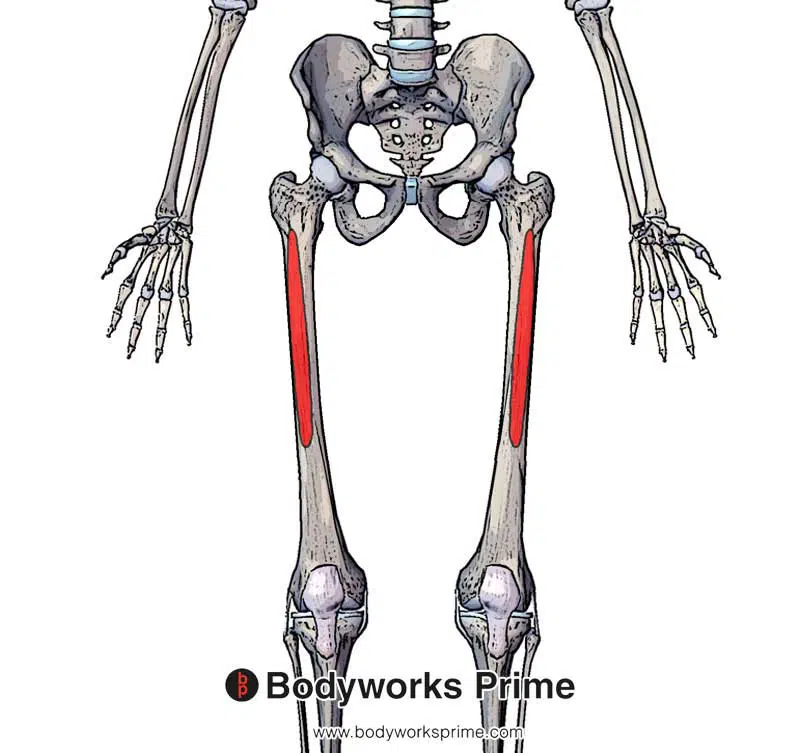
Here we can see the origin of the vastus intermedius on the anterior and lateral surface of the femur.
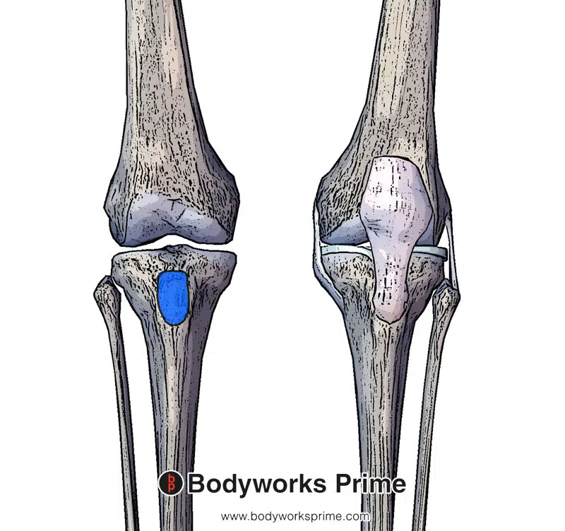
Here we can see the insertion of the patellar tendon. The vastus intermedius fibres connect into the patellar tendon.
Actions
The vastus intermedius, along with the other quadricep muscles (vastus lateralis, vastus medialis, and rectus femoris), plays an important role in extending the knee joint [16]. Knee extension is essential for various daily activities, such as walking, running, climbing stairs, and standing up from a seated position. During these activities, the vastus intermedius contracts and works synergistically with the other quadricep muscles to straighten the leg at the knee joint [17].
In addition to knee extension, the vastus intermedius is also involved with maintaining knee stability [18]. As one of the deeper quadricep muscles, the vastus intermedius provides support to the patella and helps distribute force across the knee joint. This supportive role is useful for maintaining proper knee alignment and preventing excessive stress on the joint during activities that require dynamic knee movements, such as sports and exercise [19]. Moreover, the vastus intermedius contributes to the eccentric control of the knee during activities such as descending stairs and landing from a jump. Eccentric control refers to the ability of a muscle to generate force while lengthening, which acts like a brake to slow down joint movement [20].
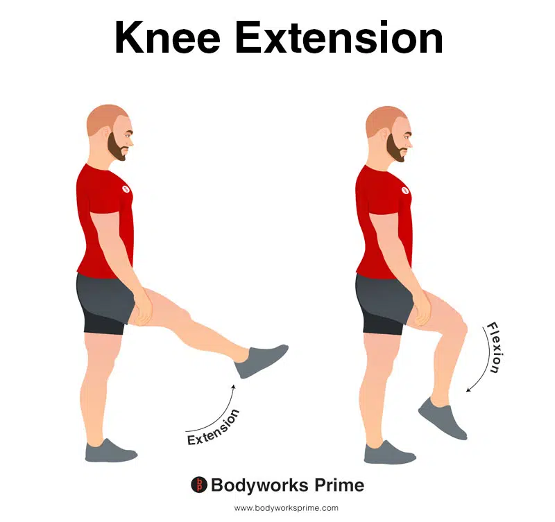
In this image, you can see an example of knee extension, which is the action of straightening out your leg at the knee. The opposite movement of knee flexion is knee extension. The main action of the vastus intermedius muscle is knee extension.
Innervation
The vastus intermedius muscle receives its innervation from the femoral nerve, which is derived from the lumbar spinal nerve roots L2, L3, and L4 [21]. The femoral nerve is a large nerve that originates from the lumbar plexus in the lower back, passes through the pelvis, and descends into the thigh. It is the primary nerve which innervates the quadriceps muscle group, including the vastus intermedius, vastus lateralis, vastus medialis, and rectus femoris muscles [22].
Proper function of the femoral nerve is vital for the coordinated movements and stability of the knee. Damage or injury to the femoral nerve can result in muscle weakness or even paralysis, affecting the ability to extend the knee and potentially leading to difficulty when walking or maintaining balance [23]
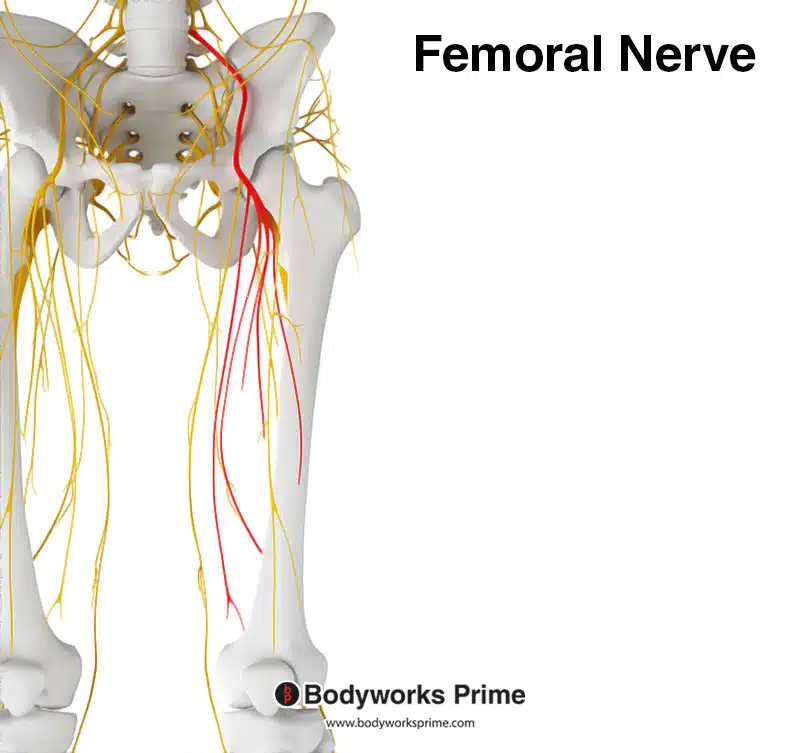
Here we can see the femoral nerve highlighted in red. The femoral nerve innervates the quadriceps muscle group, including the vastus intermedius. The femoral nerve originates from the spinal nerve roots of L2, L3, and L4.

Here we can see the innervation of the quadriceps muscle group, the femoral nerve is a major branch of the lumbar plexus.
Blood Supply
Blood is supplied to the vastus intermedius via the femoral artery. The femoral artery begins at the inguinal ligament as the continuation of the external iliac artery. It courses through the femoral triangle of the thigh, continuing down the leg and supplying the many muscles and structures within this region, including the quadricep muscles [24] [25].
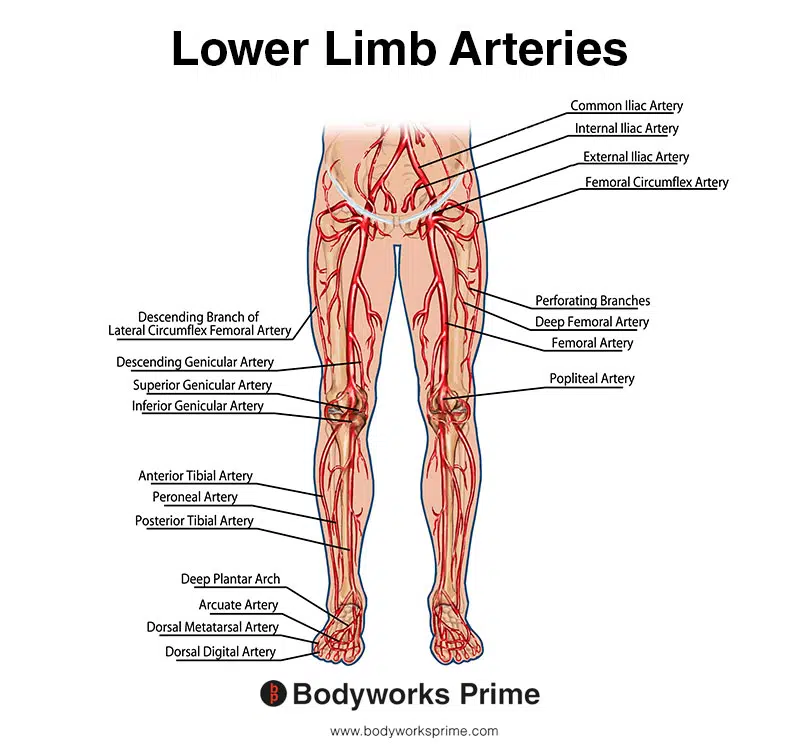
This image shows the arteries of the lower limb, including the femoral artery.
Want some flashcards to help you remember this information? Then click the link below:
Vastus Intermedius Flashcards
Support Bodyworks Prime
Running a website and YouTube channel can be expensive. Your donation helps support the creation of more content for my website and YouTube channel. All donation proceeds go towards covering expenses only. Every contribution, big or small, makes a difference!
References
| ↑1, ↑14, ↑15, ↑17, ↑20, ↑22 | Moore KL, Agur AMR, Dalley AF. Clinically Oriented Anatomy. 8th ed. Philadelphia: Lippincot Williams & Wilkins; 2017. |
|---|---|
| ↑2, ↑4 | Willan PL, Mahon M, Golland JA. Morphological variations of the human vastus lateralis muscle. J Anat. 1990 Feb;168:235-9. PMID: 2323995; PMCID: PMC1256904. |
| ↑3 | Becker I, Woodley SJ, Baxter GD. Gross morphology of the vastus lateralis muscle: An anatomical review. Clin Anat. 2009 May;22(4):436-50. doi: 10.1002/ca.20792. PMID: 19306318. |
| ↑5, ↑6, ↑7 | Escamilla RF, Fleisig GS, Zheng N, Barrentine SW, Wilk KE, Andrews JR. Biomechanics of the knee during closed kinetic chain and open kinetic chain exercises. Med Sci Sports Exerc. 1998 Apr;30(4):556-69. doi: 10.1097/00005768-199804000-00014. PMID: 9565938. |
| ↑8 | Signorile JF, Weber B, Roll B, et al. A comparative electromyographical investigation of the squat and knee extension exercises. J Strength Cond Res. 1994 Aug;8(3):178-83. doi: 10.1519/00124278-199408000-00006. |
| ↑9, ↑12 | Yoshida S, Ichimura K, Sakai T. Structural diversity of the vastus intermedius origin revealed by analysis of isolated muscle specimens. Clin Anat. 2017 Jan;30(1):98-105. doi: 10.1002/ca.22791. Epub 2016 Oct 31. PMID: 27598432. |
| ↑10 | Waligora AC, Johanson NA, Hirsch BE. Clinical anatomy of the quadriceps femoris and extensor apparatus of the knee. Clin Orthop Relat Res. 2009 Dec;467(12):3297-306. doi: 10.1007/s11999-009-1052-y. Epub 2009 Aug 19. PMID: 19690926; PMCID: PMC2772911. |
| ↑11, ↑18 | Grob K, Manestar M, Filgueira L, Kuster MS, Gilbey H, Ackland T. The interaction between the vastus medialis and vastus intermedius and its influence on the extensor apparatus of the knee joint. Knee Surg Sports Traumatol Arthrosc. 2018 Mar;26(3):727-738. doi: 10.1007/s00167-016-4396-3. Epub 2017 Jan 25. PMID: 28124107. |
| ↑13, ↑16, ↑21, ↑25 | Bordoni B, Varacallo M. Anatomy, Bony Pelvis and Lower Limb, Thigh Quadriceps Muscle. 2021 Jul 22. In: StatPearls [Internet]. Treasure Island (FL): StatPearls Publishing; 2021 Jan–. PMID: 30020706. |
| ↑19 | Neumann DA. Kinesiology of the Musculoskeletal System: Foundations for Rehabilitation. 3rd ed. St. Louis, MO: Elsevier; 2017. |
| ↑23 | Luo TD, Ashraf A, Dahm DL, Stuart MJ, McIntosh AL. Femoral nerve block is associated with persistent strength deficits at 6 months after anterior cruciate ligament reconstruction in pediatric and adolescent patients. Am J Sports Med. 2015 Feb;43(2):331-6. doi: 10.1177/0363546514559823. Epub 2014 Dec 2. PMID: 25466410. |
| ↑24 | Hammond E, Nassereddin A, Costanza M. Anatomy, Abdomen and Pelvis: External Iliac Arteries. [Updated 2022 Aug 8]. In: StatPearls [Internet]. Treasure Island (FL): StatPearls Publishing; 2023 Jan-. Available from: https://www.ncbi.nlm.nih.gov/books/NBK519552/ |


