| Origin | Greater trochanter Intertrochanteric line Lateral aspect of the linea aspera Gluteal tuberosity Lateral intermuscular septum |
| Insertion | Lateral aspect of the patella The quadriceps tendon, which inserts into the patellar tendon, which connects onto the tibial tuberosity |
| Action | Knee extension |
| Nerve | Femoral nerve (L2, L3, L4) |
| Artery | Femoral artery |
Location & Overview
The vastus lateralis is a large, thick muscle located in the anterior compartment of the thigh. It is one of the four quadricep muscles, which together make up the quadriceps femoris muscle group, responsible for extending the knee and stabilising the knee joint [1] [2]. The vastus lateralis is the most lateral and largest of these quadricep muscles, playing an important role in maintaining balance and stability during physical activities [3] The other three quadricep muscles are the vastus intermedius, vastus medialis, and rectus femoris [4] [5]. Some studies have also reported a fusion of the vastus lateralis and vastus intermedius muscles [6] [7]. In extreme cases of this fusion, the two muscle present as an almost complete fusion [8].
The vastus lateralis is not only essential for the proper functioning of the lower limb but also serves as a common site for intramuscular injections due to its large size and accessibility [9] [10].
Common pathologies involving the vastus lateralis muscle include muscle strain, tendinopathy, and muscle imbalances, which may contribute to conditions such as patellofemoral pain syndrome and iliotibial band syndrome [11] [12]. Strengthening exercises targeting the vastus lateralis can help prevent injuries and improve overall lower limb function. Research has shown that exercises such as squats, lunges, leg presses, and step-ups effectively activate the vastus lateralis and other quadriceps muscles [13] [14].
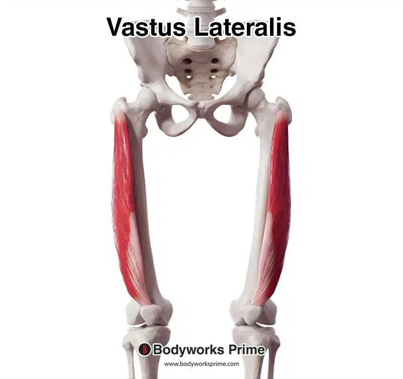
Here we can see the vastus lateralis in isolation from an anterior view.
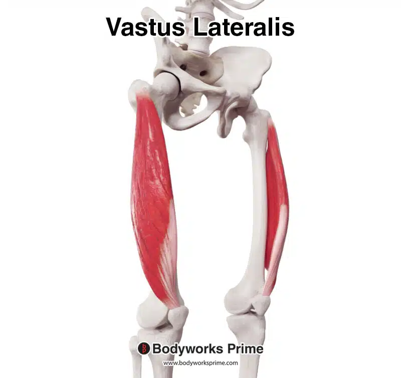
Here we can see the vastus lateralis in isolation from an anterolateral view.
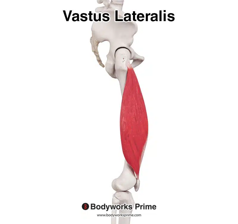
Here we can see the vastus lateralis in isolation from a lateral view.
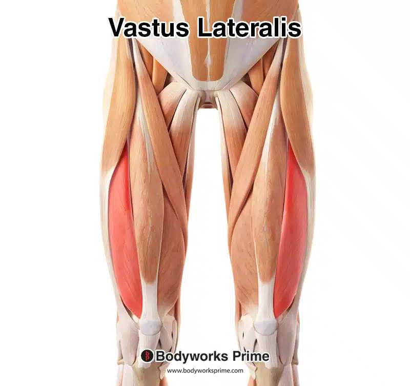
Pictured here is the vastus lateralis highlighted in red, seen from an anterior view amongst the other muscles.
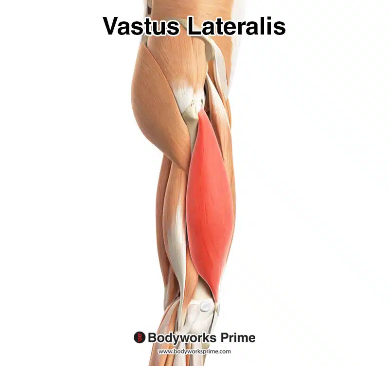
Pictured here is the vastus lateralis highlighted in red, seen from a lateral view amongst the other muscles. The iliotibial band and the tensor fascia latae muscles have been removed, due to a portion of the vastus lateralis being deep to the iliotibial band. These can still be seen in the previous picture seen from an anterior view.
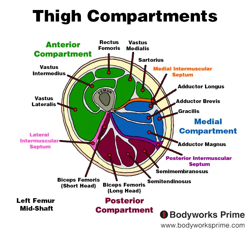
Here we can see an image of the compartments of the thigh. We can see the vastus lateralis in the anterior compartment, the section coloured in green.
Origin & Insertion
The vastus lateralis originates from multiple locations on the femur. These locations include the greater trochanter, which is a prominent bony protrusion close to the hip joint; the intertrochanteric line, a ridge that connects the greater and lesser trochanters; the lateral aspect of the linea aspera, a vertical ridge on the posterior surface of the femur; the gluteal tuberosity, a roughened area on the proximal end of the femur; and the lateral intermuscular septum [15] [16]. The lateral intermuscular septum of the thigh is a fold of deep fascia that separates the anterior compartment of the thigh from the posterior compartment. It divides the vastus lateralis and the biceps femoris muscle, providing an origin point for the vastus lateralis [17] [18].
The vastus lateralis inserts into the quadriceps tendon, which attaches to the patellar tendon. The patellar tendon inserts onto the tibial tuberosity, a bony prominence located on the front of the tibia below the knee joint. Additionally, the vastus lateralis also inserts onto the lateral aspect of the patella, the triangular-shaped bone that forms the kneecap [19] [20].
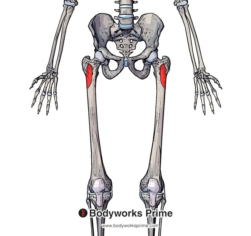
Here we can see the anterior origins of the vastus lateralis, highlighted in red, on the anterior side of the femur (greater trochanter and intertrochanteric line).
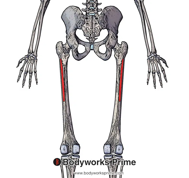
Here we can see the posterior origins of the vastus lateralis, highlighted in red, on the posterior side of the femur (lateral aspect of the linea aspera, gluteal tuberosity, and lateral intermuscular septum).
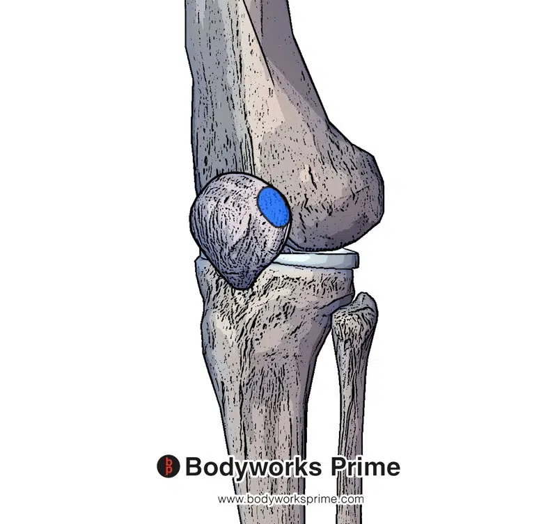
Here we can see an insertion of the vastus lateralis, highlighted in blue, on lateral side of the patellar.
Here we can see the insertion of the patellar tendon at the tibial tuberosity. The vastus lateralis inserts into the quadriceps tendon which then inserts into the patellar tendon.
Actions
The vastus lateralis muscle’s primary action is knee extension, working alongside the other muscles of the quadriceps femoris group to extend the knee during activities such as walking, running, and jumping [21]. It also plays an important role in stabilising the knee joint, working together with the vastus medialis to maintain proper tracking of the patella and prevent excessive lateral movement [22]. The vastus lateralis also plays a role in eccentric control during deceleration, helping to absorb impact forces when landing from a jump or slowing down from a sprint [23].
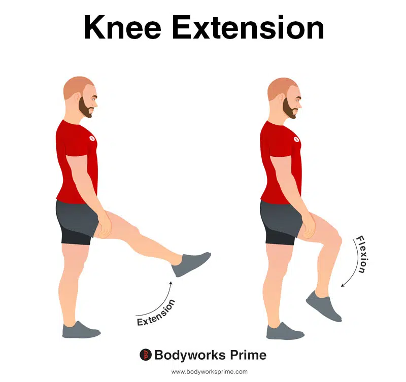
In this image, you can see an example of knee extension, which is the action of straightening out your leg at the knee joint. The opposite movement of knee flexion is knee extension. The vastus lateralis’ main function is knee extension.
Innervation
The vastus lateralis muscle is innervated by the femoral nerve, which arises from the lumbar plexus, a network of nerves formed by the anterior rami of spinal nerve roots L2, L3, and L4 [24] [25]. The femoral nerve descends through the psoas major muscle in the abdomen and enters the thigh by passing beneath the inguinal ligament, a fibrous band that runs from the anterior superior iliac spine to the pubic tubercle, forming the base of the abdominal wall [26].
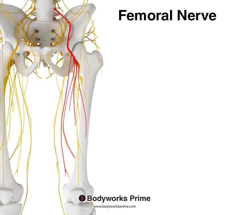
Here we can see the femoral nerve highlighted in red. The femoral nerve innervates the quadriceps muscle group, including the vastus lateralis. The femoral nerve originates from the spinal nerve roots of L2, L3, and L4.
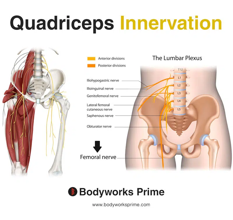
Here we can see the innervation of the quadriceps muscle group, the femoral nerve is a major branch of the lumbar plexus.
Blood Supply
The vastus lateralis primarily gets its blood supply from the lateral circumflex femoral artery. It also receives some blood from the perforating arteries of the profunda femoris [27].
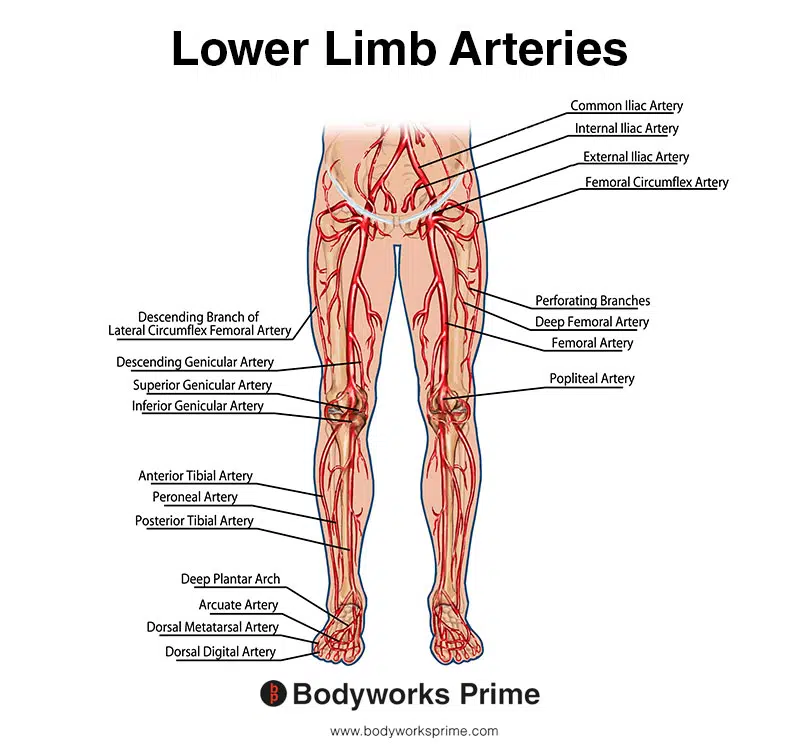
This image shows the arteries of the lower limb, including the femoral artery.
Want some flashcards to help you remember this information? Then click the link below:
Vastus Lateralis Flashcards
Support Bodyworks Prime
Running a website and YouTube channel can be expensive. Your donation helps support the creation of more content for my website and YouTube channel. All donation proceeds go towards covering expenses only. Every contribution, big or small, makes a difference!
References
| ↑1, ↑4, ↑21 | Bordoni B, Varacallo M. Anatomy, Bony Pelvis and Lower Limb, Thigh Quadriceps Muscle. 2021 Jul 22. In: StatPearls [Internet]. Treasure Island (FL): StatPearls Publishing; 2021 Jan–. PMID: 30020706. |
|---|---|
| ↑2, ↑5 | Khan A, Arain A. Anatomy, Bony Pelvis and Lower Limb, Anterior Thigh Muscles. 2021 Jul 26. In: StatPearls [Internet]. Treasure Island (FL): StatPearls Publishing; 2021 Jan–. PMID: 30860696. |
| ↑3 | Ema R, Wakahara T, Mogi Y, et al. In vivo measurement of human rectus femoris architecture by ultrasonography: validity and applicability. Clin Physiol Funct Imaging. 2013 Nov;33(3):267-73. doi: 10.1111/cpf.12018. Epub 2013 Feb 28. PMID: 23551745. |
| ↑6, ↑8 | Willan PL, Mahon M, Golland JA. Morphological variations of the human vastus lateralis muscle. J Anat. 1990 Feb;168:235-9. PMID: 2323995; PMCID: PMC1256904. |
| ↑7 | Becker I, Woodley SJ, Baxter GD. Gross morphology of the vastus lateralis muscle: An anatomical review. Clin Anat. 2009 May;22(4):436-50. doi: 10.1002/ca.20792. PMID: 19306318. |
| ↑9 | Nakajima Y, Fujii T, Mukai K, Ishida A, Kato M, Takahashi M, Tsuda M, Hashiba N, Mori N, Yamanaka A, Ozaki N, Nakatani T. Anatomically safe sites for intramuscular injections: a cross-sectional study on young adults and cadavers with a focus on the thigh. Hum Vaccin Immunother. 2020;16(1):189-196. doi: 10.1080/21645515.2019.1646576. Epub 2019 Aug 23. PMID: 31403356; PMCID: PMC7012163. |
| ↑10 | Polania Gutierrez JJ, Munakomi S. Intramuscular Injection. [Updated 2023 Feb 12]. In: StatPearls [Internet]. Treasure Island (FL): StatPearls Publishing; 2023 Jan-. Available from: https://www.ncbi.nlm.nih.gov/books/NBK556121/ |
| ↑11 | Kujala UM, Osterman K, Kormano M, Nelimarkka O, Hurme M, Taimela S. Patellofemoral relationships in recurrent patellar dislocation. J Bone Joint Surg Br. 1989 Nov;71(5):788-92. doi: 10.1302/0301-620X.71B5.2584248. PMID: 2584248. |
| ↑12 | Fredericson M, Cookingham CL, Chaudhari AM, Dowdell BC, Oestreicher N, Sahrmann SA. Hip abductor weakness in distance runners with iliotibial band syndrome. Clin J Sport Med. 2000 Jul;10(3):169-75. doi: 10.1097/00042752-200007000-00004. PMID: 10959926. |
| ↑13 | Escamilla RF, Fleisig GS, Zheng N, Lander JE, Barrentine SW, Andrews JR, Bergemann BW, Moorman CT 3rd. Effects of technique variations on knee biomechanics during the squat and leg press. Med Sci Sports Exerc. 2001 Sep;33(9):1552-66. doi: 10.1097/00005768-200109000-00020. PMID: 11528346. |
| ↑14 | Signorile, Joseph F.1; Weber, Brad2; Roll, Brad2; Caruso, John F.3; Lowensteyn, Ilka3; Perry, Arlette C.3. An Electromyographical Comparison of the Squat and Knee Extension Exercises. Journal of Strength and Conditioning Research 8(3):p 178-183, August 1994. |
| ↑15, ↑19, ↑24, ↑27 | Biondi NL, Varacallo M. Anatomy, Bony Pelvis and Lower Limb, Vastus Lateralis Muscle. [Updated 2021 Aug 13]. In: StatPearls [Internet]. Treasure Island (FL): StatPearls Publishing; 2021 Jan-. Available from: https://www.ncbi.nlm.nih.gov/books/NBK532309/ |
| ↑16, ↑18, ↑20 | Moore KL, Agur AMR, Dalley AF. Clinically Oriented Anatomy. 8th ed. Philadelphia: Lippincot Williams & Wilkins; 2017. |
| ↑17 | Hayashi A, Maruyama Y. Lateral intermuscular septum of the thigh and short head of the biceps femoris muscle: an anatomic investigation with new clinical applications. Plast Reconstr Surg. 2001 Nov;108(6):1646-54. doi: 10.1097/00006534-200111000-00032. PMID: 11711941. |
| ↑22 | Powers CM. The influence of altered lower-extremity kinematics on patellofemoral joint dysfunction: a theoretical perspective. J Orthop Sports Phys Ther. 2003 Nov;33(11):639-46. doi: 10.2519/jospt.2003.33.11.639. PMID: 14669959. |
| ↑23 | Roig M, O’Brien K, Kirk G, Murray R, McKinnon P, Shadgan B, Reid WD. The effects of eccentric versus concentric resistance training on muscle strength and mass in healthy adults: a systematic review with meta-analysis. Br J Sports Med. 2009 Aug;43(8):556-68. doi: 10.1136/bjsm.2008.051417. Epub 2008 Nov 3. PMID: 18981046. |
| ↑25 | Palastanga N, Field D, Soames R. Anatomy and Human Movement: Structure and Function. 7th ed. Edinburgh: Elsevier; 2019. |
| ↑26 | Refai NA, Black AC, Tadi P. Anatomy, Bony Pelvis and Lower Limb: Thigh Femoral Nerve. [Updated 2022 Nov 18]. In: StatPearls [Internet]. Treasure Island (FL): StatPearls Publishing; 2023 Jan-. Available from: https://www.ncbi.nlm.nih.gov/books/NBK556065/ |


