| Origin | Superficial Head: Anterior mid-shaft of the humerus and lateral intermuscular septum Deep Head: Anterior aspect of the humerus and medial intermuscular septum |
| Insertion | Superficial Head: Ulnar tuberosity Deep Head: Aponeurosis branching to the ulna and anterior joint capsule of the elbow |
| Action | Elbow Flexion |
| Nerve | Musculocutaneous nerve, C5-C6 (primary nerve supply) Radial nerve, C7 (frequently contributes) Median nerve (occasionally contributes) |
| Artery | Mainly brachial artery and radial recurrent artery tributaries Additional contributions from ulnar collateral arteries |
Location & Overview
The brachialis muscle, being a substantial constituent of the upper arm, resides in the anterior compartment of the arm alongside the coracobrachialis and the biceps brachii. The brachialis is located deep to the biceps brachii muscle. This brachialis is mainly involved with flexion of the elbow joint. It also contributes to the antecubital fossa’s superior aspect, playing a prominent role in shaping the antecubital fossa’s upper surface. The antecubital fossa, also known as the cubital fossa, is the triangular region located at the front of the elbow (where the crease of the elbow joint is). It serves as a passageway for important nerves, blood vessels, and tendons, including the median nerve, the brachial artery, and the tendon of the biceps brachii muscle [1] [2].
The brachialis muscle exhibits a unique anatomical location within the upper arm. The muscle is located deep to the biceps brachii, rendering it less visible. This positioning results in the brachialis being one of the muscles in closest proximity to the humerus. Its medial surface is contiguous with the medial intermuscular septum and it is located in close proximity to the ulnar nerve. Meanwhile, the lateral surface of the brachialis muscle is adjacent to the radial nerve and the lateral intermuscular septum [3] [4].
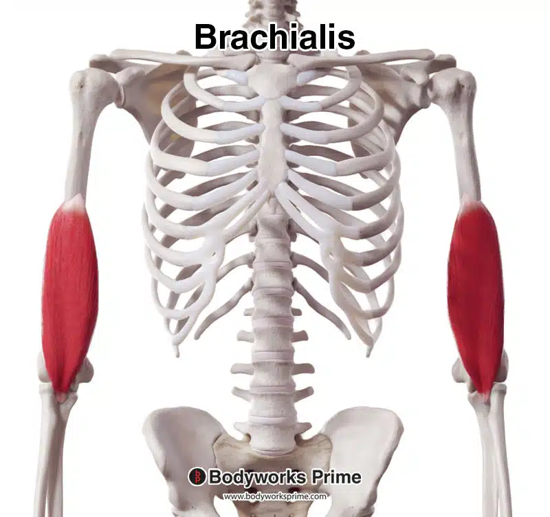
Here we can see the brachialis muscle from an anterior view.
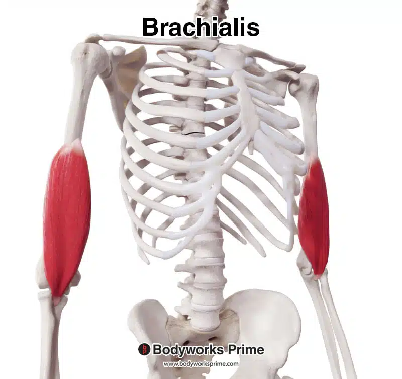
Here we can see the brachialis muscle from an anterolateral view.

Here we can see the brachialis muscle from a lateral view.
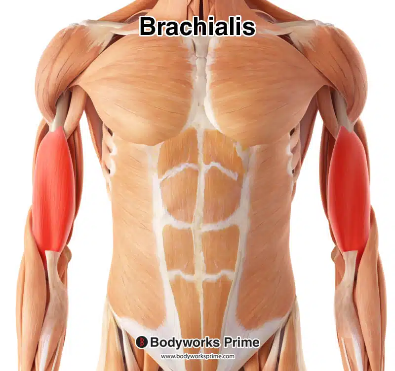
Here we can see the brachialis muscle highlighted in red amongst the other muscles of the body.
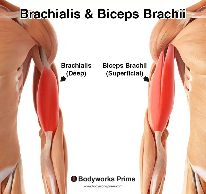
Here we can see the brachialis and biceps brachii muscles. This image demonstrates how the brachialis is deep to the biceps brachii.
Origin & Insertion
Many sources often characterise the anatomy of the brachialis as a muscle with just one head. However, scientific developments over the years have revealed that the brachialis muscle has two heads, a superficial and a deep head.
The superficial head of the brachialis muscle, which forms the larger portion of the muscle, primarily originates from the anterior mid-shaft of the humerus. More specifically, it arises from the lateral and anterior aspects of the middle and distal thirds of the humeral shaft, as well as the lateral intermuscular septum [5] [6] [7]. The lateral intermuscular septum is a thick layer of connective tissue which serves to separate and contain the muscles within their respective compartments in the arm [8].
The deep head, on the other hand, forms a smaller muscle segment and originates more medially from the anterior aspect of the humerus and the medial intermuscular septum [9]. Despite being smaller, the deep head of the brachialis still plays an important role in the muscle’s function.
The insertion points of the brachialis muscle, much like its origins, are complex and multifaceted. The main insertion point for the superficial head is the ulnar tuberosity, where the muscle attaches via a thick, rounded tendon [10] [11] [12]. This strong tendonous insertion onto the ulna allows the brachialis muscle to exert significant force in elbow flexion.
The deep head of the brachialis muscle has a slightly more intricate insertion. It primarily inserts via an aponeurosis, a broad, flat tendon, that branches to the ulna. Specifically, this aponeurosis splits into three distinct portions: medial, lateral, and muscular contractile fibers that attach directly to the ulna [13] [14].
The deep head of the brachialis also inserts onto the anterior joint capsule of the elbow [15]. Furthermore, anatomical studies have reported instances where both heads of the brachialis muscle insert separately onto the coronoid process and the tuberosity of the ulna [16].
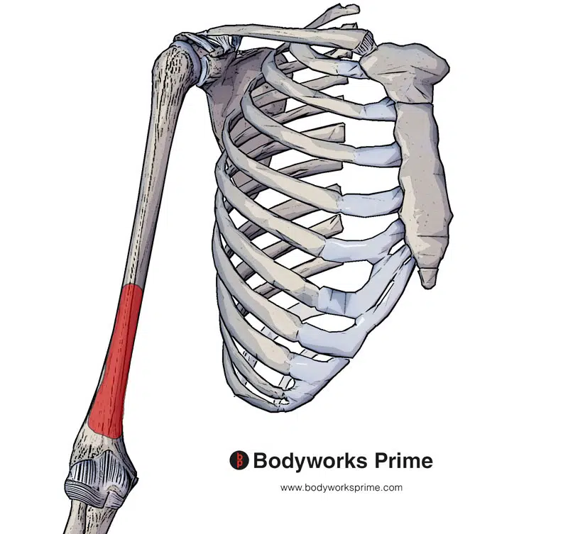
The brachialis muscle originates at the distal anterior surface of the humerus (highlighted in red).
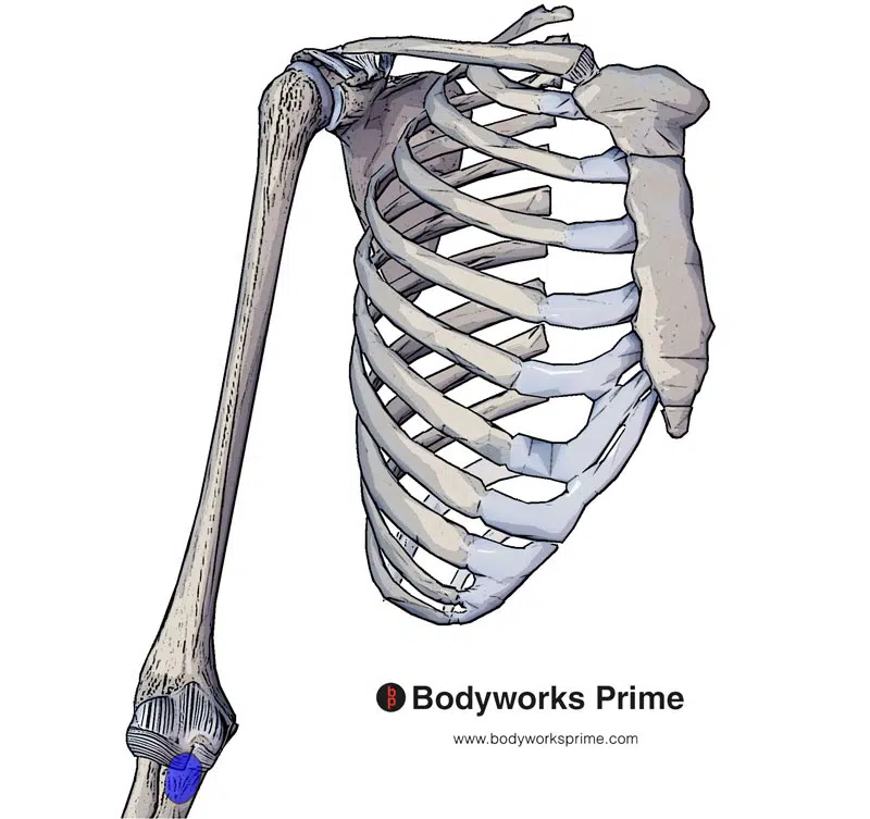
The brachialis muscle inserts at the ulnar tuberosity (highlighted in blue).
Actions
The brachialis muscle plays an essential role in elbow flexion, contributing significantly to the movement of the forearm. It is often regarded as a ‘pure flexor’ of the elbow joint, irrespective of the forearm’s orientation [17] [18] [19].
Unlike other muscles involved in elbow movements, the brachialis does not contribute to either supination or pronation of the forearm. Therefore, the brachialis muscle’s role predominantly focuses on facilitating the elbow’s flexion in various positions [20] [21] [22].
Interestingly, the brachialis muscle’s two heads contribute differently to the elbow’s flexion. The larger, superficial head, due to its mechanical advantage of a more proximal origin and more distal insertion, may provide the majority of flexion strength [23]. The more significant mechanical leverage it possesses potentially allows for it to generate a stronger contraction force during elbow flexion.
In contrast, the smaller, oblique deep head has a more anterior insertion on the coronoid process of the ulna, which may facilitate the initiation of elbow flexion from full extension [24]. This anterior insertion allows the deep head of the brachialis to exert a more specific and controlled force, contributing to the efficiency of elbow flexion movement from a fully extended position.
In summary, the brachialis is recognized as the largest contributor to elbow flexion. It provides consistent flexion strength through its superficial head, and initiates elbow flexion from full extension via its deep head.
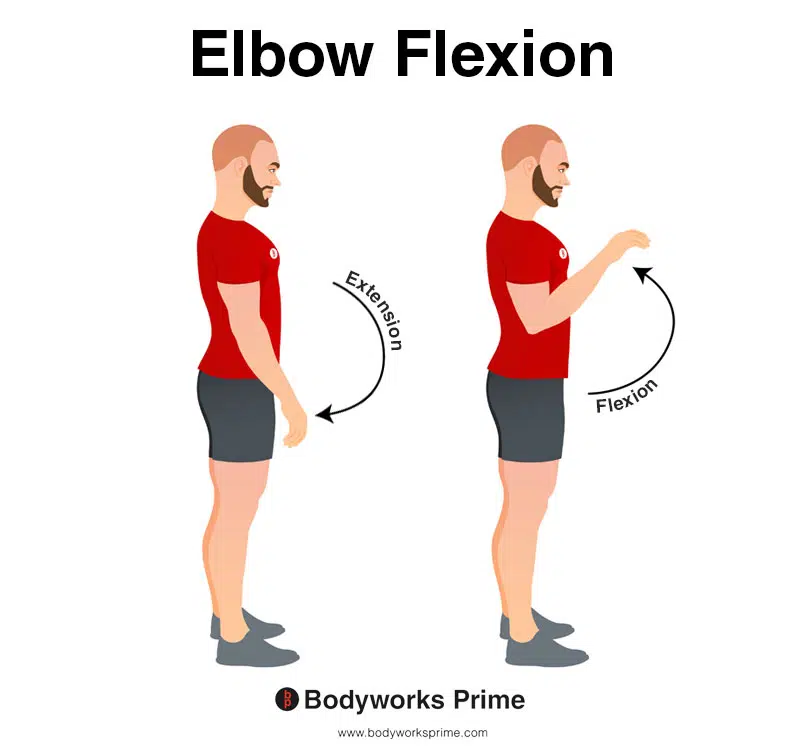
This image demonstrates elbow flexion, which involves bending elbow. The opposite movement of elbow flexion is elbow extension. The brachialis muscle is the primary flexor of the elbow joint.
Innervation
The innervation of the brachialis muscle is both fascinating and complex. Primarily, the brachialis muscle is innervated by the musculocutaneous nerve, a component of the brachial plexus [25] [26] [27]. The musculocutaneous nerve is derived from the lateral cord of the brachial plexus, and typically receives fibers from the fifth, sixth, and seventh cervical nerve roots (C5, C6, C7) [28] [29].
The brachial plexus, a web of intersecting nerves, arises from the cervical region of the spinal cord, specifically from nerve roots C5 through T1. It courses down the side of the neck, extending underneath the collarbone, and radiating into the axilla. This elaborate network ensures the innervation of the entirety of the upper limb [30] [31].
The musculocutaneous nerve penetrates the region between the biceps brachii and the brachialis muscles, where it sends out its terminal branch, the lateral antebrachial cutaneous nerve. As the main nerve that innervates the brachialis, the musculocutaneous nerve typically provides a singular branch to the muscle in about 72% of instances. However, multiple branching patterns, such as double innervation, have also been observed [32] [33].
A noteworthy aspect of the brachialis innervation is its frequent dual nerve supply. Studies have highlighted the role of the radial nerve in supplying the brachialis, alongside the musculocutaneous nerve, in a considerable number of cases, reaching up to 81.6% [34] [35] [36]. The radial nerve, another branch of the brachial plexus, typically courses between the brachialis and brachioradialis muscles on the lateral aspect of the arm after wrapping around the humerus in the spiral groove. Particularly, a branch of the radial nerve has been reported to supply the inferolateral fibers of the brachialis’ deep head [37] [38].
Moreover, variations in the innervation of the brachialis do not stop at a dual nerve supply. Research has demonstrated the occasional contribution of the median nerve to the brachialis, giving rise to an intriguing triple innervation pattern alongside the musculocutaneous nerve and radial nerve [39] [40]. However, these cases are less frequent, highlighting the variability of the brachialis’ innervation [41].
In summary, the intricate innervation of the brachialis predominantly receives innervation from the musculocutaneous nerve and often the radial nerve, with occasional contributions from the median nerve.
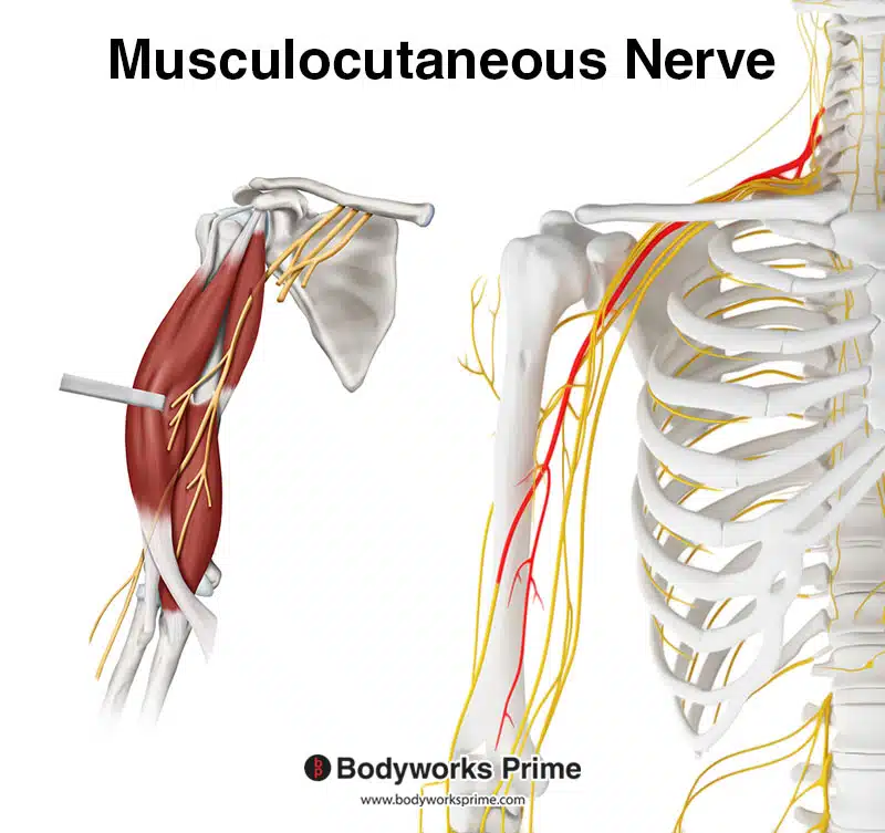
Here we can see the musculocutaneous nerve which is the primary innervation of the brachialis.
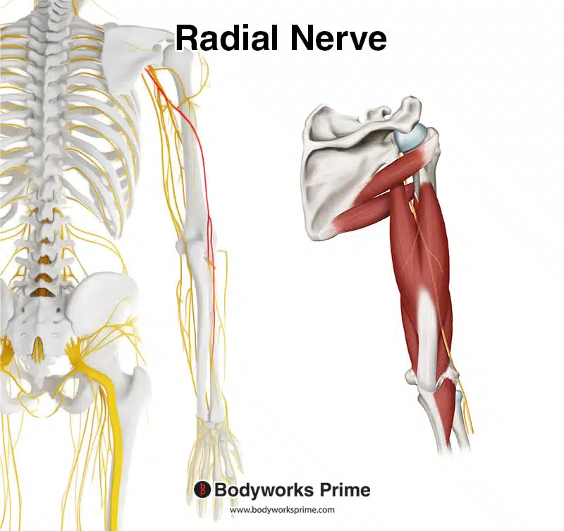
Here we can see the radial nerve which often innervates the brachialis.
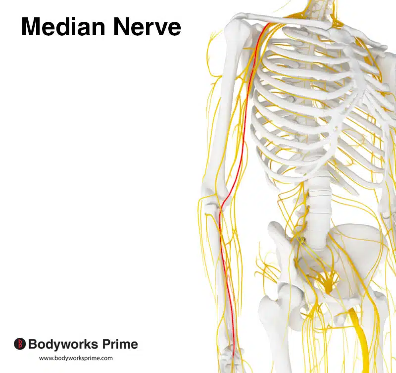
Here we can see the median nerve which occasionally innervates the brachialis in some individuals.
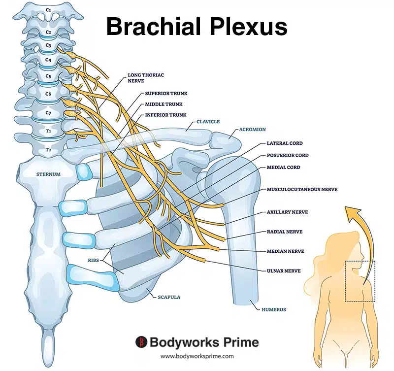
Here we can see an image of the brachial plexus. The musculocutaneous nerve, radial nerve, and median nerve are all a part of the brachial plexus.
Blood Supply
The brachialis muscle receives its primary blood supply from tributaries of the brachial artery and the radial recurrent artery [42]. The brachial artery, situated along the ventral surface of the arm, spawns several branches that contribute to the brachialis muscle’s blood supply, such as the deep brachial artery, the superior ulnar collateral artery, and the inferior ulnar collateral artery [43].
Notably, the inferior ulnar collateral artery, originating anterior to the lower segment of the humerus, traverses medially along the brachialis muscle before penetrating the medial intermuscular septum. This artery forms an anastomosis with the anterior ulnar recurrent artery and the superior ulnar collateral artery [44].
In addition, the brachialis muscle also receives vascular contributions from the radial artery, which originates in the cubital fossa at the bifurcation of the brachial artery [45].
The brachial vein is primarily responsible for venous drainage from the brachialis muscle, later merging with the basilic vein tributary to form the axillary vein. Deep lymphatic channels of the upper limb, generally accompanying the main vessels, drain into the axillary lymph nodes [46].
In essence, the brachialis muscle’s blood supply primarily involves the tributaries of the brachial artery and radial recurrent artery, with auxiliary contributions from the branches of the ulnar collateral arteries [47] [48].
Want some flashcards to help you remember this information? Then click the link below:
Brachialis Muscle flashcards
Support Bodyworks Prime
Running a website and YouTube channel can be expensive. Your donation helps support the creation of more content for my website and YouTube channel. All donation proceeds go towards covering expenses only. Every contribution, big or small, makes a difference!
References
| ↑1, ↑3, ↑8 | Moore KL, Agur AMR, Dalley AF. Clinically Oriented Anatomy. 8th ed. Philadelphia: Lippincot Williams & Wilkins; 2017. |
|---|---|
| ↑2, ↑4 | Standring S. (2015). Gray’s Anatomy: The Anatomical Basis of Clinical Practice, 41st Edn. Amsterdam: Elsevier. |
| ↑5, ↑9, ↑10, ↑13, ↑17, ↑20, ↑25, ↑30, ↑32, ↑34, ↑42, ↑46, ↑47 | Plantz MA, Bordoni B. Anatomy, Shoulder and Upper Limb, Brachialis Muscle. [Updated 2023 Feb 21]. In: StatPearls [Internet]. Treasure Island (FL): StatPearls Publishing; 2023 Jan-. Available from: https://www.ncbi.nlm.nih.gov/books/NBK551630/ |
| ↑6, ↑12, ↑15, ↑19, ↑22, ↑27 | Vadgaonkar R, Rai R, Nayak SR, D’Costa S, Saralaya V, Dhanya. An anatomical and clinical insight on brachialis with emphasis on portal’s muscle. Rom J Morphol Embryol. 2010;51(3):551-3. PMID: 20809036. |
| ↑7, ↑16, ↑26, ↑29, ↑36 | Ilayperuma I, Uluwitiya SM, Nanayakkara BG, Palahepitiya KN. Re-visiting the brachialis muscle: morphology, morphometry, gender diversity, and innervation. Surg Radiol Anat. 2019 Apr;41(4):393-400. doi: 10.1007/s00276-019-02182-2. Epub 2019 Feb 28. PMID: 30820647. |
| ↑11, ↑14, ↑18, ↑21, ↑23, ↑24, ↑37 | Leonello DT, Galley IJ, Bain GI, Carter CD. Brachialis muscle anatomy. A study in cadavers. J Bone Joint Surg Am. 2007 Jun;89(6):1293-7. doi: 10.2106/JBJS.F.00343. PMID: 17545433. |
| ↑28, ↑31, ↑33 | Pacha Vicente D, Forcada Calvet P, Carrera Burgaya A, Llusá Pérez M. Innervation of biceps brachii and brachialis: Anatomical and surgical approach. Clin Anat. 2005 Apr;18(3):186-94. doi: 10.1002/ca.20057. PMID: 15768419. |
| ↑35, ↑38, ↑39 | Mahakkanukrauh P, Somsarp V. Dual innervation of the brachialis muscle. Clin Anat. 2002 May;15(3):206-9. doi: 10.1002/ca.10014. PMID: 11948956. |
| ↑40, ↑41 | Won SY, Cho YH, Choi YJ, Favero V, Woo HS, Chang KY, Hu KS, Kim HJ. Intramuscular innervation patterns of the brachialis muscle. Clin Anat. 2015 Jan;28(1):123-7. doi: 10.1002/ca.22387. Epub 2014 Mar 4. PMID: 24596238. |
| ↑43, ↑44, ↑45, ↑48 | Epperson TN, Varacallo M. Anatomy, Shoulder and Upper Limb, Brachial Artery. [Updated 2022 Jul 25]. In: StatPearls [Internet]. Treasure Island (FL): StatPearls Publishing; 2023 Jan-. Available from: https://www.ncbi.nlm.nih.gov/books/NBK537145/ |


