| Origin | Ilium of the hip (between the anterior and inferior gluteal lines) |
| Insertion | Greater trochanter of femur (anterolateral aspect) |
| Action | Hip abduction Pelvic stability Hip medial rotation |
| Nerve | Superior gluteal nerve (L4, L5, S1) |
| Artery | Superior gluteal artery |
Location & Overview
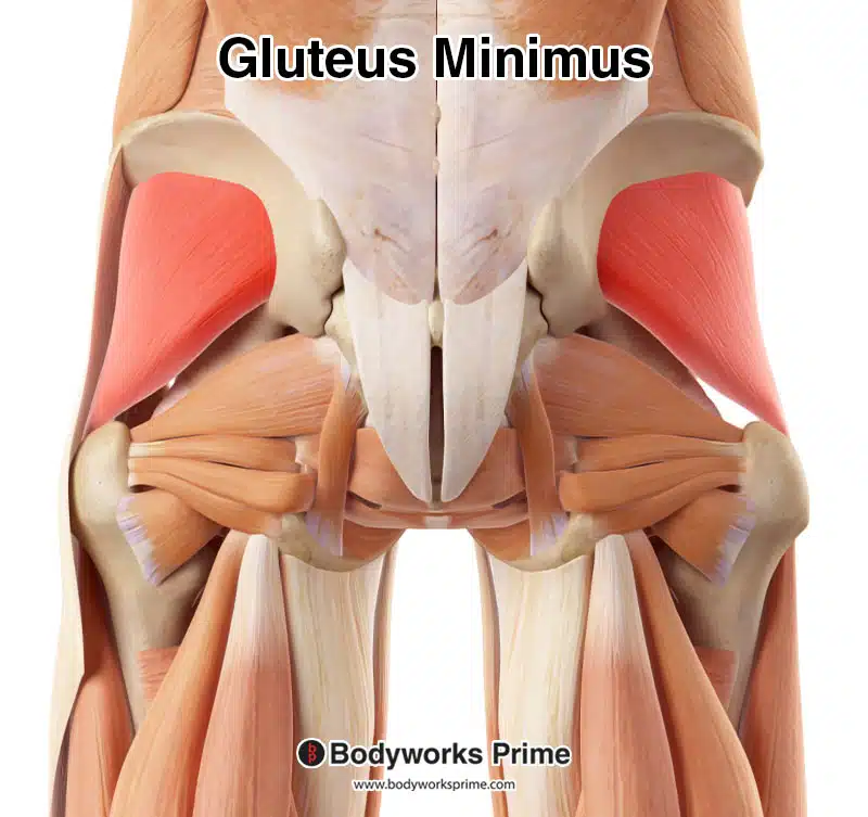
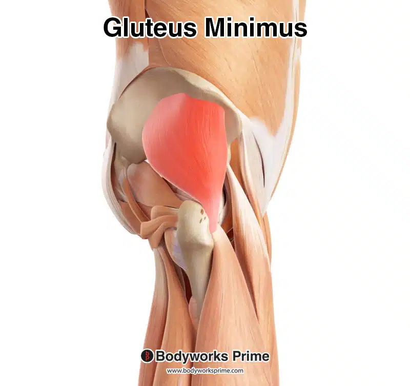
The gluteus minimus muscle is one of the three gluteal muscles, accompanied by the gluteus maximus and gluteus medius. As the smallest of the trio, the gluteus minimus plays an important role in hip joint stability and movement during activities like walking and running. Situated in the lateral part of the hip, this fan-shaped muscle is nestled deep within the gluteal region, below the gluteus medius and further beneath the gluteus maximus. The gluteus minimus shares similar characteristics with the gluteus medius, including actions, structure, innervation, and blood supply [1] [2].
Pathologies associated with the gluteus minimus muscle include gluteal tendinopathy and muscle strains. Gluteal tendinopathy occurs when the tendon connecting the muscle to the bone becomes inflamed or degenerated, causing pain in the hip and outer thigh region [3]. Muscle strains can result from overuse, trauma, or inadequate warm-up before engaging in high intensity physical activities.
To strengthen the gluteus minimus and promote its overall health, various exercises can target this muscle. Some effective exercises include side-lying leg lifts, clamshells, lateral band walks, and single-leg squats [4].
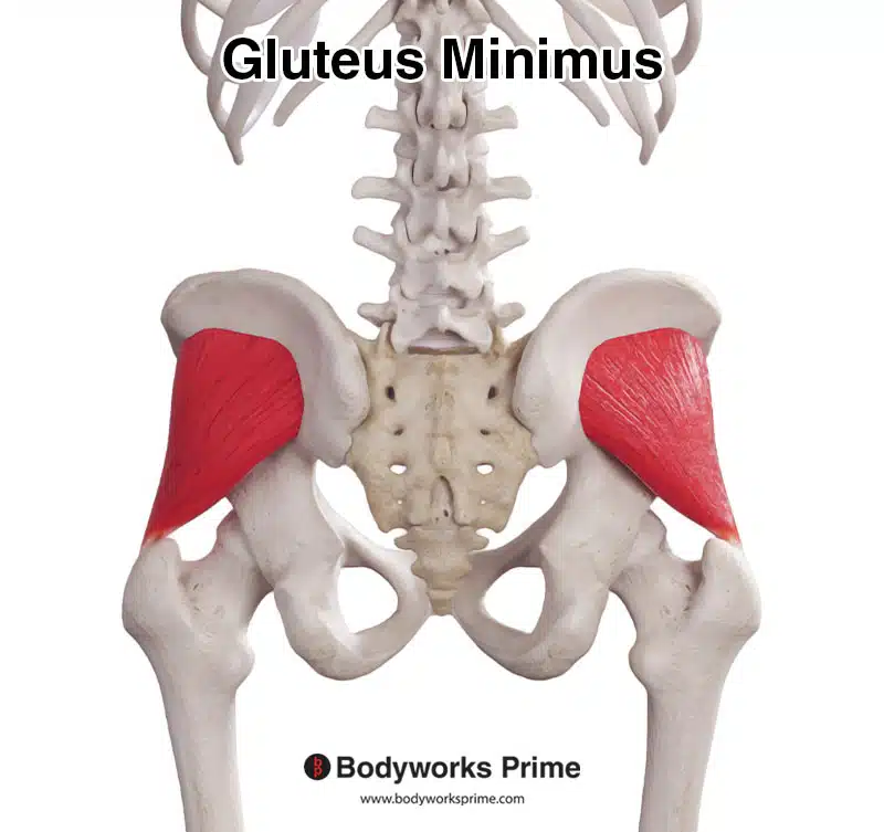
Pictured here we can see the gluteus minimus muscle from a posterior view.
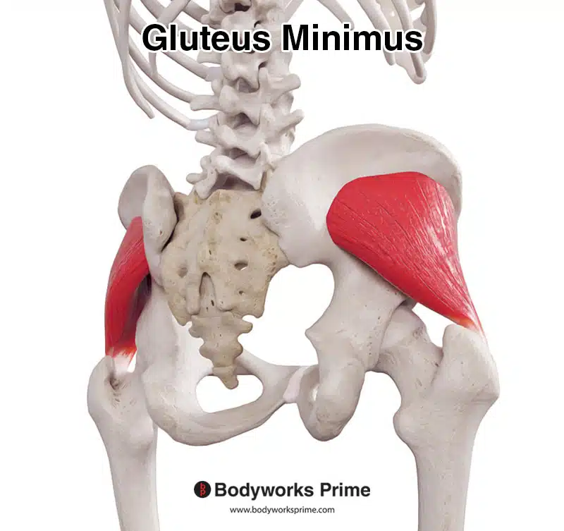
Pictured here we can see the gluteus minimus muscle from a posterolateral view.
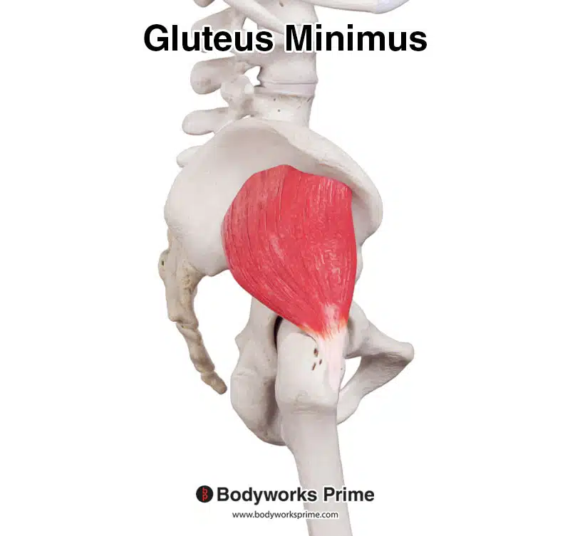
Pictured here we can see the gluteus minimus muscle from a lateral view.
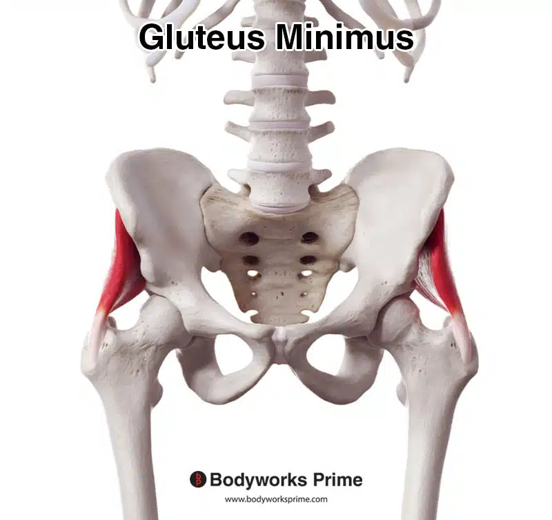
Pictured here we can see the gluteus minimus muscle from an anterior view.
Pictured here we can see the gluteus minimus muscle highlighted in red, seen from a posterior view. As the gluteus minimus is a relatively deep muscle, superficial muscles such as the gluteus maximums, gluteus minimus, tensor fascia latae and the iliotibial band have been removed to reveal the gluteus minimus.
Pictured here we can see the gluteus minimus muscle highlighted in red, seen from a lateral view. As with the previous image, muscles superficial to the gluteus minimus have been removed to reveal the gluteus minimus.
Origin & Insertion
Understanding the origin and insertion points of the gluteus minimus provide a deeper understanding of its location and functions. The gluteus minimus’ origin is on the external surface of the ilium, between the anterior and inferior gluteal lines. The ilium is a large, fan-shaped bone which forms the upper part of the hip bone, and the gluteal lines are bony ridges on the ilium’s external surface [5] [6].
The insertion point of the gluteus minimus is on the anterior border of the greater trochanter of the femur. The greater trochanter is a prominent, bony protrusion on the upper part of the femur, located on its lateral side. The anterior border refers to the front edge of this bony landmark [7] [8].
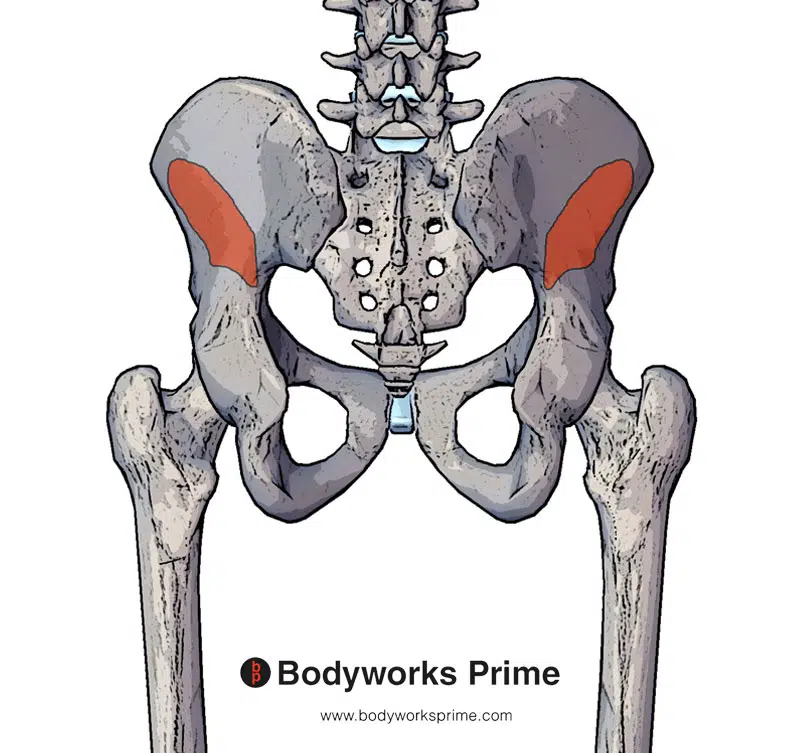
Here we can see pictured the origin of the gluteus minimus on the ilium. Specifically, between the anterior and inferior gluteal lines.
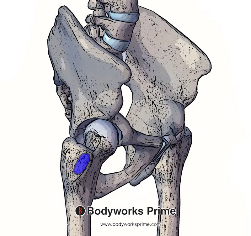
Here we can see pictured the insertion of the gluteus minimus on the greater trochanter of the femur. Specifically, on the anterolateral aspect of the greater trochanter.
Actions
The gluteus minimus muscle plays an important role in several key actions at the hip joint, primarily in abduction, stabilising the hip (such as when standing on one leg), and to a lesser degree hip internal rotation. The main actions of the gluteus minimus are abduction and stabilising the pelvis [9] [10] [11]. Abduction involves lifting the leg away from the body’s midline, which is utilised during various activities such as side-stepping or getting out of a car. Abduction is also essential for maintaining stability and balance during single-leg stances, like when walking or running.
While the role of the gluteus minimus in hip internal rotation is less well-established. However, there is evidence to suggest that the anterior portion of the gluteus minimus contributes to this action [12] [13] [14] [14] [15]. Internal rotation involves rotating the thigh bone (femur) inwards, towards the body’s midline, and is important for certain athletic movements and general mobility [16].
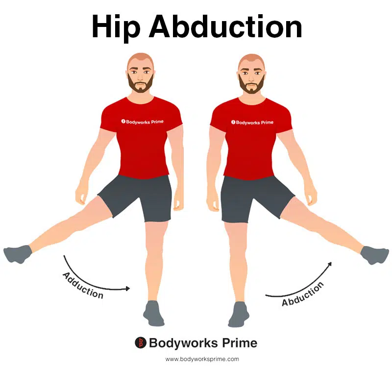
This image shows an example of hip abduction, which involves moving the leg out to the side (laterally). The opposite of hip abduction is hip adduction. The gluteus minimus performs hip abduction.
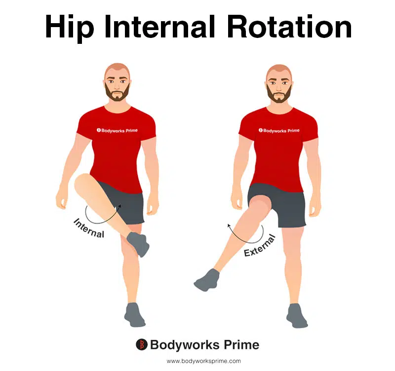
This image depicts an example of hip internal rotation, which involves rotating the leg from the hip joint inwards (medially). Internal rotation is also referred to as medial rotation. The opposite of internal rotation or medial rotation is external rotation or lateral rotation. The gluteus minimus performs hip internal rotation. However, this is not its primary action.
Innervation
The gluteus minimus is innervated by the superior gluteal nerve, which originates from the lumbosacral plexus with nerve roots L4, L5, and S1. This nerve is responsible for transmitting nerve signals between the gluteus minimus and the central nervous system, allowing for the muscle’s controlled contractions and coordinated movements. The superior gluteal nerve also innervates the gluteus medius and tensor fasciae latae muscles, which share functional similarities with the gluteus minimus, particularly in hip abduction and stabilisation [17] [18].
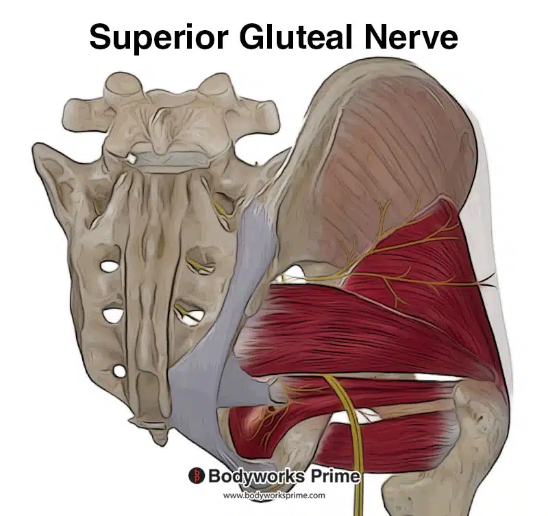
Here we can see the superior gluteal nerve which innervates the gluteus minimus with nerve roots from L4, L5, and S1.
Blood Supply
The blood supply to the gluteus minimus muscle is primarily provided by the superior gluteal artery, which is a branch of the internal iliac artery. This artery ensures that the muscle receives enough oxygen and nutrients to function effectively. In addition to supplying the gluteus minimus, the superior gluteal artery also provides blood to other nearby muscles, such as the gluteus medius and tensor fasciae latae [19] [20].
Want some flashcards to help you remember this information? Then click the link below:
Gluteus Minimus Flashcards
Support Bodyworks Prime
Running a website and YouTube channel can be expensive. Your donation helps support the creation of more content for my website and YouTube channel. All donation proceeds go towards covering expenses only. Every contribution, big or small, makes a difference!
References
| ↑1 | Greco AJ, Vilella RC. Anatomy, Bony Pelvis and Lower Limb, Gluteus Minimus Muscle. [Updated 2021 Jul 26]. In: StatPearls [Internet]. Treasure Island (FL): StatPearls Publishing; 2021 Jan-. Available from: https://www.ncbi.nlm.nih.gov/books/NBK556144/ |
|---|---|
| ↑2, ↑11, ↑16 | Moore KL, Agur AMR, Dalley AF. Clinically Oriented Anatomy. 8th ed. Philadelphia: Lippincot Williams & Wilkins; 2017. |
| ↑3 | Federer AE, Steele JR, Dekker TJ, Liles JL, Adams SB. Tendonitis and Tendinopathy: What Are They and How Do They Evolve? Foot Ankle Clin. 2017 Dec;22(4):665-676. doi: 10.1016/j.fcl.2017.07.002. Epub 2017 Sep 27. PMID: 29078821. |
| ↑4 | Selkowitz DM, Beneck GJ, Powers CM. Which exercises target the gluteal muscles while minimizing activation of the tensor fascia lata? Electromyographic assessment using fine-wire electrodes. J Orthop Sports Phys Ther. 2013 Feb;43(2):54-64. doi: 10.2519/jospt.2013.4116. Epub 2013 Jan 10. PMID: 23312684. |
| ↑5, ↑7 | Walters J, Solomons M, Davies J. Gluteus minimus: observations on its insertion. J Anat. 2001 Feb;198(Pt 2):239-42. doi: 10.1046/j.1469-7580.2001.19820239.x. PMID: 11273048; PMCID: PMC1468210. |
| ↑6, ↑8, ↑20 | Standring S. (2015). Gray’s Anatomy: The Anatomical Basis of Clinical Practice, 41st Edn. Amsterdam: Elsevier. |
| ↑9 | Flack NA, Nicholson HD, Woodley SJ. A review of the anatomy of the hip abductor muscles, gluteus medius, gluteus minimus, and tensor fascia lata. Clin Anat. 2012 Sep;25(6):697-708. doi: 10.1002/ca.22004. Epub 2011 Nov 22. PMID: 22109658. |
| ↑10, ↑15 | Peduzzi de Castro M, de Brito Fontana H, Fóes MC, Santos GM, Ruschel C, Roesler H. Activation of the gluteus maximus, gluteus medius and tensor fascia lata muscles during hip internal and external rotation exercises at three hip flexion postures. J Bodyw Mov Ther. 2021 Jul;27:487-492. doi: 10.1016/j.jbmt.2021.05.011. Epub 2021 May 14. PMID: 34391276. |
| ↑12 | Gottschalk F, Kourosh S, Leveau B. The functional anatomy of tensor fasciae latae and gluteus medius and minimus. J Anat. 1989 Oct;166:179-89. PMID: 2621137; PMCID: PMC1256751. |
| ↑13 | Whiler L, Fong M, Kim S, Ly A, Qin Y, Yeung E, Mathur S. Gluteus Medius and Minimus Muscle Structure, Strength, and Function in Healthy Adults: Brief Report. Physiother Can. 2017;69(3):212-216. doi: 10.3138/ptc.2016-16. PMID: 30275637; PMCID: PMC5963550. |
| ↑14 | Neumann DA. Kinesiology of the hip: a focus on muscular actions. J Orthop Sports Phys Ther. 2010 Feb;40(2):82-94. doi: 10.2519/jospt.2010.3025. PMID: 20118525. |
| ↑17 | Jacobs LG, Buxton RA. The course of the superior gluteal nerve in the lateral approach to the hip. J Bone Joint Surg Am. 1989 Sep;71(8):1239-43. PMID: 2777853. |
| ↑18 | Akita K, Sakamoto H, Sato T. Origin, course and distribution of the superior gluteal nerve. Acta Anat (Basel). 1994;149(3):225-30. doi: 10.1159/000147581. PMID: 7976174. |
| ↑19 | Collinge CA, Ziran NM, Coons DA. Relationship Between the Superior Gluteal Vessels and Nerve at the Greater Sciatic Notch. Orthopedics. 2015 Oct;38(10):e929-33. doi: 10.3928/01477447-20151002-62. PMID: 26488790. |


