| Origin | Posterior surface of the ilium (between anterior and posterior gluteal lines) Gluteal aponeurosis |
| Insertion | Greater trochanter of femur (lateral and superoposterior facet) |
| Action | Hip abduction Hip medial rotation (anterior fibers) Hip lateral rotation (posterior fibers) Stabilises pelvis |
| Nerve | Superior gluteal nerve (L4, L5, S1 nerve roots) |
| Artery | Superior gluteal artery |
Location & Overview
The gluteus medius is one of the three gluteal muscles, the other two are the gluteus maximus and gluteus minimus muscles. The gluteus medius is located on the lateral aspect of the upper buttocks, just below the iliac crest. The iliac crest is the curved, uppermost edge of the ilium and can be felt along the sides of your hips. The gluteus medius has a fan-like shape, with the proximal portion being wider and narrowing as it approaches its tendon insertion [1]. Its posterior third is covered by the gluteus maximus. As a primary hip abductor, the gluteus medius works together with the gluteus maximus, gluteus minimus and tensor fasciae latae in hip abduction [2].
Pathologies associated with the gluteus medius muscle are muscle strains, tendinopathies, weakness, hip instability, trochanteric bursitis, gluteus medius syndrome, and lower back pain [3]. Trochanteric bursitis occurs due to an inflammation of the hip bursa and results in lateral hip pain [4]. Gluteus medius syndrome is a condition that presents with symptoms of lower back pain, leg pain, and often weakness in the gluteus medius muscle [5]. Poor strength or dysfunction of the gluteus medius can also lead to altered gait patterns and poor balance, especially during the stance phase of gait [6].
A systematic review conducted by Macadam, Cronin, and Contreras (2015) looked at gluteal muscle activity associated with dynamic hip abduction and hip external rotation exercise. Based off this study, excellent exercise choices for the gluteus medius muscle (in no specific order) are: lateral stepping with a band at the ankle, side bridges with abduction, side lying hip abduction, clam shells, and the seated hip abduction machine. These exercises involve hip abduction and to an extent, hip external rotation movements, which have been proven to significantly enhance gluteus medius activation. Incorporating these exercises into a well-rounded workout routine can lead to improved gluteal strength, hypertrophy and functionality [7].
Here we can see the gluteus medius from a posterior view.
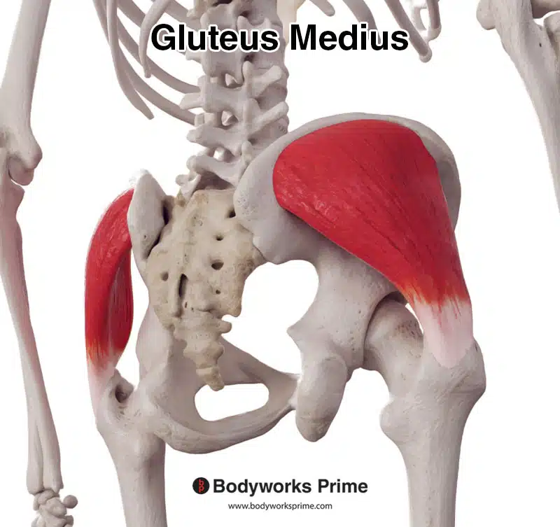
Here we can see the gluteus medius from a posterolateral view.
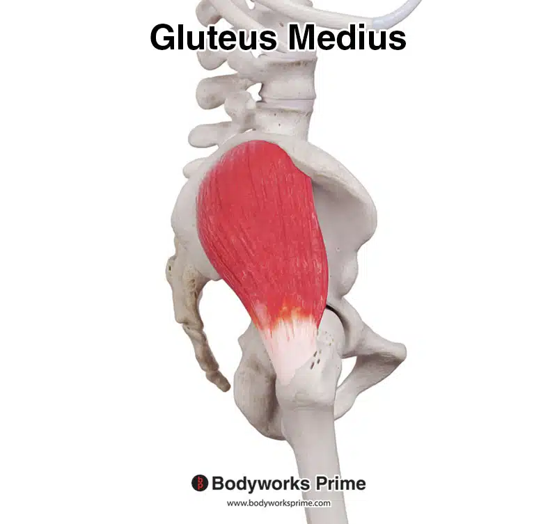
Here we can see the gluteus medius from a lateral view.
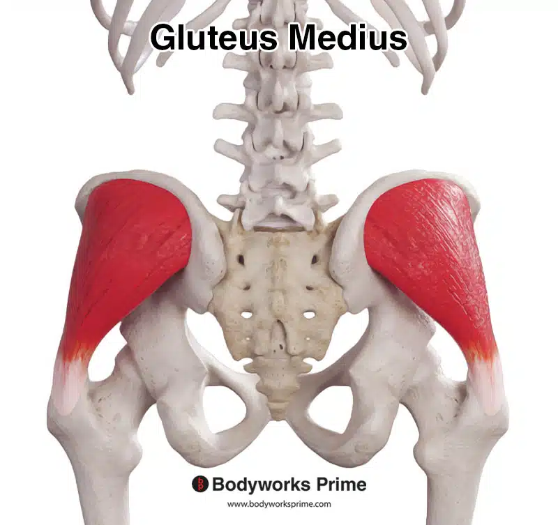
Here we can see the gluteus medius from an anterior view.
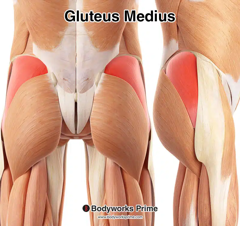
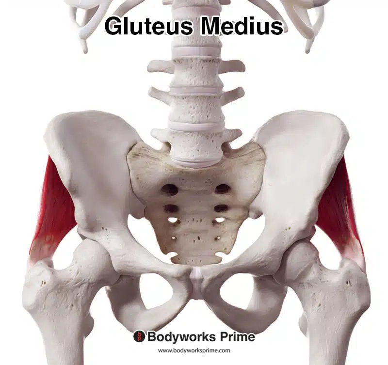
Here, the gluteus medius is highlighted in red among the other muscles in the hip and thigh region. On the left, we see a posterior view, and on the right, a lateral view.
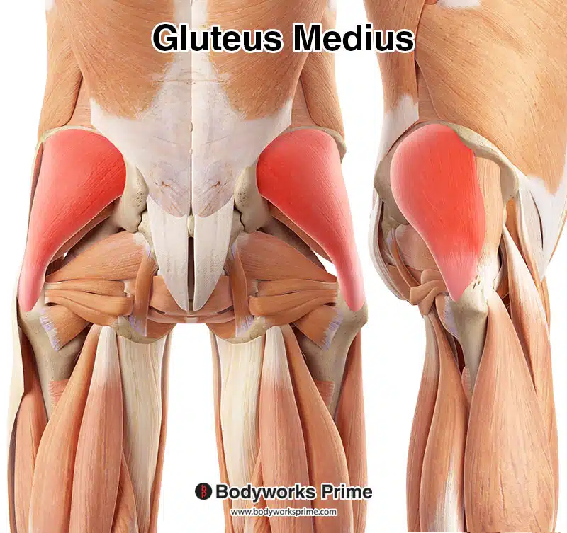
Here, we can see a more detailed view with the gluteus medius highlighted in red. Superficial structures, such as the gluteus maximus, tensor fasciae latae, and the iliotibial band, have been removed to reveal the gluteus medius. On the left, there’s a posterior view, and on the right, a lateral view.
Origin & Insertion
The gluteus medius muscle originates from the posterior surface of the ilium, specifically the area between the anterior and posterior gluteal lines. To locate these lines, first find the iliac crest, which is the curved upper edge of the ilium and can be felt along the top of your hips. The anterior gluteal line is situated just below the iliac crest and runs downward and backward, while the posterior gluteal line is slightly further down the illium (roughly a quarter of the way down). The gluteus medius originates from the outer surface of the ilium between these two lines [8] [9].
The gluteus medius also originates from the gluteal aponeurosis, which is a fibrous sheet that covers the gluteus medius and gluteus minimus muscles. This aponeurosis is an extension of the fascia lata, a deep layer of connective tissue that encircles the thigh. It serves as an attachment site for various muscles. The gluteal aponeurosis is located over the gluteus medius and minimus muscles and extends from the iliac crest down to the greater trochanter of the femur. The gluteus medius muscle fibers blend with this aponeurosis which further strengthens its origin and provides further stability to the muscle [10] [11].
The muscle fibers of the gluteus medius converge into a tendon, which inserts onto the greater trochanter of the femur. The greater trochanter is a large, bony prominence on the upper part of the femur. The greater trochanter can be felt on the lateral side of the upper thigh. The insertion of the gluteus medius is on the lateral aspect of the greater trochanter, slightly posterior and superior to the insertion of the gluteus minimus muscle [12] [13].
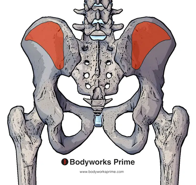
Here we can see the origin of the gluteus medius muscle marked in red on the posterior surface of the ilium (between the anterior and posterior gluteal lines).
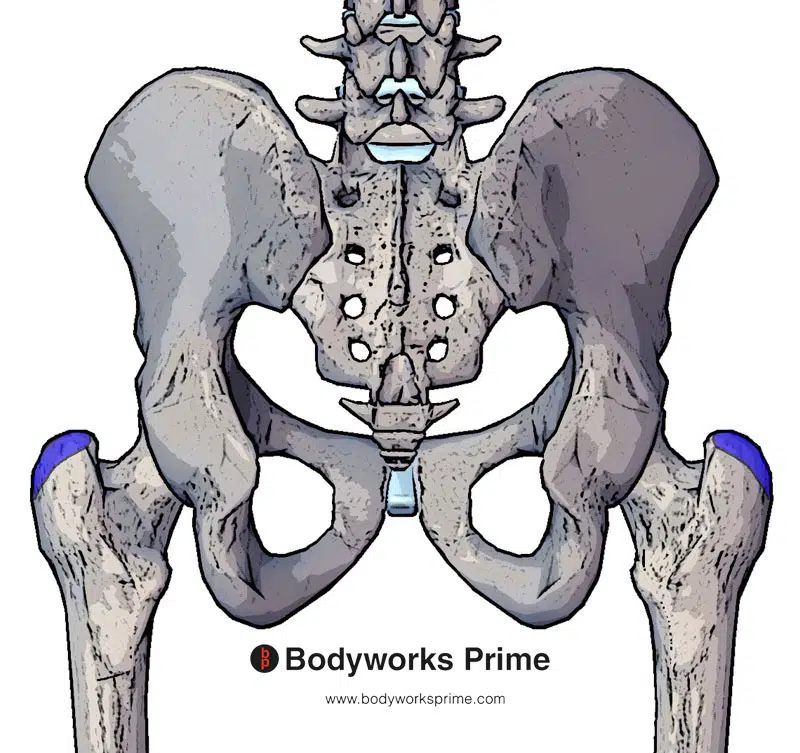
Here we can see the insertion of the gluteus medius muscle marked in blue on the greater trochanter of femur (lateral and superoposterior facet).
Actions
The gluteus medius muscle contributes an important role in the movements and stabilisation of the hip joint. The primary actions of the gluteus medius are hip abduction and stabilisation of the pelvis [14] [15]. During hip abduction, the gluteus medius works together with the gluteus minimus, gluteus maximus, and the tensor fascia latae muscles to move the leg away from the midline of the body (abduction) [16] [17]. The anterior fibers of the gluteus medius are able to assist in hip medial rotation, whereas the posterior fibers are able to assist in lateral rotation [18] [19] [20] [21]. Medial rotation of the hip occurs when the femur rotates inward towards the body’s midline and rotating the opposite way would be lateral rotation.
In addition to its actions of hip abduction and rotation, the gluteus medius also contributes to stabilisation of the hip during activities such as: walking, running, hopping, and standing on one leg [22] [23]. When standing on one leg, the gluteus medius muscle on the standing side works to maintain pelvic stability which prevents the opposite side of the pelvis from dropping down. This action is known as the ‘Trendelenburg sign’, and when the gluteus medius is weak or dysfunctional, a positive Trendelenburg sign may be present, indicating an inability of the person to maintain pelvic stability [24].
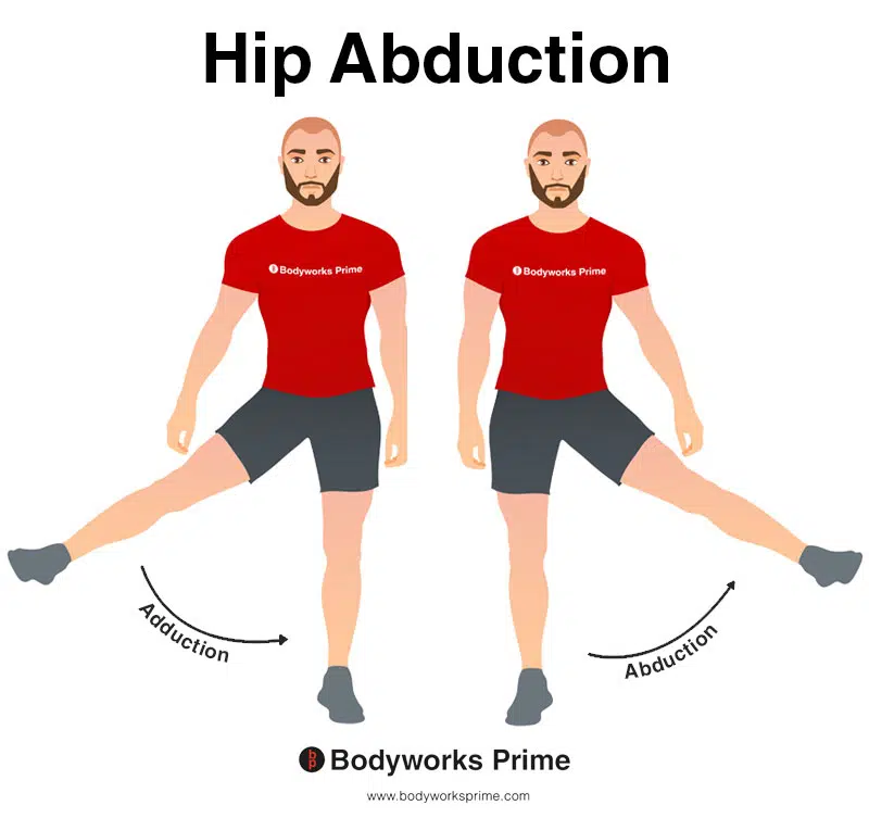
This image shows an example of hip abduction, which involves moving the leg out to the side (laterally). The opposite of hip abduction is hip adduction. The gluteus medius performs hip abduction and also stabilisation of the pelvis.
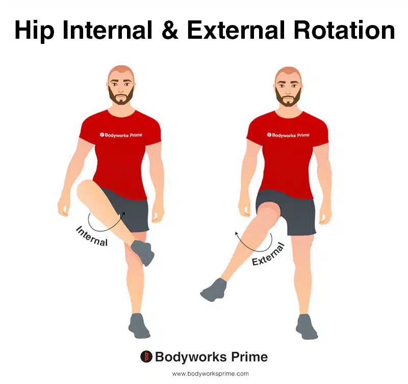
This image shows an example of hip internal (medial) rotation and external (lateral) rotation. Internal rotation involves rotating the leg from the hip joint inwards (medially). The opposite of internal rotation is external rotation, which involves rotating the leg from the hip joint outwards (laterally). The gluteus medius can assist with both external and internal rotation of the hip.
Innervation
The gluteus medius muscle is innervated by the superior gluteal nerve, which arises from the lumbar and sacral spinal nerves L4, L5, and S1. The superior gluteal nerve travels through the pelvis, exiting through the greater sciatic foramen, which is a large opening in the pelvis, allowing structures to pass between the pelvic cavity and the gluteal region. As it exits the foramen, the nerve travels above the piriformis muscle and then splits into branches which supply the gluteus medius, gluteus minimus, and tensor fascia latae muscles [25] [26].
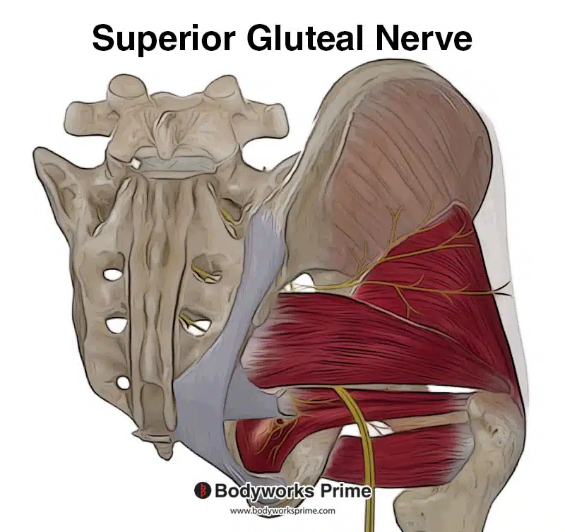
Here we can see the superior gluteal nerve which innervates the gluteus medius with nerve roots from L4, L5, and S1.
Blood Supply
The blood supply to the gluteus medius is primarily provided by the superior gluteal artery. The superior gluteal artery arises from the internal iliac artery, which is one of the main arteries supplying the pelvis and lower limbs. The superior gluteal artery leaves the pelvic cavity through the greater sciatic foramen, superior to the piriformis muscle. It then divides into deep and superficial branches that run across the gluteus medius and gluteus minimus muscles, supplying them with oxygenated blood and nutrients [27] [28].
Want some flashcards to help you remember this information? Then click the link below:
Gluteus Medius Flashcards
Support Bodyworks Prime
Running a website and YouTube channel can be expensive. Your donation helps support the creation of more content for my website and YouTube channel. All donation proceeds go towards covering expenses only. Every contribution, big or small, makes a difference!
References
| ↑1, ↑8, ↑12, ↑14, ↑16, ↑22 | Moore KL, Agur AMR, Dalley AF. Clinically Oriented Anatomy. 8th ed. Philadelphia: Lippincot Williams & Wilkins; 2017. |
|---|---|
| ↑2, ↑15, ↑17, ↑18, ↑23 | Shah A, Bordoni B. Anatomy, Bony Pelvis and Lower Limb, Gluteus Medius Muscle. In: StatPearls [Internet]. Treasure Island (FL): StatPearls Publishing; 2021 Jan-. Available from: https://www.ncbi.nlm.nih.gov/books/NBK557509/ |
| ↑3, ↑5 | Kameda M, Tanimae H, Kihara A, Matsumoto F. Does low back pain or leg pain in gluteus medius syndrome contribute to lumbar degenerative disease and hip osteoarthritis and vice versa? A literature review. J Phys Ther Sci. 2020 Feb;32(2):173-191. doi: 10.1589/jpts.32.173. Epub 2020 Feb 14. PMID: 32158082; PMCID: PMC7032979. |
| ↑4 | Seidman AJ, Taqi M, Varacallo M. Trochanteric Bursitis. [Updated 2022 Nov 20]. In: StatPearls [Internet]. Treasure Island (FL): StatPearls Publishing; 2023 Jan-. Available from: https://www.ncbi.nlm.nih.gov/books/NBK538503/ |
| ↑6 | Nunes GS, Pizzari T, Neate R, Barton CJ, Semciw A. Gluteal muscle activity during running in asymptomatic people. Gait Posture. 2020 Jul;80:268-273. doi: 10.1016/j.gaitpost.2020.06.008. Epub 2020 Jun 6. PMID: 32563058. |
| ↑7, ↑21 | Macadam P, Cronin J, Contreras B. AN EXAMINATION OF THE GLUTEAL MUSCLE ACTIVITY ASSOCIATED WITH DYNAMIC HIP ABDUCTION AND HIP EXTERNAL ROTATION EXERCISE: A SYSTEMATIC REVIEW. Int J Sports Phys Ther. 2015 Oct;10(5):573-91. PMID: 26491608; PMCID: PMC4595911. |
| ↑9, ↑13 | Shah A, Bordoni B. Anatomy, Bony Pelvis and Lower Limb, Gluteus Medius Muscle. 2023 Feb 17. In: StatPearls [Internet]. Treasure Island (FL): StatPearls Publishing; 2023 Jan–. PMID: 32491441. |
| ↑10 | Flack NA, Nicholson HD, Woodley SJ. The anatomy of the hip abductor muscles. Clin Anat. 2014 Mar;27(2):241-53. doi: 10.1002/ca.22248. Epub 2013 Apr 27. PMID: 23625344. |
| ↑11 | Hwang SW, Nam YS, Hwang K, Han SH. Thickness and tension of the gluteal aponeurosis and the implications for subfascial gluteal augmentation. J Anat. 2012 Jul;221(1):69-72. doi: 10.1111/j.1469-7580.2012.01510.x. Epub 2012 May 14. PMID: 22578136; PMCID: PMC3390535. |
| ↑19 | Conneely MO Sullivan K Edmondston S. Dissection of gluteus maximus and medius with respect to their suggested roles in pelvic and hip stability: implications for rehabilitation? Phys Ther in Sport. 2006;7:176‐178. |
| ↑20 | Nakagawa TH, Muniz TB, Baldon Rde M, Dias Maciel C, de Menezes Reiff RB, Serrão FV. The effect of additional strengthening of hip abductor and lateral rotator muscles in patellofemoral pain syndrome: a randomized controlled pilot study. Clin Rehabil. 2008 Dec;22(12):1051-60. doi: 10.1177/0269215508095357. PMID: 19052244. |
| ↑24 | Gogu S, Gandbhir VN. Trendelenburg Sign. [Updated 2022 Nov 14]. In: StatPearls [Internet]. Treasure Island (FL): StatPearls Publishing; 2023 Jan-. Available from: https://www.ncbi.nlm.nih.gov/books/NBK555987/ |
| ↑25 | Jacobs LG, Buxton RA. The course of the superior gluteal nerve in the lateral approach to the hip. J Bone Joint Surg Am. 1989 Sep;71(8):1239-43. PMID: 2777853. |
| ↑26 | Gottschalk F, Kourosh S, Leveau B. The functional anatomy of tensor fasciae latae and gluteus medius and minimus. J Anat. 1989 Oct;166:179-89. PMID: 2621137; PMCID: PMC1256751. |
| ↑27 | Ebraheim NA, Olexa TA, Xu R, Georgiadis G, Yeasting RA. The quantitative anatomy of the superior gluteal artery and its location. Am J Orthop (Belle Mead NJ). 1998 Jun;27(6):427-31. PMID: 9652885. |
| ↑28 | Mu LH, Yan YP, Luan J, Fan F, Li SK. [Anatomy study of superior and inferior gluteal artery perforator flap]. Zhonghua Zheng Xing Wai Ke Za Zhi. 2005 Jul;21(4):278-80. Chinese. PMID: 16248524. |


