Regarding the above video, current research doesn’t support adduction as an action of the gluteus maximus’ inferior fibers. The muscle’s role is more likely in controlling and stabilising the hip during adduction, rather than directly causing it. Some popular anatomy websites and textbooks may suggest otherwise, but the evidence supporting adduction is insufficient to make such a conclusion.
| Origin | Posterior gluteal line of the ilium Thoracolumbar fascia Sacrotuberous ligament Posterior surface of the sacrum Posterior surface of the coccyx |
| Insertion | Gluteal tuberosity of the femur Iliotibial band (which inserts into the lateral condyle of the tibia) |
| Action | Hip extension Hip lateral rotation Contributes weakly to hip abduction |
| Nerve | Inferior gluteal nerve (L4, L5, S1, S2) |
| Artery | Superior and inferior gluteal arteries |
Location & Overview
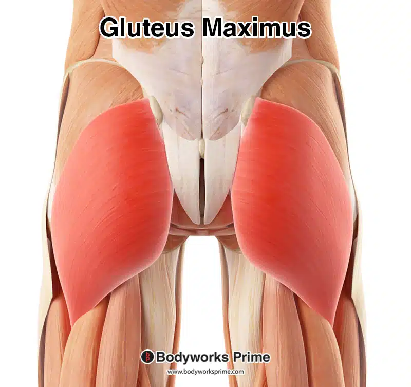
The gluteus maximus is the largest and heaviest muscle in the human body. It forms the most superficial layer of the gluteal muscle group, which also includes the gluteus medius and gluteus minimus muscles. The gluteus maximus is located in the posterior part of the hip and buttock region and it covers the other two gluteal muscles, except for the anterior superior third of the gluteus medius [1] [2]. Its primary function is hip extension, and it is involved in activities such as: walking, running, jumping, weightlifting and climbing stairs.
Exercises that target the gluteus maximus muscle are important for improving muscle strength, hypertrophy, and functionality. The barbell hip thrust, popularised by Bret Contreras, has been demonstrated to be one of the best exercises to develop the size and strength of the gluteus maximus, above even the barbell squat [3]. Other exercises for strengthening the gluteus maximus include: Romanian deadlifts, step-ups, lunges, and barbell glute bridges [4] [5] [6].
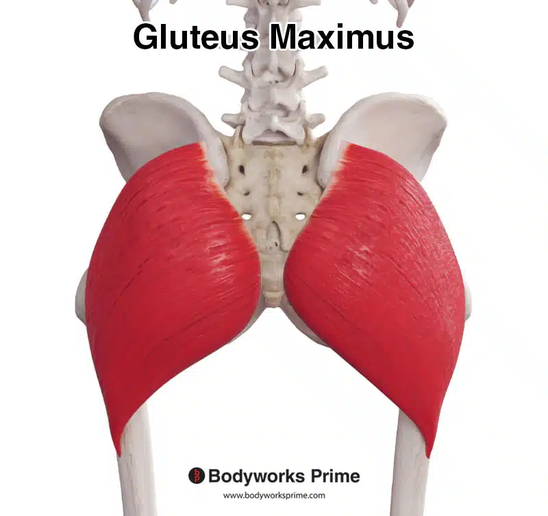
Here we can see the gluteus maximus from a posterior view.
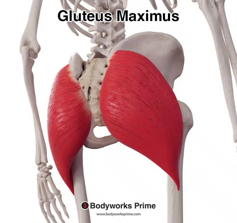
Here we can see the gluteus maximus from a posterolateral view.
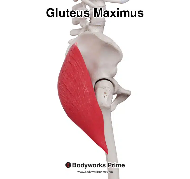
Here we can see the gluteus maximus from a lateral view.
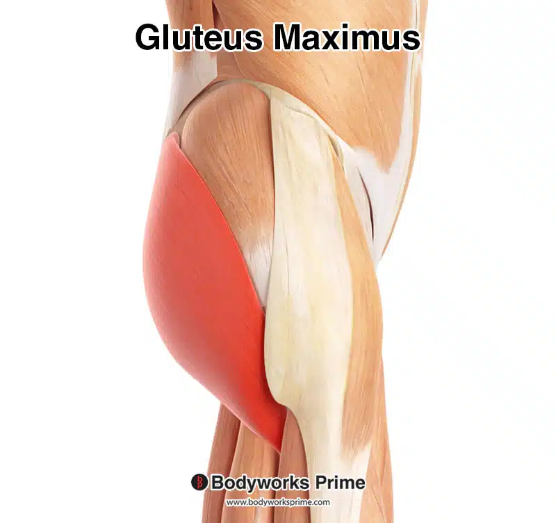
Here we can see the gluteus maximus highlighted in red, seen from a posterior view.
Here we can see the gluteus maximus highlighted in red, seen from a lateral view.
Origin & Insertion
The gluteus maximus muscle originates from several places on the pelvic bones and the lower back. The first origin point is on the posterior gluteal line of the ilium. The ilium is the largest bone of the pelvis located at the top of the hip. The posterior gluteal line is a bony ridge on the outer surface of the ilium. The gluteus maximus also originates from the posterior surface of the sacrum and the coccyx, which are two bones at the base of the spine [7][8] [9].
Furthermore, the gluteus maximus originates from the thoracolumbar fascia. The thoracolumbar fascia is a broad sheet of connective tissue that covers the lower back muscles. Another origin point is the sacrotuberous ligament, which is a strong band of fibrous tissue that connects the sacrum to the ischial tuberosity. The ischial tuberosity is a bony prominence located on the lower part of the pelvis [10][11] [12].
The muscle fibers of the gluteus maximus merge together to form a tendon that inserts onto two main locations. The insertions of the gluteus maximus are the gluteal tuberosity of the femur and the iliotibial tract (also known as the iliotibial band or IT band). The femur is the long bone of the thigh, and the gluteal tuberosity is a roughened area on its lateral surface, near the upper part of the bone. The iliotibial tract is a thick band of fibrous connective tissue that runs down the lateral aspect of the thigh, from the ilium to the tibia [13][14] [15].
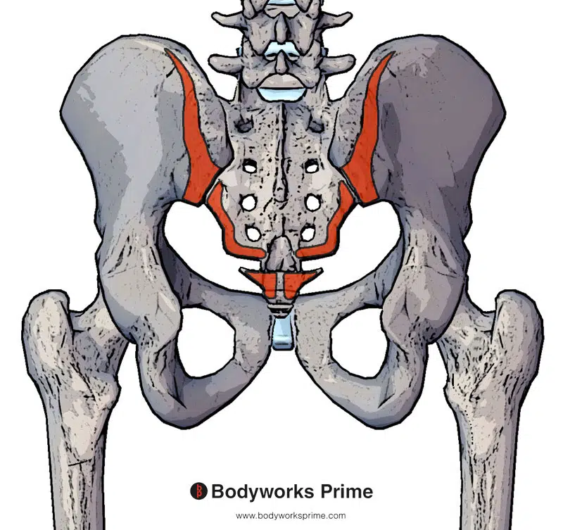
Here we can see pictured the bony origins of the gluteus maximus muscle marked in red. These are on the posterior gluteal line of the ilium, posterior surface of the sacrum and the posterior surface of the coccyx.
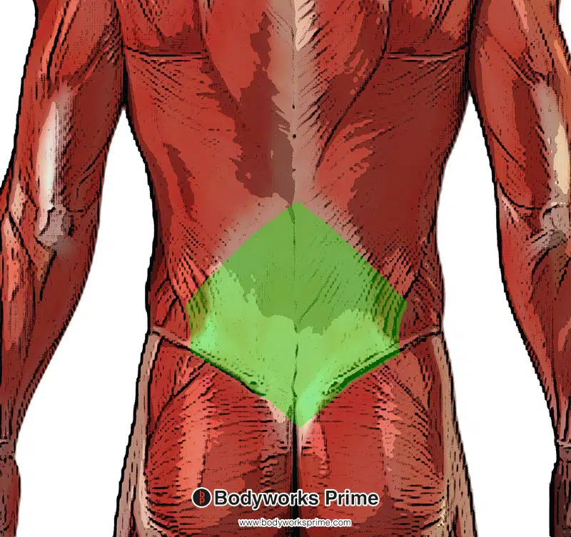
Here we can see pictured another origin of the gluteus maximus muscle, which is the thoracolumbar fascia (highlighted in green).
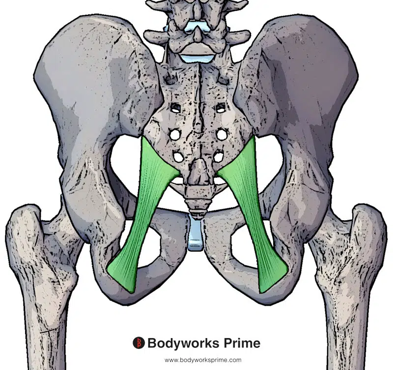
Here we can see pictured the last origin of the gluteus maximus muscle, which is the sacrotuberous ligament (highlighted in green).
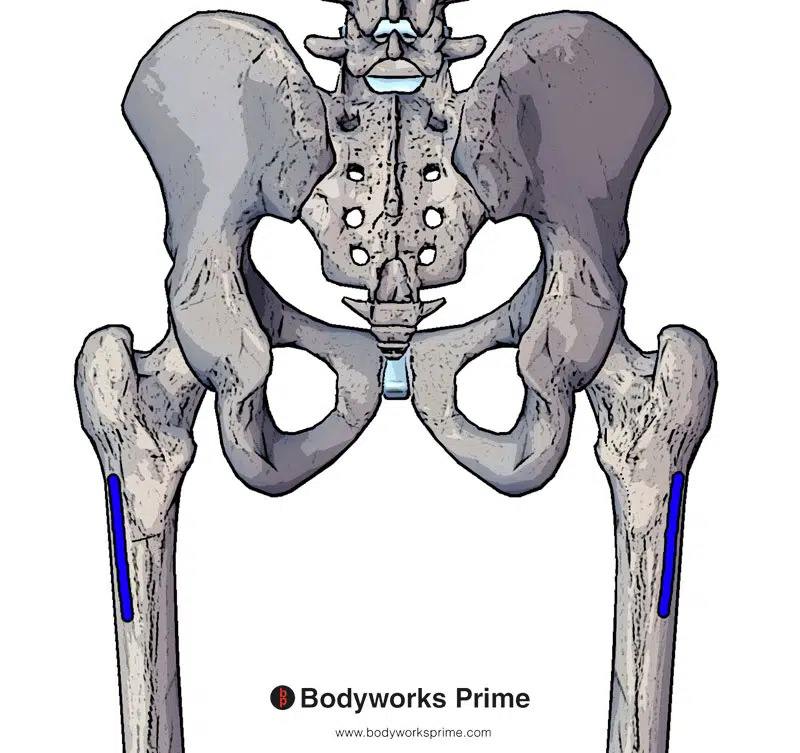
Here we can see an insertion of the gluteus maximus muscle on the gluteal tuberosity of the femur (marked in blue).
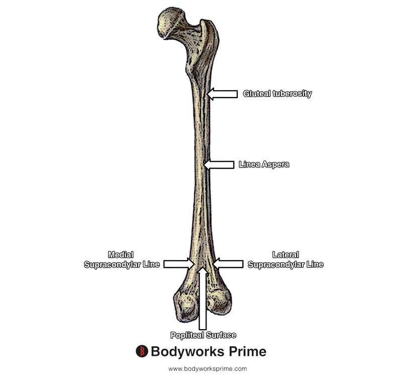
Here we can see the gluteal tuberosity of the femur amongst other bony landmarks of the femur.
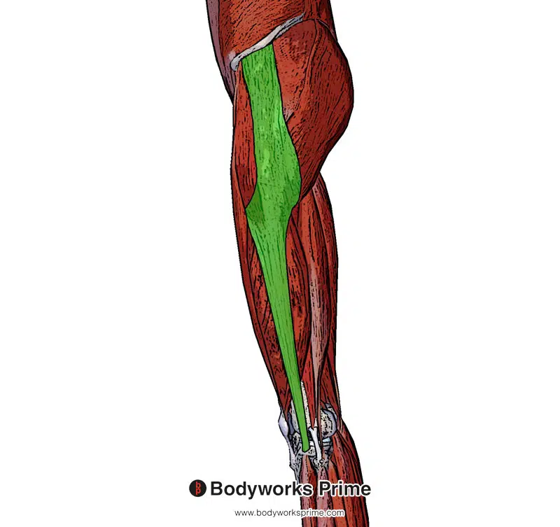
Here we can see pictured another insertion of the gluteus maximus muscle, which is the iliotibial band (highlighted in green).
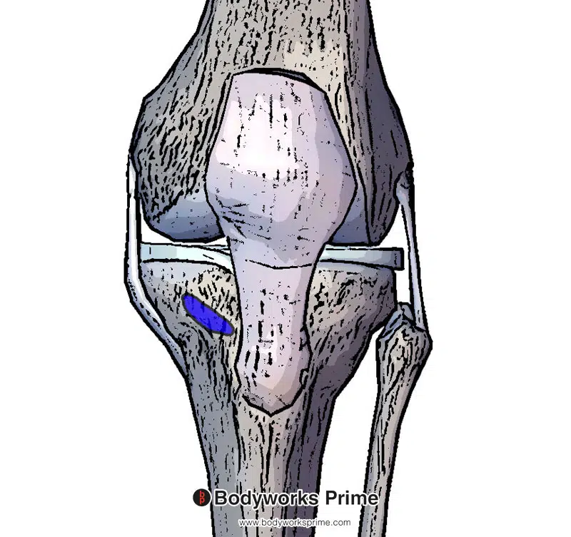
Here we can see the insertion of the iliotibial band on the tibia (highlighted in blue).
Actions
The gluteus maximus muscle has actions involving the hip joint. Its primary function is hip extension, which is the movement of straightening out the hip joint from a flexed position. This action is useful for activities such as standing up from a seated position, walking up stairs, or running. In addition to hip extension, the gluteus maximus also assists with lateral rotation of the hip. Lateral (external) rotation is the rotation of the thigh outwards, relative to the hip joint. This movement is important for stabilising the hip during weight-bearing activities [16] [17] [18].
Furthermore, the gluteus maximus is able to to weakly contribute to hip abduction [19] [20], which involves the movement of the leg away from the midline of the body. The upper fibers contribute more in frontal plane hip abduction, and all of the fibers contribute in the transverse plane.
Some online sources and even textbooks suggest that the inferior part of the gluteus maximus muscle contributes to hip adduction, there isn’t sufficient evidence to support this claim. It is more likely that the role which the gluteus maximus plays during adduction is controlling and stabilising the hip, rather than directly causing adduction. The glute max can abduct but(t) not adduct [21] [22].
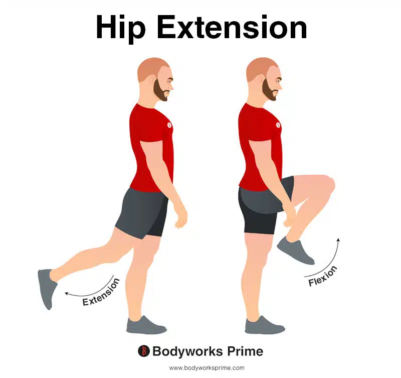
In this image, you can see an example of hip extension. Hip extension is the movement of the thigh or the upper leg backward, away from the body, in the sagittal plane. This action involves increasing the angle between the thigh and the pelvis, causing the hip joint to straighten or extend. The opposite movement of hip extension is hip flexion. The primary action of the gluteus maximus is hip extension.
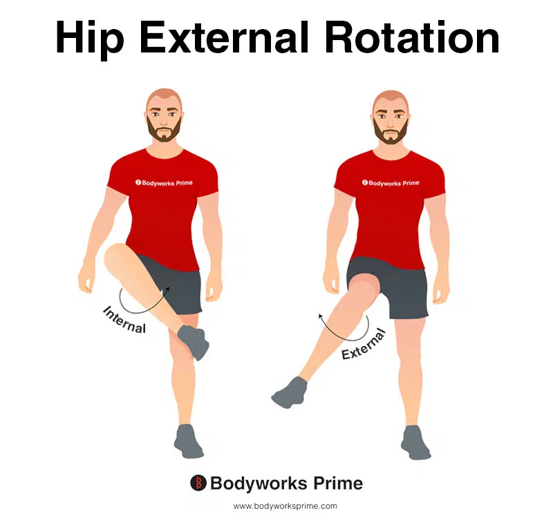
This image depicts an example of hip external rotation, which involves rotating the leg from the hip joint outwards (laterally). External rotation is also referred to as lateral rotation. The opposite of external rotation (lateral rotation) is internal rotation (medial rotation). A secondary action of the gluteus maximus is external rotation of the hip joint.
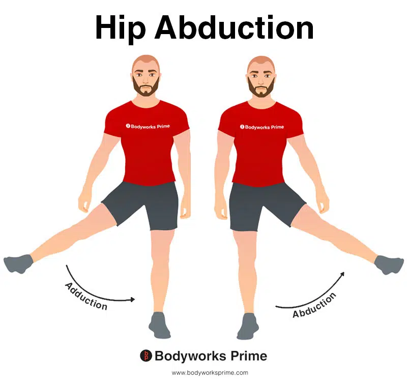
This image shows an example of hip abduction, which involves moving the leg out to the side (laterally). The opposite of hip abduction is hip adduction. The gluteus maximus contributes weakly to hip abduction.
Innervation
The gluteus maximus muscle is innervated by the inferior gluteal nerve, which arises from the lumbar and sacral segments of the spinal cord, specifically L5, S1, and S2 nerve roots. These nerve roots exit the spinal cord and combine to form the sacral plexus. The sacral plexus is a network of nerves located within the pelvis [23] [24].
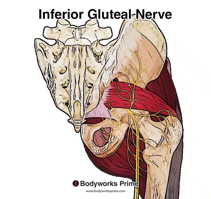
Here we can see the inferior gluteal nerve, which innervates the gluteus maximus muscle from L5, S1, and S2 nerve roots.
The inferior gluteal nerve leaves the sacral plexus through the greater sciatic foramen, a space between the pelvic bones that provides passage for various nerves and blood vessels. It then passes deep to the gluteus maximus muscle, supplying it with motor innervation. This allows the muscle to contract and perform its various functions, such as hip extension, lateral rotation, and potentially hip abduction [25] [26].
Blood Supply
The gluteus maximus muscle receives its blood supply from two main sources: the superior gluteal artery and the inferior gluteal artery. Both of these arteries are branches of the internal iliac artery, which is a major blood vessel in the pelvic region [27].
The superior gluteal artery exits the pelvis through the greater sciatic foramen, which is just above the piriformis muscle. It then divides into superficial and deep branches, which supply the gluteus maximus and other nearby muscles, as well as the nearby skin and connective tissues [28].
The inferior gluteal artery, on the other hand, exits the pelvis through the greater sciatic foramen, below the piriformis muscle. It also gives off branches that supply the gluteus maximus muscle and neighboring structures. Both the superior and inferior gluteal arteries provide oxygenated blood and nutrients to the gluteus maximus muscle [29].
Want some flashcards to help you remember this information? Then click the link below:
Gluteus Maximus Muscle Flashcards
Support Bodyworks Prime
Running a website and YouTube channel can be expensive. Your donation helps support the creation of more content for my website and YouTube channel. All donation proceeds go towards covering expenses only. Every contribution, big or small, makes a difference!
References
| ↑1 | Jeong, Ju-Ri & Lee, Su-Jin & Lee, Wan-Hee. (2017). Reliability of rehabilitative ultrasound imaging for measuring the gluteus maximus muscle at rest and during contraction. Physical Therapy Rehabilitation Science. 6. 7-13. 10.14474/ptrs.2017.6.1.7. |
|---|---|
| ↑2, ↑23, ↑25 | Moore KL, Agur AMR, Dalley AF. Clinically Oriented Anatomy. 8th ed. Philadelphia: Lippincot Williams & Wilkins; 2017. |
| ↑3 | Contreras B, Vigotsky AD, Schoenfeld BJ, Beardsley C, Cronin J. A Comparison of Gluteus Maximus, Biceps Femoris, and Vastus Lateralis Electromyography Amplitude for the Barbell, Band, and American Hip Thrust Variations. J Appl Biomech. 2016 Jun;32(3):254-60. doi: 10.1123/jab.2015-0091. Epub 2015 Dec 22. PMID: 26695353. |
| ↑4 | Delgado J, Drinkwater EJ, Banyard HG, Haff GG, Nosaka K. Comparison Between Back Squat, Romanian Deadlift, and Barbell Hip Thrust for Leg and Hip Muscle Activities During Hip Extension. J Strength Cond Res. 2019 Oct;33(10):2595-2601. doi: 10.1519/JSC.0000000000003290. PMID: 31356511. |
| ↑5, ↑17 | Neto WK, Soares EG, Vieira TL, Aguiar R, Chola TA, Sampaio VL, Gama EF. Gluteus Maximus Activation during Common Strength and Hypertrophy Exercises: A Systematic Review. J Sports Sci Med. 2020 Feb 24;19(1):195-203. PMID: 32132843; PMCID: PMC7039033. |
| ↑6 | Kennedy D, Casebolt JB, Farren GL, Fiaud V, Bartlett M, Strong L. Electromyographic differences of the gluteus maximus, gluteus medius, biceps femoris, and vastus lateralis between the barbell hip thrust and barbell glute bridge. Sports Biomech. 2022 May 19:1-15. doi: 10.1080/14763141.2022.2074875. Epub ahead of print. PMID: 35586943. |
| ↑7, ↑10, ↑13 | Moore KL, Agur AMR, Dalley AF. Clinically Oriented Anatomy. 8th ed. Philadelphia: Lippincot Williams & Wilkins; 2017. |
| ↑8, ↑11, ↑14 | Barker, Priscilla & Hapuarachchi, Kamal & Ross, J.A. & Sambaiew, E & Ranger, T.A. & Briggs, Christopher. (2014). Anatomy and Biomechanics of Gluteus Maximus and the Thoracolumbar Fascia at the Sacroiliac Joint. Clinical anatomy (New York, N.Y.). 27. 10.1002/ca.22233. |
| ↑9, ↑12, ↑15, ↑18 | Elzanie A, Borger J. Anatomy, Bony Pelvis and Lower Limb, Gluteus Maximus Muscle. 2022 Mar 28. In: StatPearls [Internet]. Treasure Island (FL): StatPearls Publishing; 2023 Jan–. PMID: 30855781. |
| ↑16, ↑22 | Neumann DA. Kinesiology of the hip: a focus on muscular actions. J Orthop Sports Phys Ther. 2010 Feb;40(2):82-94. doi: 10.2519/jospt.2010.3025. PMID: 20118525. |
| ↑19 | Selkowitz DM, Beneck GJ, Powers CM. Which exercises target the gluteal muscles while minimizing activation of the tensor fascia lata? Electromyographic assessment using fine-wire electrodes. J Orthop Sports Phys Ther. 2013 Feb;43(2):54-64. doi: 10.2519/jospt.2013.4116. Epub 2012 Nov 16. PMID: 23160432. |
| ↑20, ↑21 | Lyons K, Perry J, Gronley JK, Barnes L, Antonelli D. Timing and relative intensity of hip extensor and abductor muscle action during level and stair ambulation. An EMG study. Phys Ther. 1983 Oct;63(10):1597-605. doi: 10.1093/ptj/63.10.1597. PMID: 6622534. |
| ↑24, ↑26 | Sumalatha S, D Souza AS, Yadav JS, Mittal SK, Singh A, Kotian SR. An unorthodox innervation of the gluteus maximus muscle and other associated variations: A case report. Australas Med J. 2014 Oct 31;7(10):419-22. doi: 10.4066/AMJ.2014.2225. PMID: 25379064; PMCID: PMC4221778. |
| ↑27 | Elzanie A, Borger J. Anatomy, Bony Pelvis and Lower Limb, Gluteus Maximus Muscle. [Updated 2021 Jul 19]. In: StatPearls [Internet]. Treasure Island (FL): StatPearls Publishing; 2021 Jan-. Available from: https://www.ncbi.nlm.nih.gov/books/NBK538193/ |
| ↑28, ↑29 | Standring S. (2015). Gray’s Anatomy: The Anatomical Basis of Clinical Practice, 41st Edn. Amsterdam: Elsevier. |


