| Origin | Spinous processes of C7-T1 and the nuchal ligament |
| Insertion | Medial border of the scapula |
| Actions | Scapula retraction (adduction) Scapula elevation Scapula downward rotation |
| Nerve | Dorsal scapular nerve (C5) |
| Artery | Dorsal scapular artery |
Location & Overview
The rhomboid minor is a small, slender, somewhat cylindrical shaped and it forms part of the intricate network of muscles in the upper back and shoulder girdle. It is located between the scapula and the vertebral column. The rhomboid minor is one of the rhomboid muscles, which consist of the rhomboid minor and the rhomboid major [1] [2].
The rhomboid minor is located superior to the larger rhomboid major, immediately beneath the levator scapulae, and deep to the trapezius muscle. This placement situates it between and beneath the more superficial layers of muscle, positioning it in close proximity to the vertebral column [3] [4].
To emphasize the important role of the rhomboid major in scapular function and shoulder stability, it’s worth pointing out the potential issues arising from weakness, dysfunction, or nerve complications. One such issue is scapular winging, where the scapula protrudes away from the rib cage. This can occur if the rhomboids are unable to maintain their usual tension and control. Similarly, dysfunction in the rhomboids could lead to rotation of the inferior angle of the scapula, which can cause discomfort and limit shoulder mobility [5].
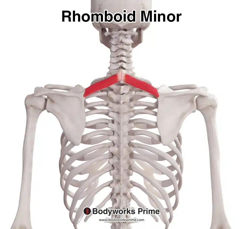
Here we can see the rhomboid minor from a posterior view.
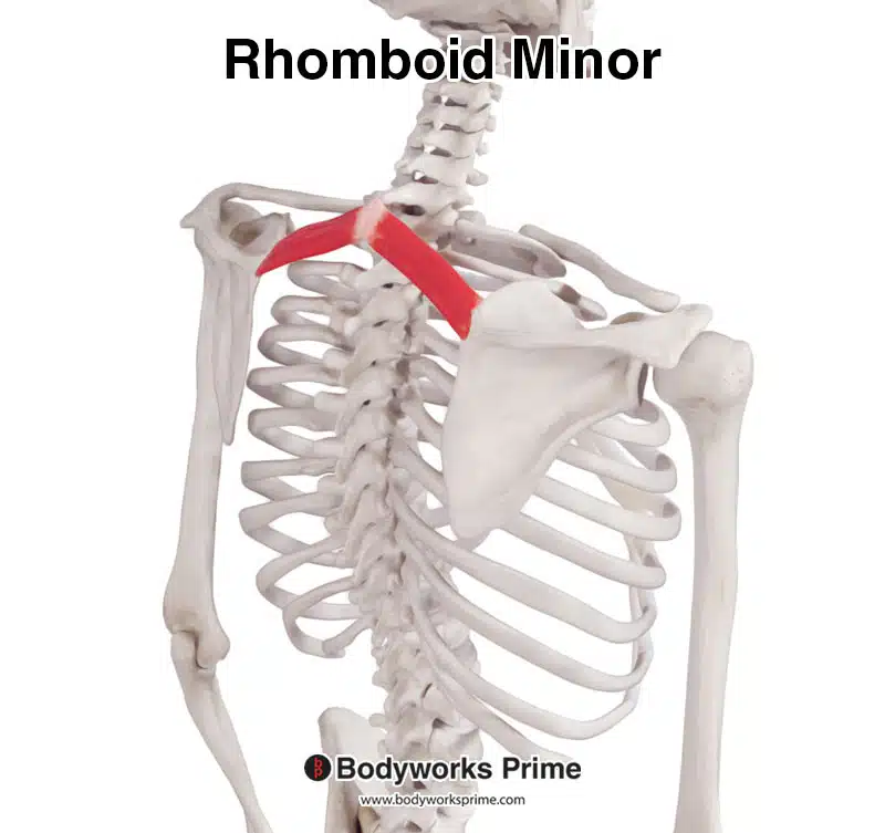
Here we can see the rhomboid minor from a posterolateral view.
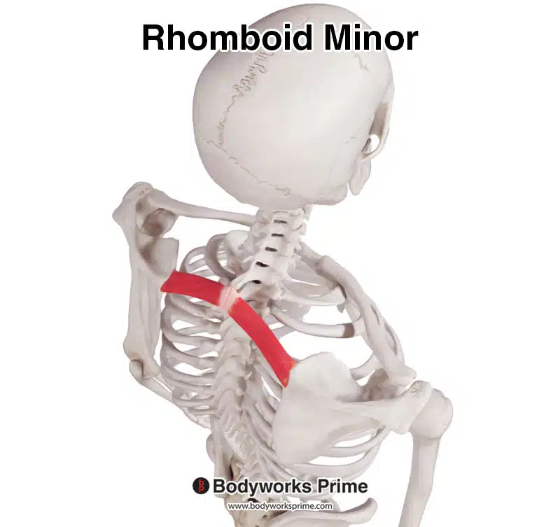
Here we can see the rhomboid minor from a superior view.
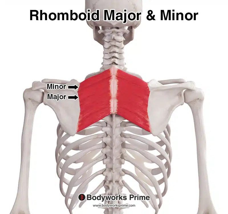
Here we can see the rhomboid minor and major together, seen from a posterior view.
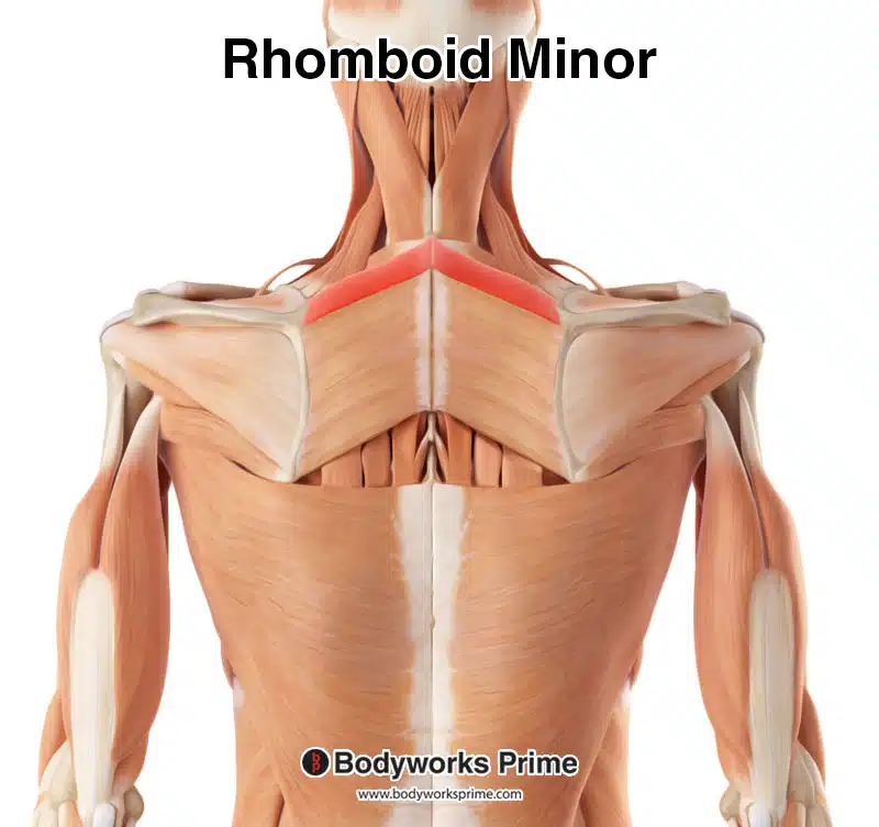
Here we can see the rhomboid minor highlighted in red amongst the other muscles of the body. Superficial muscles, such as the trapezius, have been removed to reveal the rhomboid minor.
Origin & Insertion
The rhomboid minor originates from the spinous processes of the C7 and T1 vertebrae. These points of origin are located in the lower part of the neck, with the C7 being the last cervical vertebra that forms the base of the neck and T1 being the first thoracic vertebra, right below the C7. In addition to these origins, the rhomboid minor also originates from the nuchal ligament [6] [7].
The nuchal ligament is a thick, fibrous and elastic structure that stretches from the external occipital protuberance to the spinous process of the seventh cervical vertebra (C7). The external occipital protuberance is a noticeable bony prominence located at the base of the skull, on the back of the head. The median nuchal line is a slight ridge which runs down the midline of the back of the skull [8] [9].
The insertion point of the rhomboid minor muscle is the medial border of the scapula, which is the bone that forms the shoulder blade. The insertion is found superior to the point where the rhomboid major muscle inserts [10] [11].
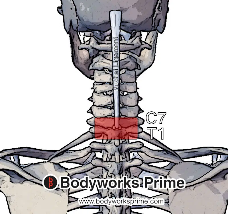
Pictured here we can see the rhomboid minor muscle and nuchal ligament. The rhomboid muscle originates at C7 and T1 vertebra and the nuchal ligament (origin highlighted in red).
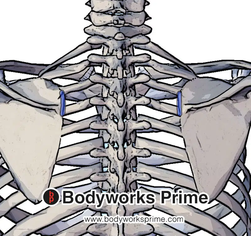
Pictured here we can see the rhomboid minor muscle insertion at the superior medial border of the scapula (highlighted in blue). The insertion is superior to the insertion of the rhomboid major.
Actions
The rhomboid minor muscle, though modest in size, contributes to several important scapula movements. Its primary action is the retraction of the scapula, essentially pulling it towards the midline of the body, or the vertebral column. To imagine this action, think about squeezing the shoulder blades together [12] [13] [14].
In addition to retraction, the rhomboid minor assists with the elevation of the scapula. To imagine this action, think about shrugging the shoulders upward [15] [16] [17].
Lastly, the rhomboid minor contributes to the medial rotation of the scapula. This rotation causes the glenoid cavity (the socket of the shoulder joint) to be depressed downwards. This action is important for enabling a wide range of arm movements and maintaining glenohumeral joint stability [18] [19] [20].
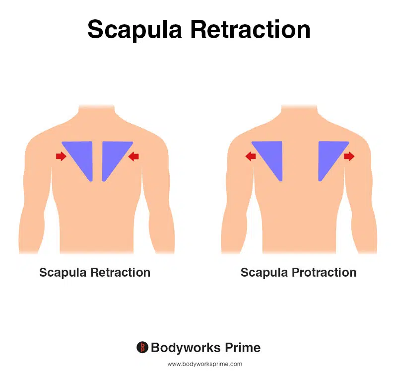
This image shows scapula retraction, also known as scapula adduction, where the shoulder blades move towards the spine, as in the action of pinching the shoulder blades together. The opposite of scapula retraction is scapula protraction, also known as scapula abduction, where the shoulder blades move away from the spine, as in the action of reaching forward. Both the rhomboid minor and major retract the scapula.
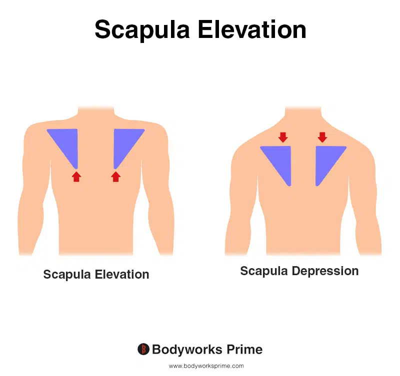
This image shows scapula elevation, where the shoulder blade moves upward, drawing the shoulder joint superiorly. This movement happens when shrugging the shoulders or lifting a weight overhead. The opposite of scapula elevation is scapula depression. Both the rhomboid minor and major contribute to the elevation of the scapula.
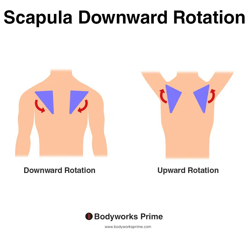
This image shows scapula downward rotation, where the shoulder blade moves medially and inferiorly, tilting the glenoid cavity downward. This movement is typical when lowering the arm from an overhead position, bringing it back to the side. The opposite of scapula downward rotation is scapula upward rotation. Both the rhomboid minor and major assist with downward rotation of the scapula.
Innervation
The rhomboid minor muscle derives its innervation predominantly from the dorsal scapular nerve, generally emerging from the C5 spinal nerve root. This root is an integral component of the brachial plexus – an intricate and highly structured aggregation of nerves situated in the cervical region. The primary function of the brachial plexus is to facilitate the transmission of neural impulses from the spinal cord to the superior extremities, including the rhomboid minor [21] [22].
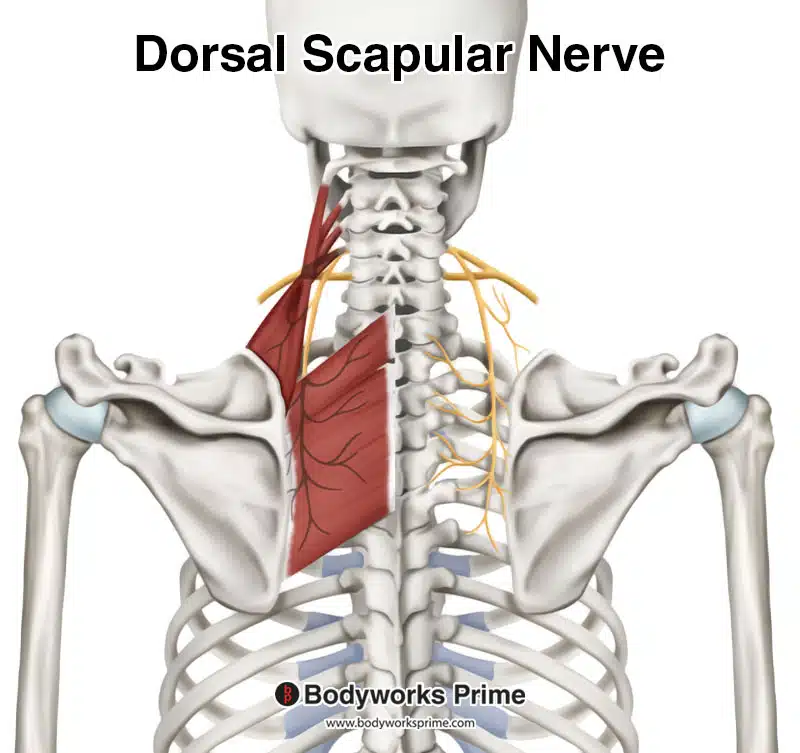
Pictured here we can see the innervation of the rhomboid minor, the dorsal scapular nerve. This nerve generally originates from C5.
Blood Supply
The rhomboid minor muscle’s blood supply is principally maintained by the dorsal scapular artery. The dorsal scapular artery typically originates from either the second or third portion of the subclavian artery. The subclavian artery is a major blood vessel responsible for supplying blood to the upper body. In some people, dorsal scapular artery can arise from the thyrocervical trunk, which is an artery located in the neck [23] [24].
The dorsal scapular artery also courses through the brachial plexus, an intricate network of nerves situated in the neck region. Here, it aligns and travels alongside the dorsal scapular nerve. Together, the nerve and artery proceed downwards to supply the rhomboid muscle group, including the rhomboid minor [25] [26].
In addition to the dorsal scapular artery, the rhomboid minor can occasionally receive blood from a deep branch of the transverse cervical artery. This artery is part of the network of blood vessels serving the neck and shoulder region [27] [28].
Want some flashcards to help you remember this information? Then click the link below:
Rhomboid Minor Flashcards
Support Bodyworks Prime
Running a website and YouTube channel can be expensive. Your donation helps support the creation of more content for my website and YouTube channel. All donation proceeds go towards covering expenses only. Every contribution, big or small, makes a difference!
References
| ↑1, ↑3, ↑8, ↑21, ↑23, ↑25 | Standring S. (2015). Gray’s Anatomy: The Anatomical Basis of Clinical Practice, 41st Edn. Amsterdam: Elsevier. |
|---|---|
| ↑2, ↑4, ↑9, ↑12, ↑15, ↑18, ↑22 | Moore KL, Agur AMR, Dalley AF. Clinically Oriented Anatomy. 8th ed. Philadelphia: Lippincot Williams & Wilkins; 2017. |
| ↑5, ↑6, ↑10 | Farrell C, Kiel J. Anatomy, Back, Rhomboid Muscles. [Updated 2022 May 20]. In: StatPearls [Internet]. Treasure Island (FL): StatPearls Publishing; 2023 Jan-. Available from: https://www.ncbi.nlm.nih.gov/books/NBK534856/ |
| ↑7, ↑11, ↑28 | Mitchell B, Imonugo O, Tripp JE. Anatomy, Back, Extrinsic Muscles. [Updated 2022 Aug 25]. In: StatPearls [Internet]. Treasure Island (FL): StatPearls Publishing; 2023 Jan-. Available from: https://www.ncbi.nlm.nih.gov/books/NBK537216/ |
| ↑13, ↑16, ↑19 | Martin RM, Fish DE. Scapular winging: anatomical review, diagnosis, and treatments. Curr Rev Musculoskelet Med. 2008 Mar;1(1):1-11. doi: 10.1007/s12178-007-9000-5. PMID: 19468892; PMCID: PMC2684151. |
| ↑14, ↑17, ↑20 | Paine R, Voight ML. The role of the scapula. Int J Sports Phys Ther. 2013 Oct;8(5):617-29. PMID: 24175141; PMCID: PMC3811730. |
| ↑24, ↑26, ↑27 | Ikka L, Mihalea C, Achour NB, Khalek HA, Vacher C. The origin of the dorsal scapular artery: anatomic variations and surgical applications. Surg Radiol Anat. 2016 Nov;38(9):1021-1027. doi: 10.1007/s00276-016-1656-3. Epub 2016 Feb 26. PMID: 26920558. |


