| Origin | Spinous process of T2 to T5 vertebrae |
| Insertion | Medial border of the scapula |
| Actions | Scapula retraction (adduction) Scapula elevation Scapula downward rotation |
| Nerve | Dorsal scapular nerve (C5) |
| Artery | Dorsal scapular artery |
Location & Overview
The rhomboid major is located in the upper back and contributes a key role in connecting the vertebrae of the spine to the scapula (shoulder blade). The rhomboid major is part of the pair of muscles known as the rhomboids, which also includes the rhomboid minor. The rhomboid major holds a significant responsibility in maintaining the stability of the shoulder girdle and the scapula [1] [2].
Distinctively rhomboid in shape, hence the name, the rhomboid major is larger and typically stronger than its minor counterpart. It is positioned inferiorly to the rhomboid minor and located deep to the broad and superficial trapezius muscle. As one of the deep back muscles, it lies against the thoracic vertebrae and the adjacent ribs. Lateral to the rhomboid major, you will find the serratus anterior, another muscle which is involved with scapula movement and stabilisation. Anatomical variations of the rhomboid major and minor can occur, such as the fusion of the rhomboid major and minor into a single muscle [3] [4].
Highlighting the significance of the rhomboid major in the stability of the shoulder and functioning of the scapula, it becomes essential to consider the potential problems that might arise from any muscular weakness, functional anomalies, or nerve issues. A noteworthy manifestation of such problems is the occurrence of scapular winging, a condition in which the scapula deviates away from the rib cage. This can transpire when the rhomboids fail to exert their typical tension and control. Similarly, the inferior angle of the scapula could undergo rotation as a result of rhomboid dysfunction, potentially leading to discomfort and a reduction in the range of shoulder movements [5].
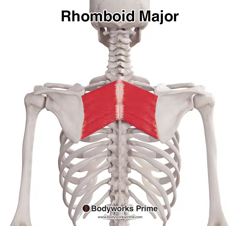
Here we can see the rhomboid major from a posterior view.
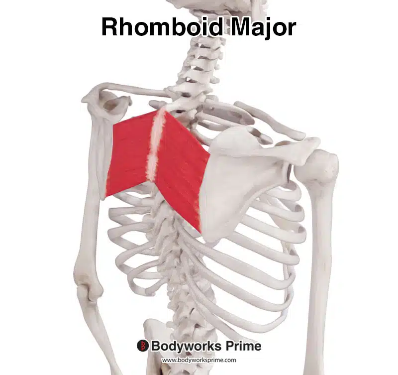
Here we can see the rhomboid major from a posterolateral view.
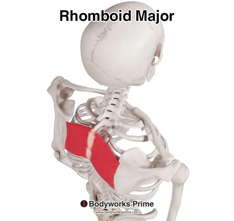
Here we can see the rhomboid major from a superior view.
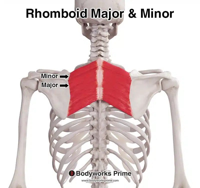
Here we can see the rhomboid major and minor together, seen from a posterior view.
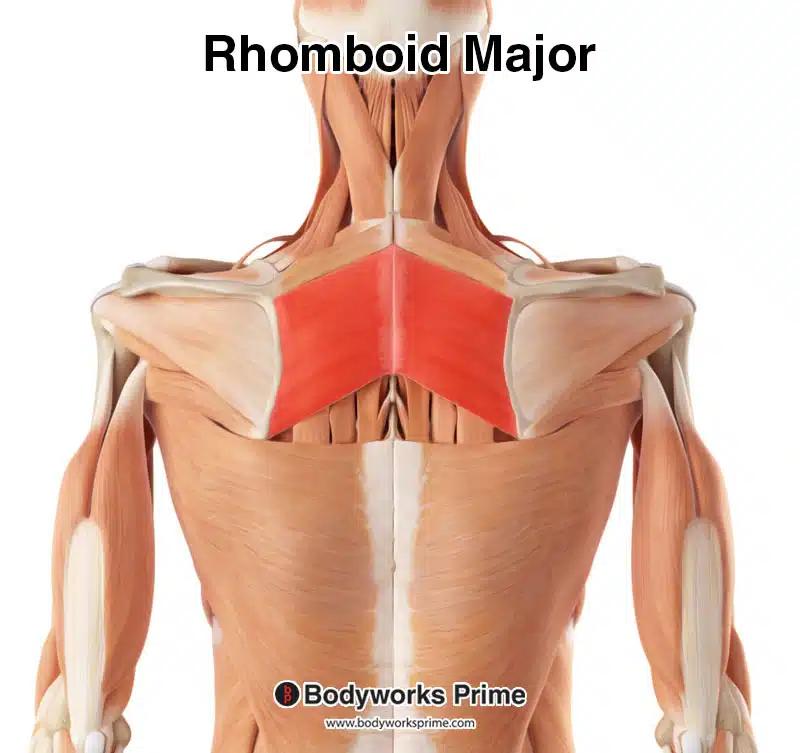
Here we can see the rhomboid major highlighted in red amongst the other muscles of the body. Superficial muscles, such as the trapezius, have been removed to reveal the rhomboid major.
Origin & Insertion
The rhomboid major originates from the spinous processes of T2 through T5, which are located in the upper part of the thoracic spine. This region of the spine extends from the base of the neck down to the middle of the back. The second thoracic vertebra (T2) is found directly below the first thoracic vertebra (T1) and there are twelve thoracic vertebrae in total. The spinous processes of these vertebrae are projections from the posterior of the vertebra, they are the pointed sections of bone that you can feel when you run your finger down the middle of your back [6] [7] [8].
The rhomboid major inserts onto the medial border of the scapula, just below the insertion point of the rhomboid minor muscle. This location is the inner edge of the shoulder blade, closest to the vertebral column. Its insertion point is below the spine of the scapula. The spine of the scapula is a prominent bony ridge that runs horizontally across the back side of the scapula [9] [10] [11].
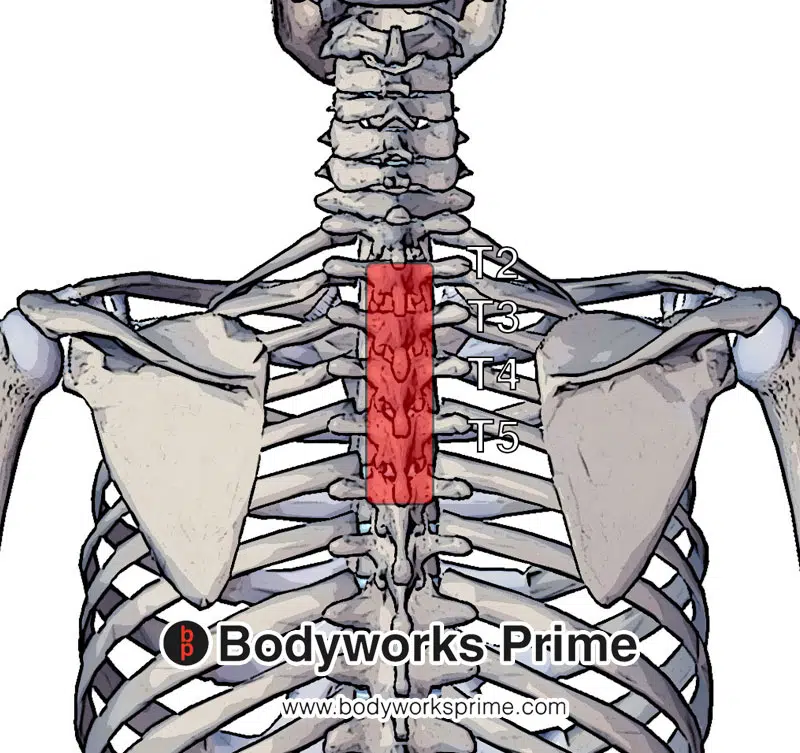
Pictured here we can see the origin of the rhomboid major muscle. Spinous process of T2 to T5 vertebrae highlighted in red.
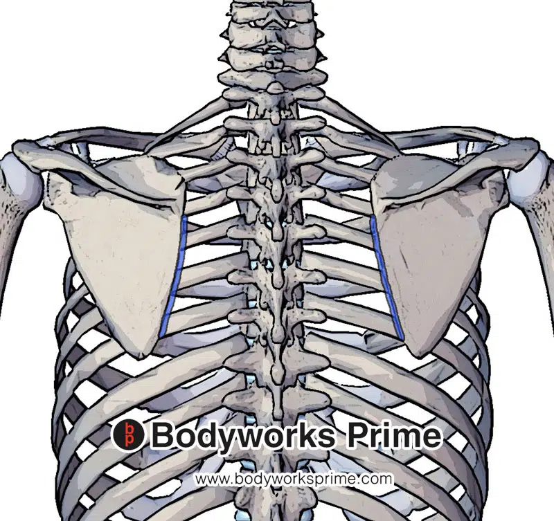
Here we can see the insertion of the rhomboid major muscle highlighted in blue. It inserts on the medial border of the scapula.
Actions
The rhomboid major, like its minor counterpart, plays an important role in movements of the scapula. One of its main functions is to retract the scapula, drawing it inwards towards the spinal column. This is the action of bringing the shoulder blades together, towards the midline of the body [12] [13] [14].
Additionally, the rhomboid major assists in elevating the scapula, the equivalent of lifting the shoulders in a shrug [15] [16] [17].
Lastly, the muscle plays a role in the medial (downward) rotation of the scapula. This function positions the glenoid cavity, the cup-shaped socket in the shoulder joint, downward. The depression of the glenoid cavity through this rotation is essential for enabling a variety of arm movements and maintaining stability in the shoulder joint [18] [19] [20].
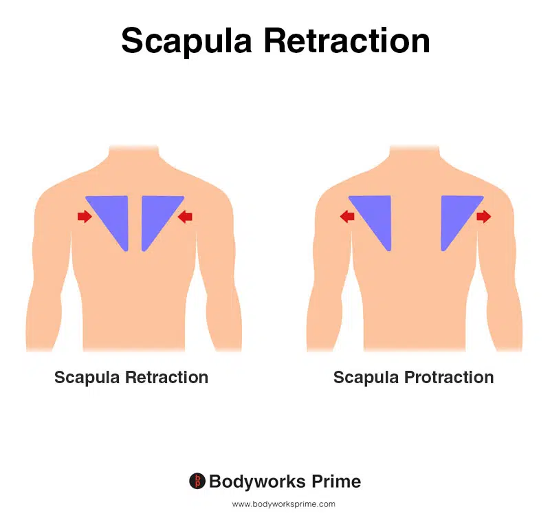
This image shows scapula retraction, also known as scapula adduction, where the shoulder blades move towards the spine, as in the action of pinching the shoulder blades together. The opposite of scapula retraction is scapula protraction, also known as scapula abduction, where the shoulder blades move away from the spine, as in the action of reaching forward. Both the rhomboid major and minor retract the scapula.
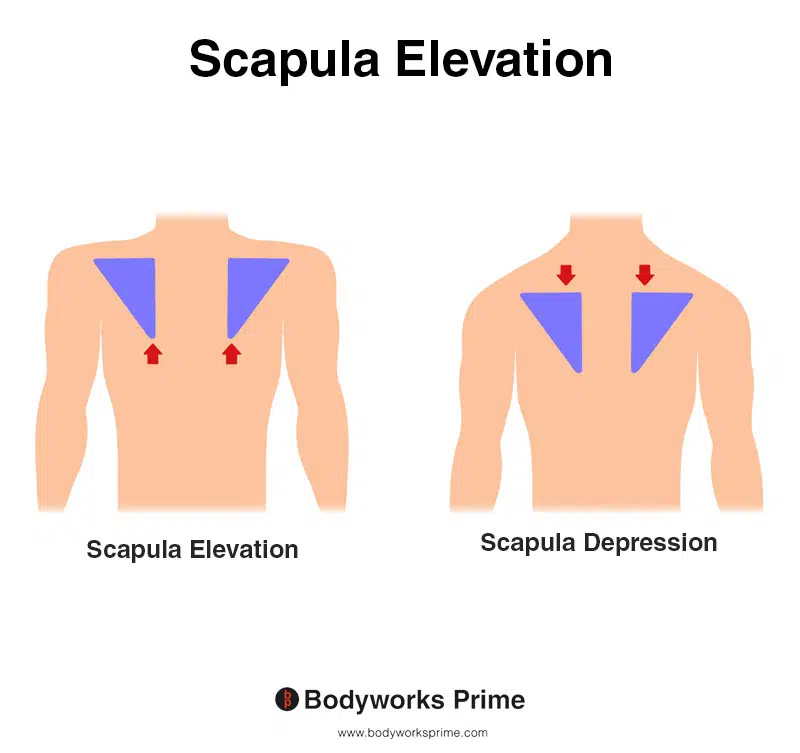
This image shows scapula elevation, where the shoulder blade moves upward, drawing the shoulder joint superiorly. This movement happens when shrugging the shoulders or lifting a weight overhead. The opposite of scapula elevation is scapula depression. Both the rhomboid major and minor contribute to the elevation of the scapula.
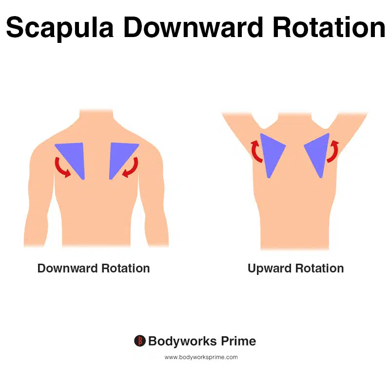
This image shows scapula downward rotation, where the shoulder blade moves medially and inferiorly, tilting the glenoid cavity downward. This movement is typical when lowering the arm from an overhead position, bringing it back to the side. The opposite of scapula downward rotation is scapula upward rotation. Both the rhomboid major and minor assist with downward rotation of the scapula.
Innervation
The rhomboid major muscle receives its main neural supply from the dorsal scapular nerve. The dorsal scapular nerve is a branch of the brachial plexus. The dorsal scapular nerve originates from the spinal nerve root of C5. Located in the neck region, the brachial plexus is a complex arrangement of intersecting nerves. It has the important task of facilitating the transmission of neural signals between the spinal cord and the upper extremities, including the rhomboids [21] [22].
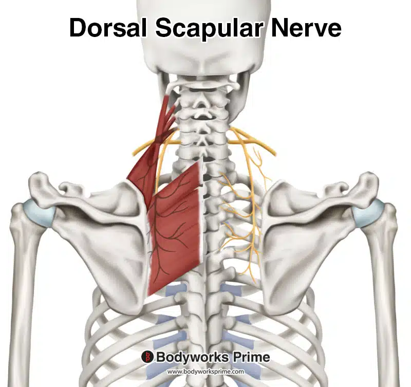
Pictured here we can see the innervation of the rhomboid major, the dorsal scapular nerve. This nerve generally originates from C5.
Blood Supply
The blood supply of the rhomboid major muscle is predominantly supplied by the dorsal scapular artery. This artery usually takes its origin from the second or third segment of the subclavian artery, a primary artery involved in delivering blood to the upper part of the body. However, in some individuals, there exists a variation where the origin of the dorsal scapular artery arises from the thyrocervical trunk instead. The thyrocervical trunk is another important artery positioned in the neck region [23] [24].
The dorsal scapular artery passes through the brachial plexus. The brachial plexus is a complex set of nerves found in the neck. Together, the dorsal scapular artery and dorsal scapular nerve run inferiorly together to innervate and supply the rhomboid muscles with blood [25] [26].
Furthermore, there exists an anatomical variation in some individuals, where the rhomboid major muscle can be supplied by a deep branch of the transverse cervical artery. This is another artery located in the neck and shoulder area [27] [28].
Want some flashcards to help you remember this information? Then click the link below:
Rhomboid major flashcards
Support Bodyworks Prime
Running a website and YouTube channel can be expensive. Your donation helps support the creation of more content for my website and YouTube channel. All donation proceeds go towards covering expenses only. Every contribution, big or small, makes a difference!
References
| ↑1, ↑3, ↑8, ↑12, ↑15, ↑18, ↑21, ↑23, ↑25 | Standring S. (2015). Gray’s Anatomy: The Anatomical Basis of Clinical Practice, 41st Edn. Amsterdam: Elsevier. |
|---|---|
| ↑2, ↑4, ↑11, ↑22 | Moore KL, Agur AMR, Dalley AF. Clinically Oriented Anatomy. 8th ed. Philadelphia: Lippincot Williams & Wilkins; 2017. |
| ↑5, ↑6, ↑9 | Farrell C, Kiel J. Anatomy, Back, Rhomboid Muscles. [Updated 2022 May 20]. In: StatPearls [Internet]. Treasure Island (FL): StatPearls Publishing; 2023 Jan-. Available from: https://www.ncbi.nlm.nih.gov/books/NBK534856/ |
| ↑7, ↑10, ↑28 | Mitchell B, Imonugo O, Tripp JE. Anatomy, Back, Extrinsic Muscles. [Updated 2022 Aug 25]. In: StatPearls [Internet]. Treasure Island (FL): StatPearls Publishing; 2023 Jan-. Available from: https://www.ncbi.nlm.nih.gov/books/NBK537216/ |
| ↑13, ↑16, ↑19 | Martin RM, Fish DE. Scapular winging: anatomical review, diagnosis, and treatments. Curr Rev Musculoskelet Med. 2008 Mar;1(1):1-11. doi: 10.1007/s12178-007-9000-5. PMID: 19468892; PMCID: PMC2684151. |
| ↑14, ↑17, ↑20 | Paine R, Voight ML. The role of the scapula. Int J Sports Phys Ther. 2013 Oct;8(5):617-29. PMID: 24175141; PMCID: PMC3811730. |
| ↑24, ↑26, ↑27 | Ikka L, Mihalea C, Achour NB, Khalek HA, Vacher C. The origin of the dorsal scapular artery: anatomic variations and surgical applications. Surg Radiol Anat. 2016 Nov;38(9):1021-1027. doi: 10.1007/s00276-016-1656-3. Epub 2016 Feb 26. PMID: 26920558. |


