| Origin | Humeral head: Medial epicondyle of humerus via common flexor tendon Ulnar head: Olecranon and upper 2/3 of posterior border of ulna |
| Insertion | Pisiform bone Hamate bone Base of fifth metacarpal bone |
| Action | Flexes hand at wrist joint Adduction of hand at wrist joint Very weak flexion of the elbow joint |
| Nerve | Ulnar nerve (C7,C8, T1) |
| Artery | Ulnar artery Ulnar recurrent artery |
Location & Overview
The flexor carpi ulnaris muscle is the most powerful wrist flexor. It has two heads, a humeral head and an ulnar head, which are linked by a tendinous arch. It is located in the anterior compartment of the forearm. It is one of the superficial flexor muscles of the forearm and the most medial of these superficial flexor muscles. The other superficial forearm flexors are: the palmaris longus, pronator teres, flexor carpi radialis, and the flexor digitorum superficialis [1].
The flexor carpi ulnaris muscle plays an important role in various activities. For instance, it is helpful in weightlifting exercises that involve wrist flexion and adduction. In racquet sports such as tennis or badminton, the flexor carpi ulnaris stabilises the wrist during hitting and provides the required force for wrist flexion and adduction during the swing. Similarly, in golf, it assists with generating the necessary force for wrist flexion and adduction during the swing, particularly in the follow-through. The flexor carpi ulnaris muscle also assists in everyday activities such as typing, writing by hand, and DIY home improvements by aiding in stabilising and holding tools like hammers and saws.
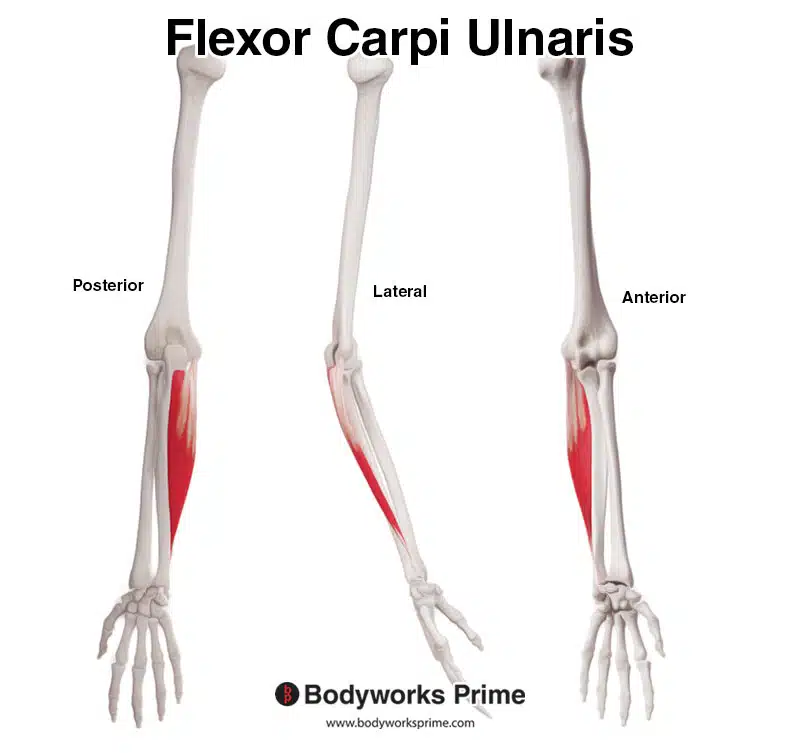
Here we can see the flexor carpi ulnaris from a posterior, lateral, and anterior view.
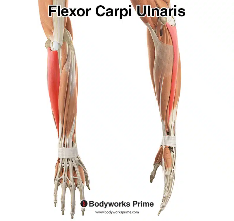
Here we can see the flexor carpi ulnaris from a superficial view amongst the other muscles of the arm. The flexor carpi ulnaris is highlighted in red.
Origin & Insertion
The flexor carpi ulnaris muscle has two heads, a humeral head and an ulnar head. These two heads arise at the origin points of this muscle. The humeral head originates from the common flexor tendon, on the medial epicondyle of the humerus. The ulnar head originates from the olecranon and the upper two thirds of the posterior border of the ulna. The two heads converge via a tendonous arch. The muscle then continues down the forearm crossing the wrist joint to insert onto the fifth metacarpal bone, the hook of hamate, and the pisiform bone of the wrist. The flexor carpi ulnaris inserts into the hook of hamate through the pisohamate ligament and inserts into the fifth metacarpal bone through the pisometacarpal ligament [2] [3].
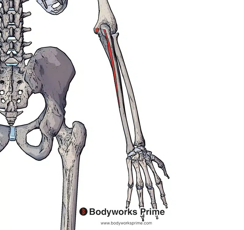
Here we can see the origin points of the flexor carpi ulnaris. The origin is highlighted in red on the olecranon of the ulna and medial epicondyle of the humerus (via the common flexor tendon).
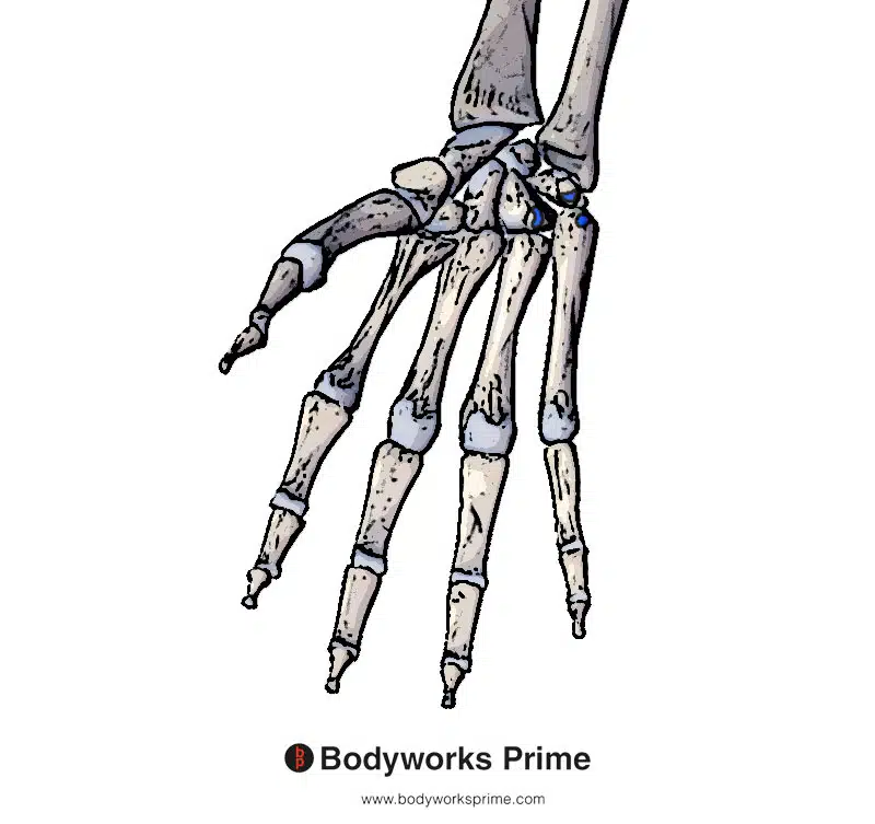
Here we can see the insertion points of the flexor carpi ulnaris. The insertions are highlighted in blue on the pisiform, hamate and fifth metacarpal bones.
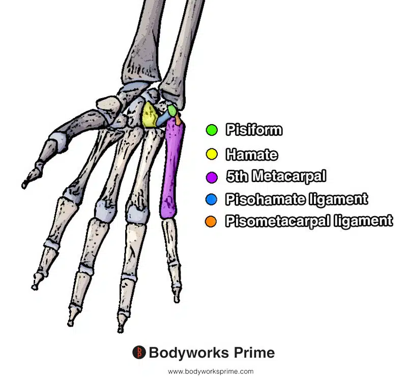
Here we can see the bones which the flexor carpi ulnaris inserts onto. These bones are colour coded for easy identification. We can also see the pisohamate ligament and the pisometacarpal ligament. The flexor carpi ulnaris inserts into the hamate through the pisohamate ligament and inserts into the fifth metacarpal bone through the pisometacarpal ligament.
This is why most depictions of this muscle do not show it connecting onto the hamate or the fifth metacarapal. The flexor carpi ulnaris does not directly connect to them. Rather, it is those two ligaments (which are is close proximity to the flexor carpi ulnaris) that are connecting these bones together and by proxy allowing the flexor carpi ulnaris to interact with them. However, these insertions on the hamate and fifth metacarpal are still considered insertions of the flexor carpi ulnaris.
Actions
The actions of the flexor carpi ulnaris are flexion and adduction of the hand at the wrist joint (radiocarpal joint). In addition to flexion and adduction, the flexor carpi ulnaris can also flex the elbow joint. However, it is very weak when it comes to elbow flexion and this is by no means a main action of the flexor carpi ulnaris [4] [5].
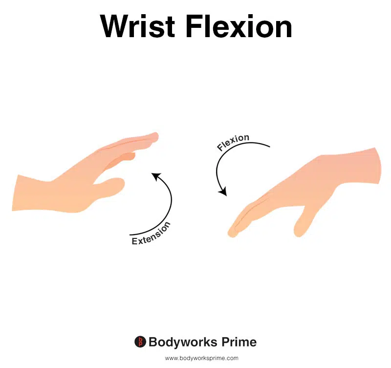
This image shows wrist flexion, which is the movement of bending the wrist in the direction of the underneath of the forearm (anterior surface of the forearm). The flexor carpi ulnaris is a wrist flexor. The opposite of wrist flexion would be wrist extension which involves extending the wrist in the direction of the top of the forearm (posterior surface of the forearm).
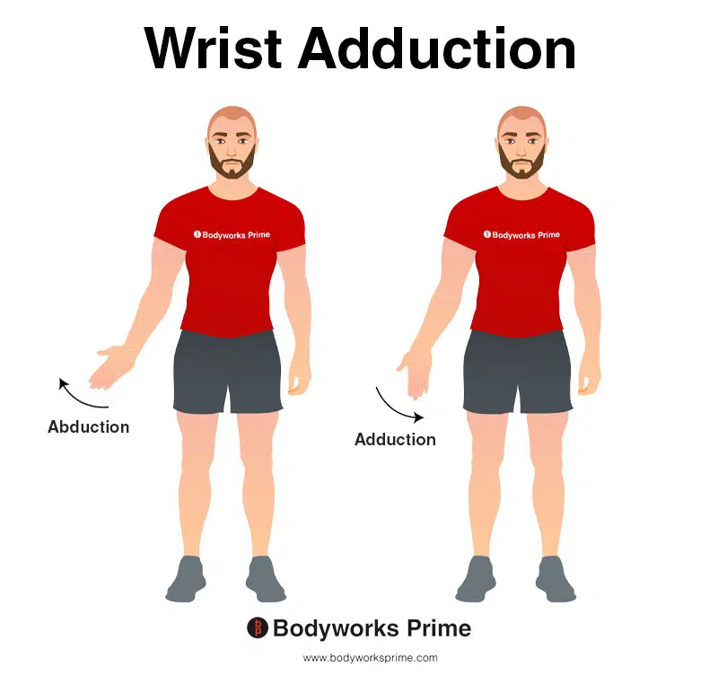
This image shows wrist adduction, which is the movement of bending the wrist to the side. The flexor carpi ulnaris is a wrist adductor. During wrist adduction, the wrist bends away from the thumb, which would be towards the body when the wrist is in the anatomical position. The opposite movement of wrist adduction is wrist abduction.
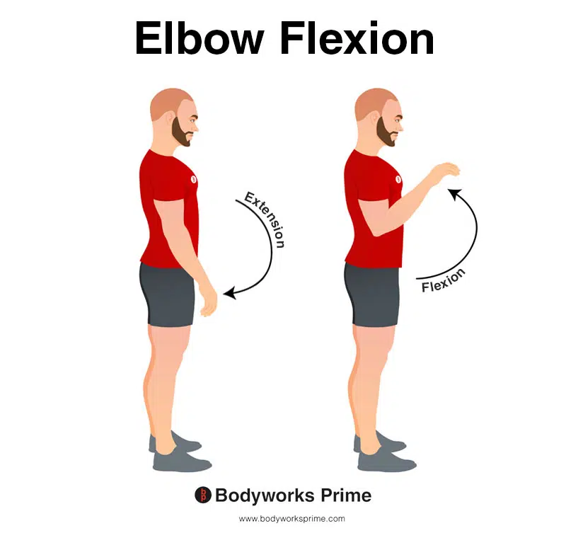
This image demonstrates elbow flexion, which involves bending elbow. The opposite movement of elbow flexion is elbow extension. The flexor carpi ulnaris can assist very weakly in elbow flexion.
Innervation
The flexor carpi ulnaris is innervated by the muscular branch of the ulnar nerve (C7, C8, T1). The ulnar nerve passes between the two heads of the flexor carpi ulnaris [6] [7] [8] [9].
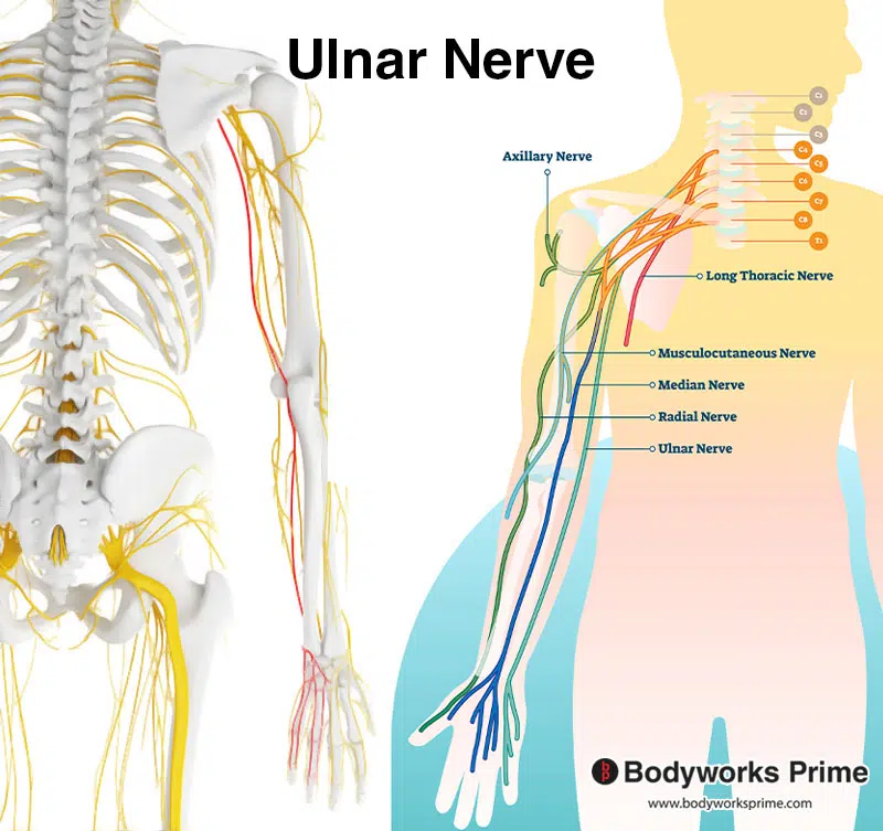
Pictured here we can see the ulnar nerve which innervates the flexor carpi ulnaris. The ulnar nerve is highlighted in red on the left of the picture.
Blood Supply
Blood is supplied to the flexor carpi ulnaris primarily via the ulnar artery (86% of cases) and the recurrent ulnar artery (14% of cases). The ulnar artery arises from the brachial artery [10] [11].
Want some flashcards to help you remember this information? Then click the link below:
Flexor Carpi Ulnaris Flashcards
Support Bodyworks Prime
Running a website and YouTube channel can be expensive. Your donation helps support the creation of more content for my website and YouTube channel. All donation proceeds go towards covering expenses only. Every contribution, big or small, makes a difference!
References
| ↑1, ↑2, ↑4, ↑6 | Lung BE, Siwiec RM. Anatomy, Shoulder and Upper Limb, Forearm Flexor Carpi Ulnaris Muscle. [Updated 2022 Jun 28]. In: StatPearls [Internet]. Treasure Island (FL): StatPearls Publishing; 2022 Jan-. Available from: https://www.ncbi.nlm.nih.gov/books/NBK526051/ |
|---|---|
| ↑3 | Maroukis BL, Ogawa T, Rehim SA, Chung KC. Guyon canal: the evolution of clinical anatomy. J Hand Surg Am. 2015 Mar;40(3):560-5. doi: 10.1016/j.jhsa.2014.09.026. Epub 2014 Oct 29. PMID: 25446410; PMCID: PMC4791630. |
| ↑5 | Chaudhry M, Aminullah H, Sinkler MA, Arain A. Anatomy, Shoulder and Upper Limb, Forearm Compartments. 2022 Aug 1. In: StatPearls [Internet]. Treasure Island (FL): StatPearls Publishing; 2022 Jan–. PMID: 30969606. |
| ↑7 | Anderson TB, Bordoni B. Anatomy, Shoulder and Upper Limb, Forearm Nerves. 2022 Jul 25. In: StatPearls [Internet]. Treasure Island (FL): StatPearls Publishing; 2022 Jan–. PMID: 32119401. |
| ↑8 | Pyun SB, Kang S, Kwon HK. Anatomical and electrophysiological myotomes corresponding to the flexor carpi ulnaris muscle. J Korean Med Sci. 2010 Mar;25(3):454-7. doi: 10.3346/jkms.2010.25.3.454. Epub 2010 Feb 17. PMID: 20191047; PMCID: PMC2826750. |
| ↑9, ↑10 | Sharpe F, Barry P, Lin SD, Stevanovic M. Anatomic study of the flexor carpi ulnaris muscle and its application to soft tissue coverage of the elbow with clinical correlation. J Shoulder Elbow Surg. 2014 Jan;23(1):82-90. doi: 10.1016/j.jse.2013.07.057. PMID: 24331124. |
| ↑11 | Shen S, Pang J, Seneviratne S, Ashton MW, Corlett RJ, Taylor GI. A comparative anatomical study of brachioradialis and flexor carpi ulnaris muscles: implications for total tongue reconstruction. Plast Reconstr Surg. 2008 Mar;121(3):816-829. doi: 10.1097/01.prs.0000299920.14548.2a. PMID: 18317131. |


