| Origin | Medial epicondyle of humerus (via common flexor tendon) |
| Insertion | Palmar aponeurosis and flexor retinaculum of hand |
| Action | Flexion at the wrist joint Stabilizes palmar aponeurosis |
| Nerve | Median nerve (C7-C8) |
| Artery | Median artery Anterior branch of ulnar recurrent artery |
Location & Overview
The palmaris longus muscle is situated in the superficial anterior compartment of the forearm and is a narrow, fusiform-shaped muscle that is wider in the middle and tapers towards its ends. It lies between the flexor carpi radialis muscle and the flexor carpi ulnaris muscle, while being superficial to the flexor digitorum superficialis. A part of the palmaris longus muscle also overlaps the median nerve [1].
The palmaris longus exhibits variations among individuals, which may include fusion with another muscle, an additional muscle belly, multiple tendinous insertions, or complete absence from one or both arms. The percentage of the population lacking this muscle varies across studies, with estimates ranging from 2.5% to 26%. The proportion of people missing the muscle also depends on the demographic of the population studied, such as ethnicity or gender [2] [3] [4] [5].
The palmaris longus is thought to have been more important in earlier primates for their lifestyle of living in trees, aiding in activities such as climbing and grasping branches. As human ancestors evolved to become more terrestrial and bipedal (walking on two legs), the reliance on the palmaris longus muscle decreased. This is thought to have lead to its reduced prevalence and variability in modern humans [6] [7].
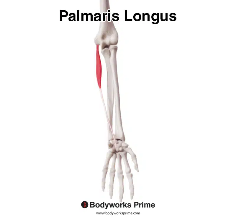
Here we can see the palmaris longus by itself from an anterior view.
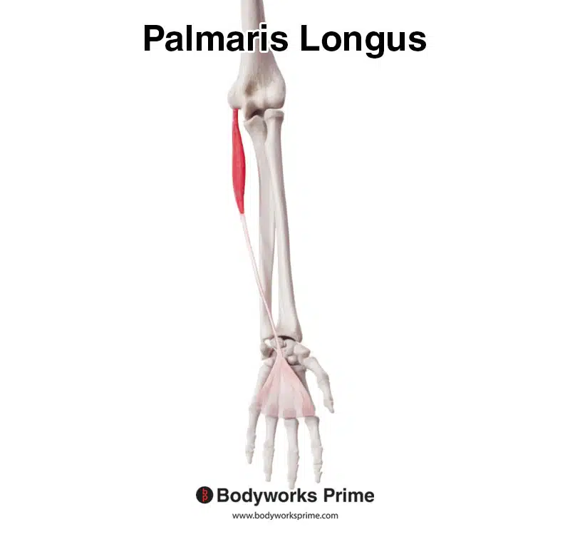
Here we can see the palmaris longus and the palmar aponeurosis which it connects to.

Here we can see the palmaris longus and the palmar aponeurosis from a medial view.
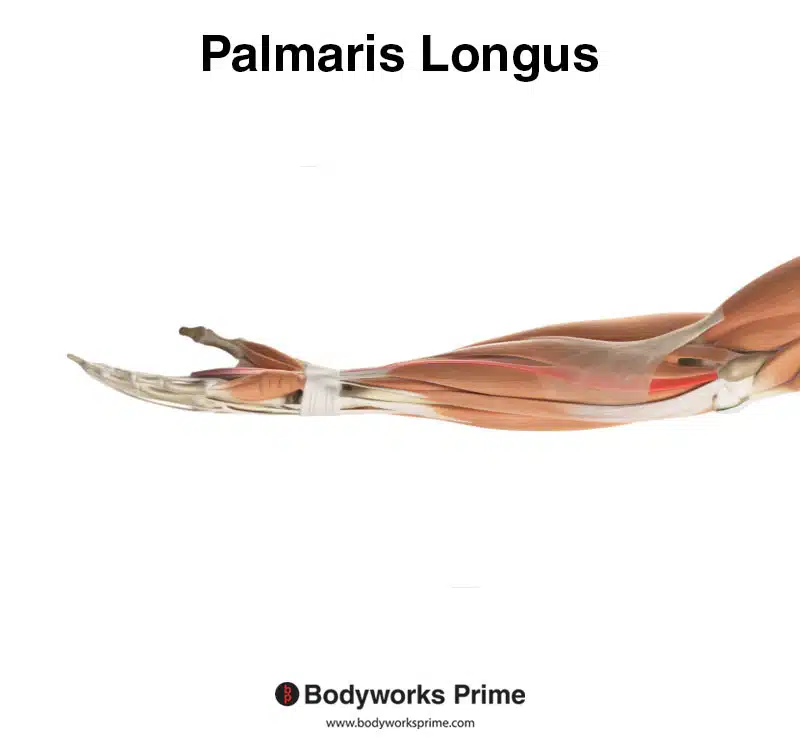
From a medial view, we can observe the palmaris longus, highlighted in red, among other muscles of the arm. The tissue covering the palmaris longus’ proximal portion is the bicipital aponeurosis.
Origin & Insertion
The palmaris longus muscle originates from the medial epicondyle of the humerus through the common flexor tendon. This common flexor tendon serves as a convergence point for the tendons of five muscles, including the flexor carpi ulnaris, palmaris longus, flexor carpi radialis, pronator teres, and flexor digitorum superficialis. The combined tendons of these five muscles are what make the common flexor tendon. The palmaris longus then continues down the arm and inserts into the palmar aponeurosis and flexor retinaculum of the hand. The flexor retinaculum helps form of the carpal tunnel by creating the structure that essentially functions as the ‘roof’ of the tunnel [8] [9].
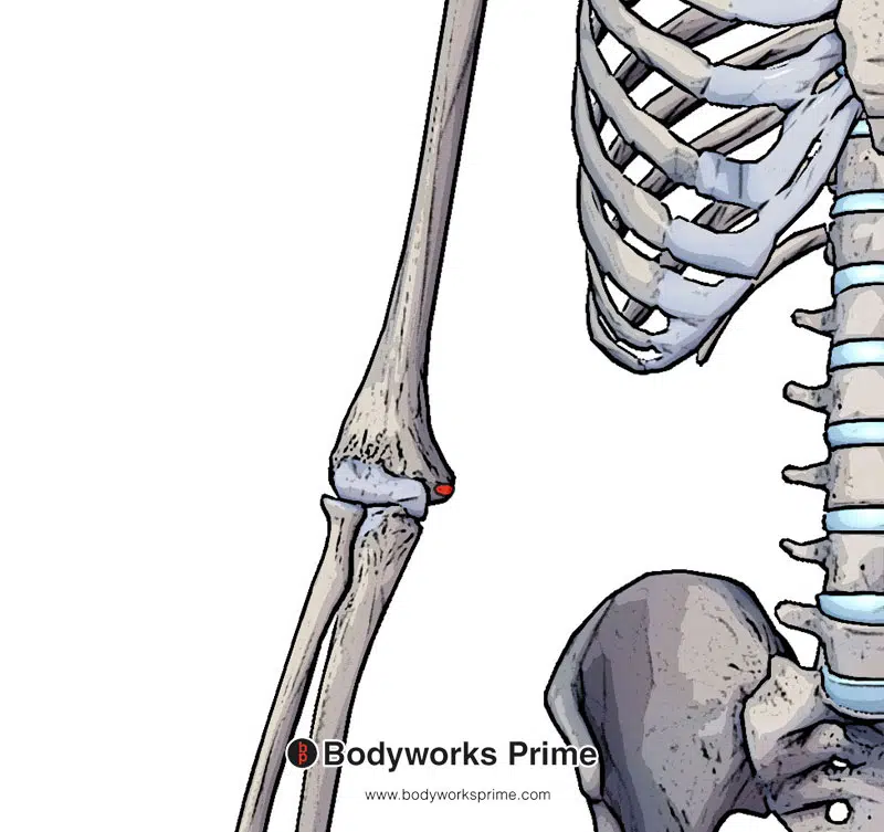
Here we can see the origin of the palmaris longus on the medial epicondyle of the humerus (highlighted in red).
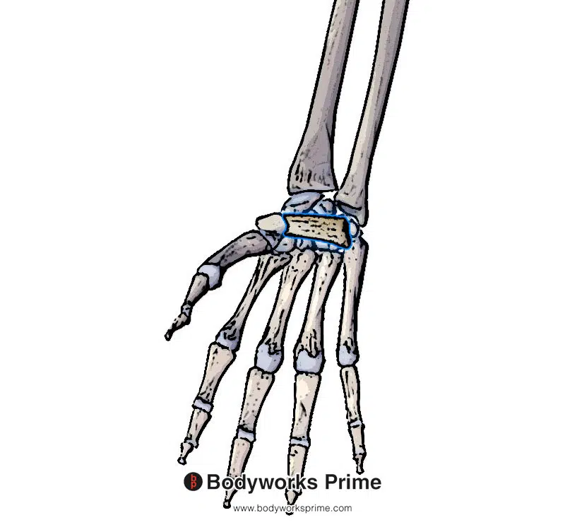
Here we can see the first insertion of the palmaris longus which is the flexor retinaculum of the hand (highlighted in blue).

Here we can see the second insertion of the palmaris longus which is the palmar aponeurosis (highlighted in blue).
Actions
The primary functions of the palmaris longus muscle include facilitating flexion of the hand at the radiocarpal (wrist) joint and stabilising the palmar aponeurosis, which in turn enhances the stability of the overlying palmar skin. Although its contribution to wrist flexion is minimal, the palmaris longus is regarded as a supportive accessory muscle during wrist flexion [10].
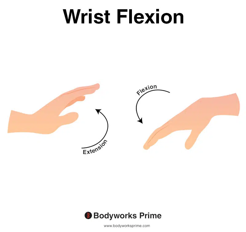
This image shows wrist flexion, a motion that involves bending the wrist towards the anterior surface, or the underside, of the forearm. Conversely, wrist extension refers to the movement of extending the wrist towards the posterior surface, or the top side, of the forearm. The palmaris longus can weakly assist in wrist flexion. The palmaris longus also stabilises the palmar aponeurosis of the hand.
Innervation
The palmaris longus is innervated by the median nerve (C7-C8). The median nerve is a part of the brachial plexus [11] [12] [13].
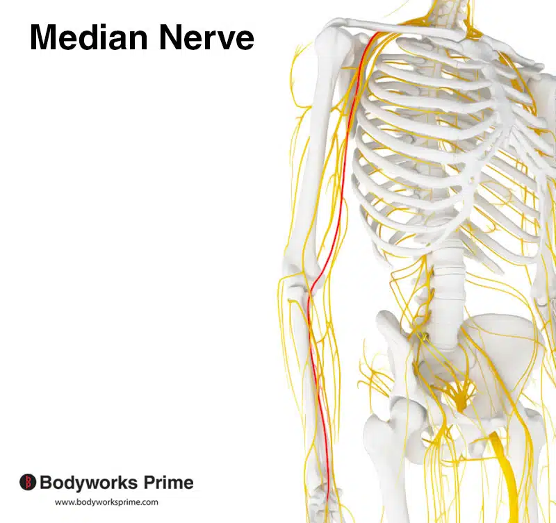
Pictured here we can see the median nerve highlighted in red. The median nerve innervates the palmaris longus.
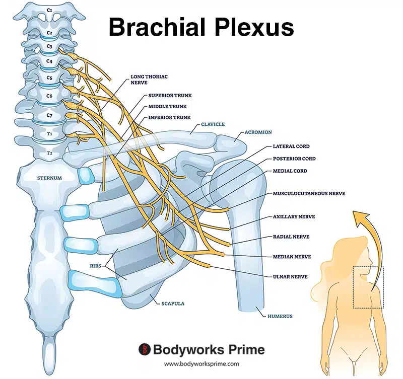
Here we can see a labelled diagram of the brachial plexus. The median nerve, which innervates the palmaris longus, is a part of the brachial plexus.
Blood Supply
Blood is supplied to the palmaris longus via the median artery and the anterior branch of ulnar recurrent artery [14].
Want some flashcards to help you remember this information? Then click the link below:
Palmaris Longus Flashcards
Support Bodyworks Prime
Running a website and YouTube channel can be expensive. Your donation helps support the creation of more content for my website and YouTube channel. All donation proceeds go towards covering expenses only. Every contribution, big or small, makes a difference!
References
| ↑1, ↑2, ↑9, ↑13 | Olewnik Ł, Wysiadecki G, Polguj M, Podgórski M, Jezierski H, Topol M. Anatomical variations of the palmaris longus muscle including its relation to the median nerve – a proposal for a new classification. BMC Musculoskelet Disord. 2017;18(1):539. Published 2017 Dec 19. doi:10.1186/s12891-017-1901-x |
|---|---|
| ↑3 | Georgiev GP, Iliev AA, Dimitrova IN, Kotov GN, Malinova LG, Landzhov BV. Palmaris Longus Muscle Variations: Clinical Significance and Proposal of New Classifications. Folia Med (Plovdiv). 2017 Sep 1;59(3):289-297. doi: 10.1515/folmed-2017-0035. PMID: 28976893. |
| ↑4, ↑8, ↑10, ↑12, ↑14 | Cooper DW, Burns B. Anatomy, Shoulder and Upper Limb, Hand Palmaris Tendon. 2021 Sep 3. In: StatPearls [Internet]. Treasure Island (FL): StatPearls Publishing; 2022 Jan–. PMID: 30137801. |
| ↑5 | Ioannis D, Anastasios K, Konstantinos N, Lazaros K, Georgios N. Palmaris Longus Muscle’s Prevalence in Different Nations and Interesting Anatomical Variations: Review of the Literature [published correction appears in J Clin Med Res. 2016 Jan;8(1):62]. J Clin Med Res. 2015;7(11):825-830. doi:10.14740/jocmr2243w |
| ↑6 | Sichting F, Holowka NB, Ebrecht F, Lieberman DE. Evolutionary anatomy of the plantar aponeurosis in primates, including humans. J Anat. 2020 Jul;237(1):85-104. doi: 10.1111/joa.13173. Epub 2020 Feb 26. PMID: 32103502; PMCID: PMC7309290. |
| ↑7 | Tocheri MW, Orr CM, Jacofsky MC, Marzke MW. The evolutionary history of the hominin hand since the last common ancestor of Pan and Homo. J Anat. 2008 Apr;212(4):544-62. doi: 10.1111/j.1469-7580.2008.00865.x. PMID: 18380869; PMCID: PMC2409097. |
| ↑11 | Moore KL, Agur AMR, Dalley AF. Clinically Oriented Anatomy. 8th ed. Philadelphia: Lippincot Williams & Wilkins; 2017. |


