| Origin | Lateral supracondylar ridge (humerus) |
| Insertion | Lateral surface of the styloid process (radius) |
| Action | Flexion of the forearm at the elbow joint Supination and pronation of the forearm |
| Nerve | Radial nerve (C5, C6, C7) |
| Artery | Radial recurrent artery |
Location & Overview
The brachioradialis is a prominent muscle located on the lateral side of the forearm, extending across the elbow joint. This superficial muscle is easily palpable in the anterolateral region of the forearm and plays an important role in forearm stability and function. Along with the wrist extensors, the brachioradialis forms one of the lateral boundaries of the cubital fossa, an important anatomical area containing structures such as the biceps tendon, brachial artery, and median nerve. The cubital fossa, also known as the antecubital fossa, is a triangular-shaped depression located on the anterior aspect of the elbow joint [1] [2] [3].
The brachioradialis muscle belongs to the group of muscles known as the ‘mobile wad’ or ‘radial group’, which also includes the extensor carpi radialis longus and extensor carpi radialis brevis. Its positioning and tough tendon make it highly effective in providing mechanical advantage during various forearm movements. As a powerful forearm muscle, the brachioradialis not only contributes to the overall appearance and definition of the forearm but also plays a key role in many daily activities and sports that involve gripping, lifting, and rotating [4].
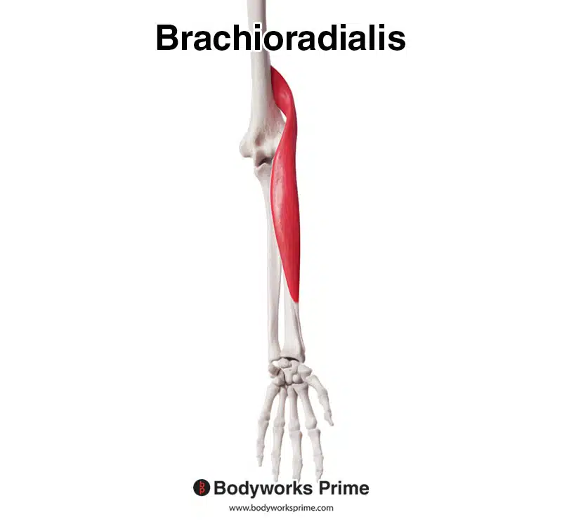
Here we can see the brachioradialis muscle from an anterior view.

Here we can see the brachioradialis muscle from a lateral view.
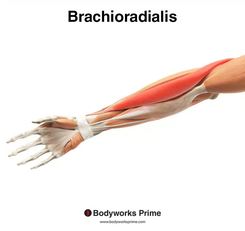
Here we can see the brachioradialis muscle from an anterior and superficial view, amongst the other muscles of the arm.
Origin & Insertion
The brachioradialis originates across the proximal two-thirds of the humerus’ lateral supracondylar ridge. Distally, it inserts onto the lateral surface of the radius’ styloid process [5] [6].
The lateral supracondylar ridge of the humerus (brachioradialis origin) is a bony ridge found on the distal, posterior aspect of the humerus, just above the lateral epicondyle. It extends proximally from the lateral epicondyle, running diagonally along the posterior surface of the humerus. The styloid process of the radius is a slender, cone-shaped projection which is located at the distal end of the radius bone, on its lateral side. The lateral surface of the radius’ styloid process (brachioradialis insertion) is smooth and rounded, facing the lateral aspect of the wrist joint [7].
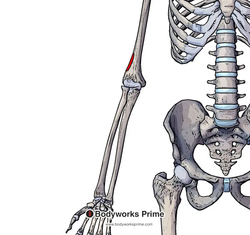
Highlighted in red is the origin of the brachioradialis muscle on the lateral supracondylar ridge of the humerus.
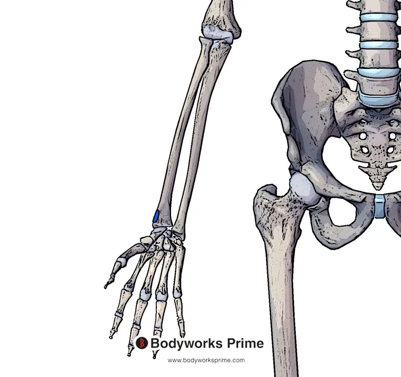
Highlighted in blue is the insertion of the brachioradialis muscle on the lateral surface of the styloid process.
Actions
The primary action of the brachioradialis is flexing the forearm at the elbow. It is also able to supinate or pronate depending on the rotation/orientation of the forearm. When the forearm is in a pronated position, the brachioradialis assists in supination as it contracts. When the forearm is in a supinated position, the brachioradialis assists in pronation as it contracts [8].
The primary action of the brachioradialis is flexion at the elbow joint. Therefore exercises involving flexion with the forearm in a pronated or hammer grip are going to target the brachioradialis most. Exercise examples would be: reverse curls (barbell, dumbbell, ez bar, or even kettlebell), hammer grip curls, or cable curls using the rope attachment.
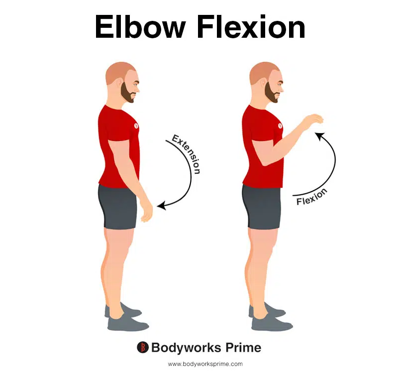
This image demonstrates elbow flexion, which involves bending elbow. The opposite movement of elbow flexion is elbow extension. The primary action of the brachioradialis is elbow flexion.
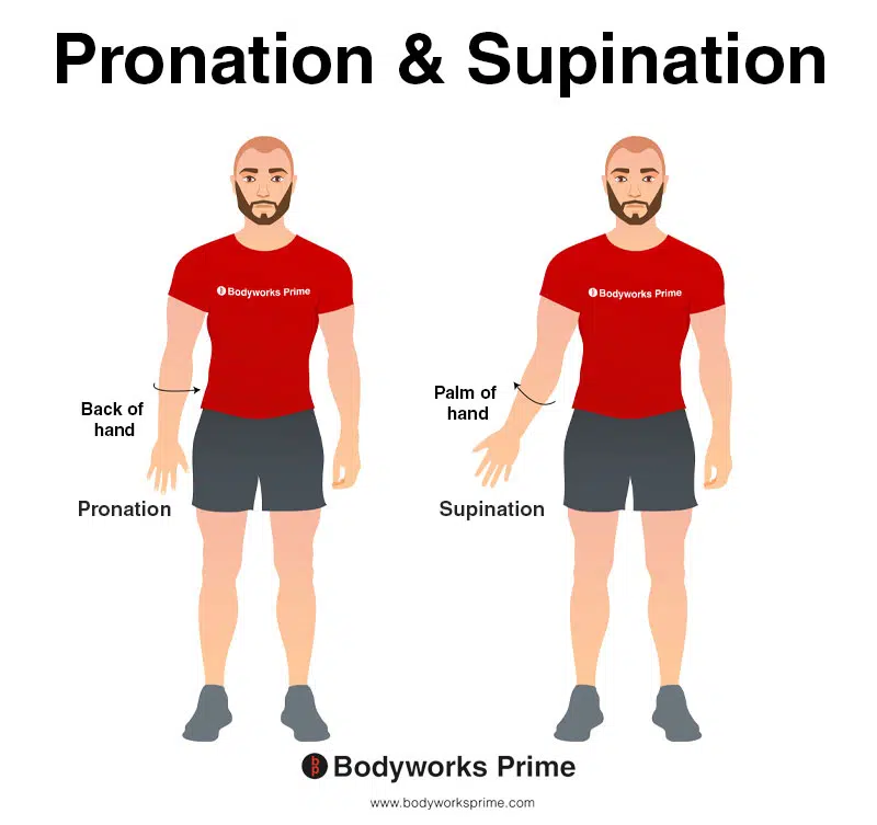
This image illustrates the movements of pronation and supination. In the anatomical position, pronation occurs when the hand is rotated, bringing the back of the hand to face the front of the body. Conversely, supination is the opposite movement, where the palm of the hand faces the front of the body while in the anatomical position. Both pronation and supination are actions of the brachioradialis. When the forearm is in a pronated position, the brachioradialis assists in supination as it contracts. When the forearm is in a supinated position, the brachioradialis assists in pronation as it contracts.
Innervation
The brachioradialis muscle is innervated by the radial nerve (C5, C6, C7). The spinal nerve roots of C5 and C6 are the nerve roots which provide the primary innervation of the brachioradialis. The radial nerve is a part of the brachial plexus [9] [10] [11] [12].
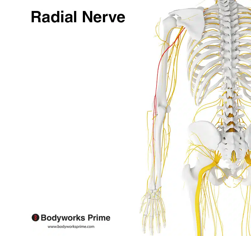
Here we can see the radial nerve highlighted in red. The brachioradialis muscle is innervated by the radial nerve from the spinal nerve roots of C5, C6, and C7.
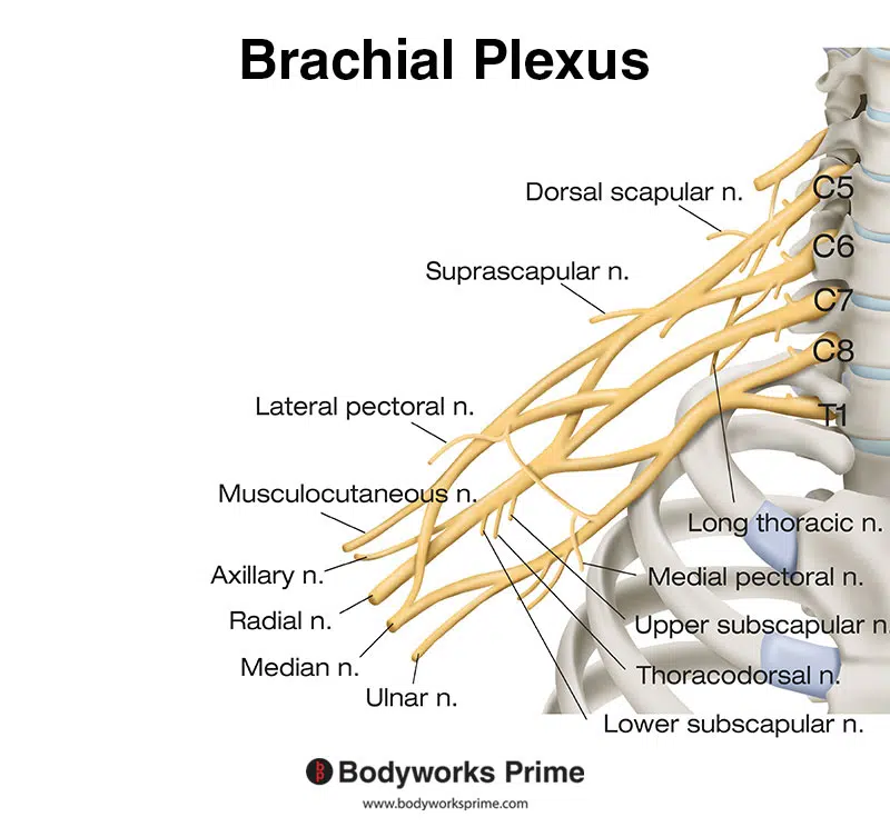
Here we can see the brachial plexus. The radial nerve is a part of the brachial plexus.
Blood Supply
Blood is supplied to the brachioradialis muscle via the radial recurrent artery which is a branch of the radial artery [13] [14].
Want some flashcards to help you remember this information? Then click the link below:
Brachioradialis Flashcards
Support Bodyworks Prime
Running a website and YouTube channel can be expensive. Your donation helps support the creation of more content for my website and YouTube channel. All donation proceeds go towards covering expenses only. Every contribution, big or small, makes a difference!
References
| ↑1, ↑5, ↑8, ↑9 | Lung BE, Ekblad J, Bisogno M. Anatomy, Shoulder and Upper Limb, Forearm Brachioradialis Muscle. [Updated 2021 Jul 31]. In: StatPearls [Internet]. Treasure Island (FL): StatPearls Publishing; 2022 Jan-. Available from: https://www.ncbi.nlm.nih.gov/books/NBK526110/ |
|---|---|
| ↑2 | Voin V, Iwanaga J, Sardi JP, Fisahn C, Loukas M, Oskouian RJ, Tubbs RS. Relationship of the Median and Radial Nerves at the Elbow: Application to Avoiding Injury During Venipuncture or Other Invasive Procedures of the Cubital Fossa. Cureus. 2017 Mar 13;9(3):e1094. doi: 10.7759/cureus.1094. PMID: 28413740; PMCID: PMC5391251. |
| ↑3, ↑4, ↑6, ↑7 | Moore KL, Agur AMR, Dalley AF. Clinically Oriented Anatomy. 8th ed. Philadelphia: Lippincot Williams & Wilkins; 2017. |
| ↑10 | Zhang L, Zhang CG, Dong Z, Gu YD. Spinal nerve origins of the muscular branches of the radial nerve: an electrophysiological study. Neurosurgery. 2012 Jun;70(6):1438-41; discussion 1441. doi: 10.1227/NEU.0b013e3182486b35. PMID: 22227484. |
| ↑11 | Catala M, Kubis N. Gross anatomy and development of the peripheral nervous system. Handb Clin Neurol. 2013;115:29-41. doi: 10.1016/B978-0-444-52902-2.00003-5. PMID: 23931773. |
| ↑12 | Caufriez B, Dugailly PM, Brassinne E, Schuind F. The Role of the Muscle Brachioradialis in Elbow Flexion: An Electromyographic Study. J Hand Surg Asian Pac Vol. 2018 Mar;23(1):102-110. doi: 10.1142/S2424835518500145. PMID: 29409427. |
| ↑13 | Leversedge FJ, Casey PJ, Payne SH, Seiler JG 3rd. Vascular anatomy of the brachioradialis rotational musculocutaneous flap. J Hand Surg Am. 2001 Jul;26(4):711-21. doi: 10.1053/jhsu.2001.26200. PMID: 11466649. |
| ↑14 | Zampeli F, Spyridonos S, Fandridis E. Brachioradialis muscle flap for posterior elbow defects: a simple and effective solution for the upper limb surgeon. J Shoulder Elbow Surg. 2019 Aug;28(8):1476-1483. doi: 10.1016/j.jse.2019.03.020. Epub 2019 Jun 18. PMID: 31227467. |


