| Origin | Posterior surface of tibia Posterior surface of fibula Posterior interosseous membrane |
| Insertion | Navicular bone tubercle Medial cuneiform bone Intermediate cuneiform bone Lateral cuneiform bone Cuboid bone Bases of the 2nd, 3rd and 4th metatarsal bones |
| Action | Foot inversion Foot plantar flexion Supports medial arch of foot |
| Nerve | Tibial Nerve (L4, L5) |
| Artery | Posterior tibial artery |
Location & Overview
The tibialis posterior muscle is the deepest muscle in the posterior compartment of the lower leg. The other deep muscles in the posterior compartment of the lower leg are: the flexor digitorum longus, the flexor hallucis longus and the popliteus. The tibialis posterior is not able to be palpated as it is covered by the large superficial muscles of the lower leg which are the gastrocnemius and soleus [1] [2] [3].
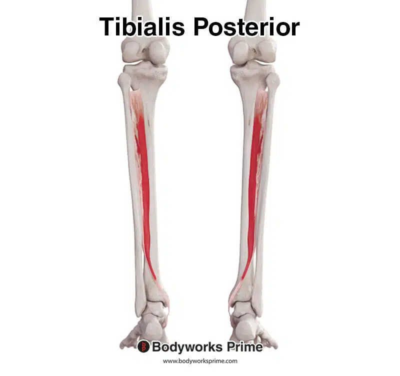
Here we can see the tibialis posterior muscle seen from a posterior view.
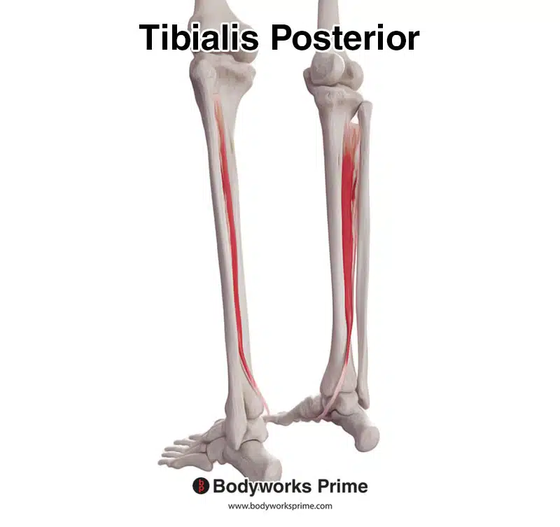
Here we can see the tibialis posterior muscle from a posterolateral view.
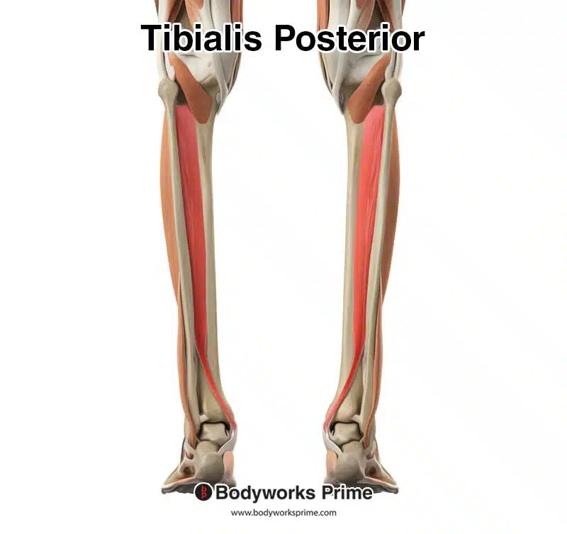
Here we can see the tibialis posterior amongst the other muscles of the leg. Superficial muscles such as the calves have been removed from this image to reveal the tibialis posterior, because it is a deep muscle.
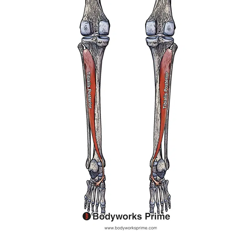
Here we can see pictured the tibialis posterior from a posterior view. This picture displays the insertion points underneath the foot due to the feet being in a plater flexed position. The next section (origin and insertion) will discuss this in further detail.
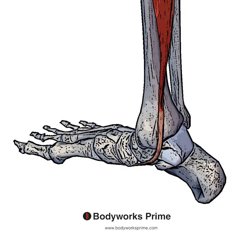
Here we can see a picture of the distal tendon of the tibialis posterior from a medial view (the inner side of the leg).
Origin & Insertion
The tibialis posterior originates from the posterior surface of the upper half of the tibia, the posterior surface of the upper half of the fibula, and the posterior side of the interosseus membrane. The tibialis posterior then continues in an inferior direction down the leg crossing the ankle joint (posterior to the medial malleolus) and inserts onto the bones underneath of the foot (on the plantar aspect). The insertion locations on the plantar aspect of the foot are: the navicular bone tubercle, medial cuneiform bone, intermediate cuneiform bone, lateral cuneiform bone, cuboid bone; and the bases of the 2nd, 3rd and 4th metatarsal bones [4] [5] [6]. These origins and insertions are displayed in the below images.
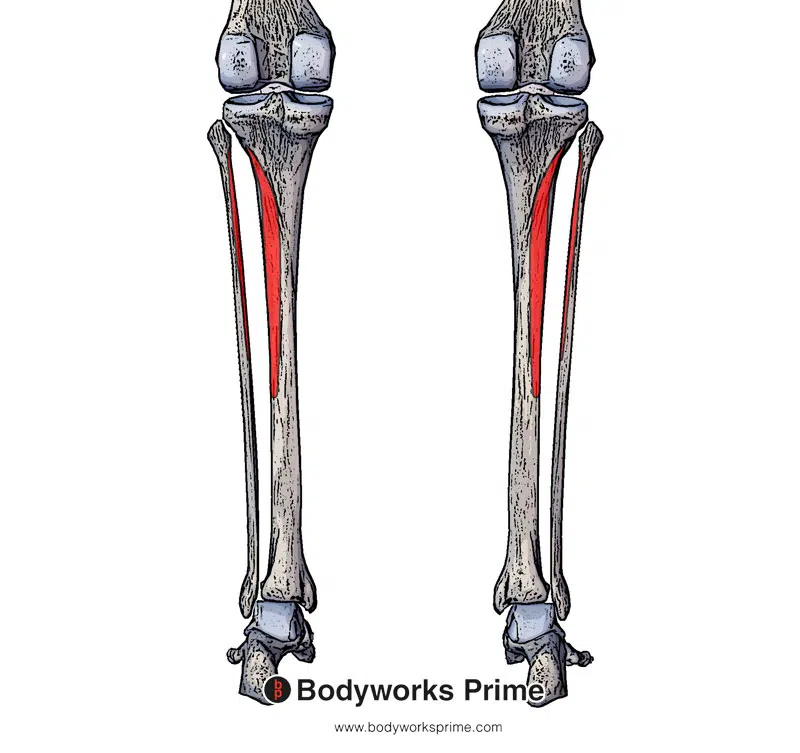
Here we can see two of the origins of the tibialis posterior muscle marked out in red. It is originating on the posterior surface of tibia and the posterior fibula.
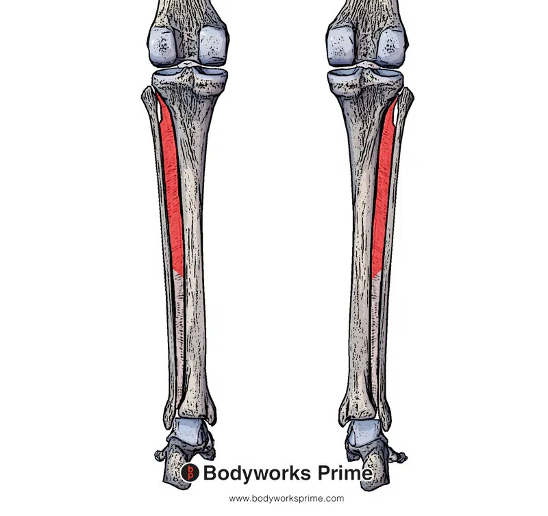
Here we can see another origin of the tibialis posterior muscle marked out in red. It is originating on the interosseous membrane which is a thick dense fibrous sheet of connective tissue that spans between the tibia and fibula.
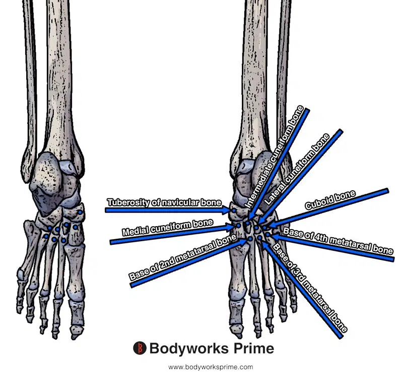
Here we can see the insertion points of the tibialis posterior underneath the foot. These insertion points are on the navicular bone tubercle, medial cuneiform bone, intermediate cuneiform bone, lateral cuneiform bone, cuboid bone, and the bases of the 2nd, 3rd and 4th metatarsal bones.
Actions
The tibialis posterior is a primary inverter of the foot. The other primary foot inverter of the foot is the tibialis anterior. The tibialis posterior is also able to assist the gastrocnemius, soleus, and plantaris muscles in causing plantar flexion of the foot. The tibialis posterior is also able to support the medial arch of the foot (the tibialis anterior is also able to assist in supporting this medial arch too) [7] [8] [9].
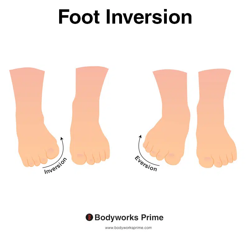
This picture demonstrates foot inversion, which means rotating the ankle inwards, causing the sole of the foot to turn towards the body. The opposite of inversion is eversion. Inversion of the foot is the primary action of the tibialis posterior muscle. The tibialis posterior also supports the medial arch of foot.
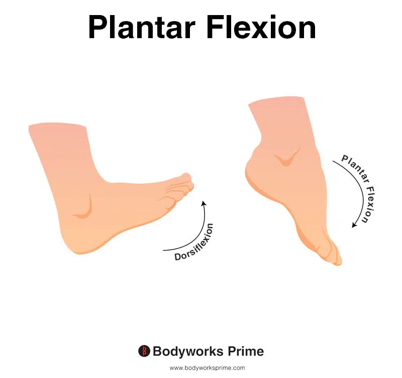
This image shows an example of foot plantar flexion, which involves extending the ankle. The opposite of plantar flexion is dorsiflexion. Plantar flexion is an action of the tibialis posterior muscle.
Innervation
The tibialis posterior muscle is innervated by the tibial nerve. The tibial nerve is a component of the sciatic nerve originating from the ventral rami of spinal roots L4-S3 [10] [11].
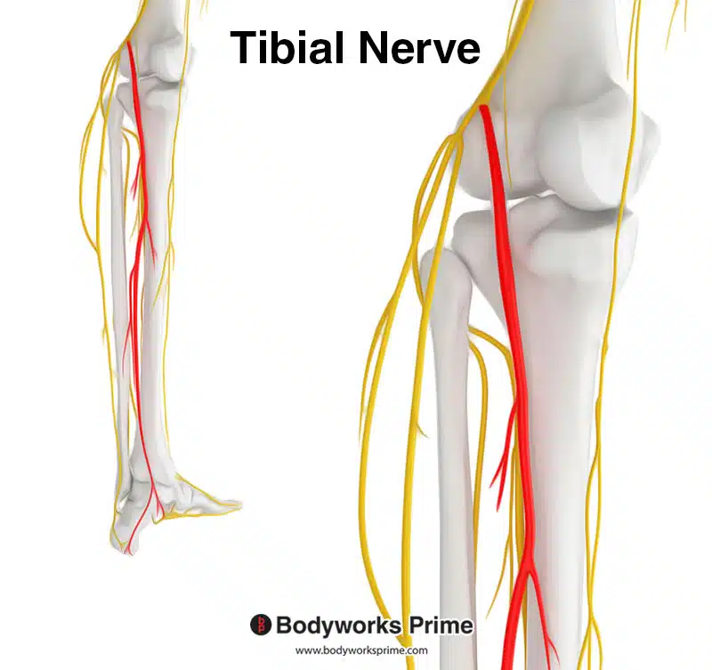
Highlighted in red is the tibial nerve which is a branch of the sciatic nerve. The tibial nerve innervates the tibialis posterior muscle.
Blood Supply
Blood is supplied to the tibialis posterior primarily from the posterior tibial artery. The posterior tibial artery arises from the popliteal artery [12].
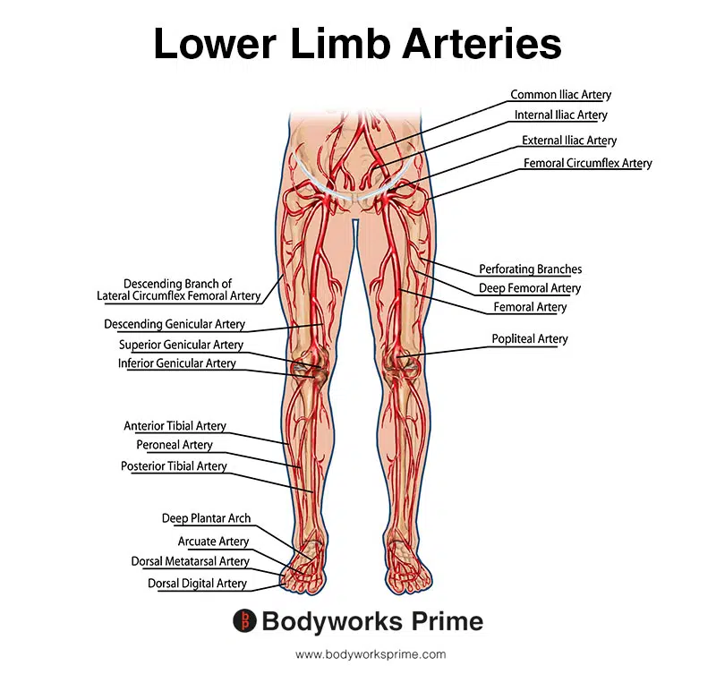
This image shows the arteries of the lower limb, including the posterior tibial artery.
Want some flashcards to help you remember this information? Then click the link below:
Tibialis Posterior Flashcards
Support Bodyworks Prime
Running a website and YouTube channel can be expensive. Your donation helps support the creation of more content for my website and YouTube channel. All donation proceeds go towards covering expenses only. Every contribution, big or small, makes a difference!
References
| ↑1, ↑4, ↑7, ↑10, ↑12 | Corcoran NM, Varacallo M. Anatomy, Bony Pelvis and Lower Limb, Tibialis Posterior Muscle. [Updated 2021 Aug 17]. In: StatPearls [Internet]. Treasure Island (FL): StatPearls Publishing; 2022 Jan-. Available from: https://www.ncbi.nlm.nih.gov/books/NBK539913/ |
|---|---|
| ↑2, ↑5, ↑8 | Park JH, Kim D, Kwon HW, Lee M, Choi YJ, Park KR, Youn KH, Cho J. A New Anatomical Classification for Tibialis Posterior Tendon Insertion and Its Clinical Implications: A Cadaveric Study. Diagnostics (Basel). 2021 Sep 4;11(9):1619. doi: 10.3390/diagnostics11091619. PMID: 34573961; PMCID: PMC8466387. |
| ↑3, ↑6, ↑9 | Moore KL, Agur AMR, Dalley AF. Clinically Oriented Anatomy. 8th ed. Philadelphia: Lippincot Williams & Wilkins; 2017. |
| ↑11 | Moore KL, Agur AMR, Dalley AF. Clinically Oriented Anatomy. 8th ed. Philadelphia: Lippincot Williams & Wilkins; 2017 |


