| Origin | Head of fibula & posterior surface of fibula Soleal line & medial border of tibia |
| Insertion | Posterior surface of the calcaneus via the calcaneal tendon |
| Action | Plantar flexes foot at ankle joint |
| Nerve | Tibial nerve (S1- S2) |
| Artery | Popliteal artery Fibular artery Posterior tibial artery |
Location & Overview
The soleus muscle is a muscle of the posterior lower leg and has the primary role of plantar flexion (extending the ankle). It is large and flat, located deep to the gastrocnemius muscle. It belongs to a duo of muscles referred to as the ‘calf muscles’. The official anatomical term for the calf muscles is the ‘triceps surae’. The triceps surae is comprised of the gastrocnemius muscle and the soleus muscle. Some people also consider the plantaris muscle as part of the calf muscles too. The muscle bellies of the soleus and gastrocnemius muscle merge together to form the calcaneal tendon as they approach the foot [1] [2].
The calcaneal tendon which is formed by the soleus and gastrocnemious is also known as the ‘Achilles tendon’. It is the thickest tendon in the human body. The plantaris muscle, though not a major contributor, can also be involved in the formation of the calcaneal tendon. In some people, the plantaris tendon is attached to the calcaneal tendon or runs alongside it before inserting onto the calcaneus (heel bone). However, the plantaris muscle is highly variable and may be absent entirely in some people (around 7–20% of people) [3] [4].
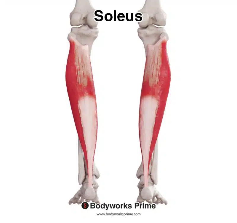
Here we can see the soleus muscle from an anterior view.
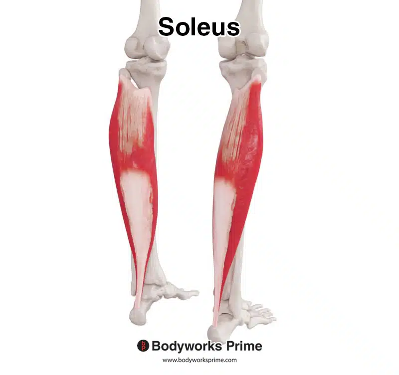
Here we can see the soleus from a posterolateral view.
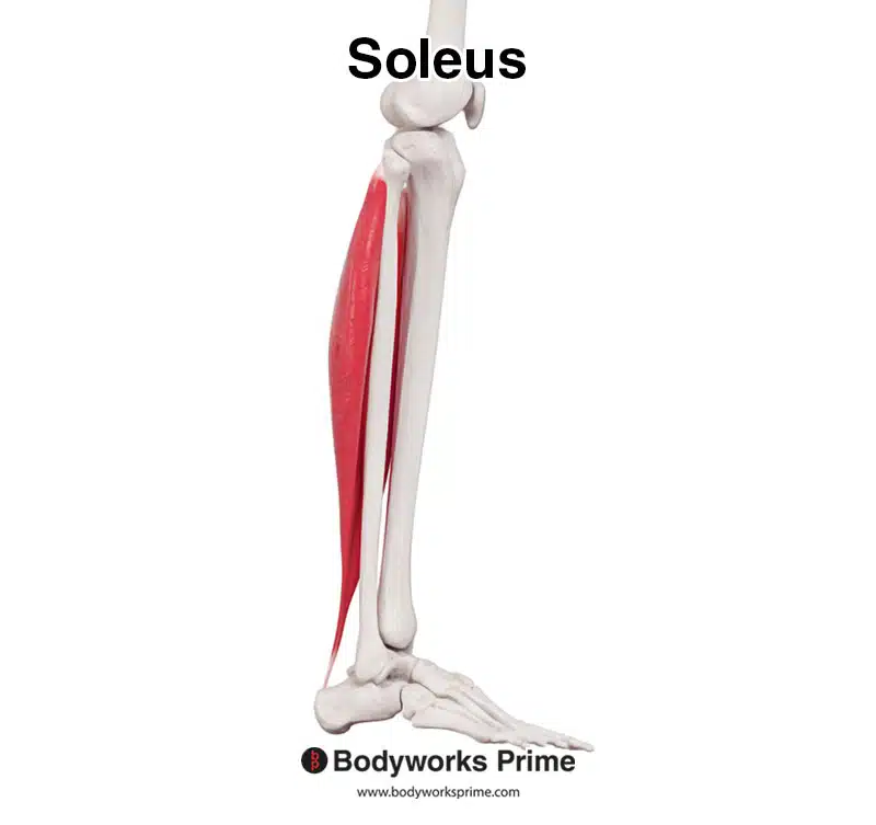
Here we can see the soleus from a lateral view.
Here we can see the soleus from a medial view.
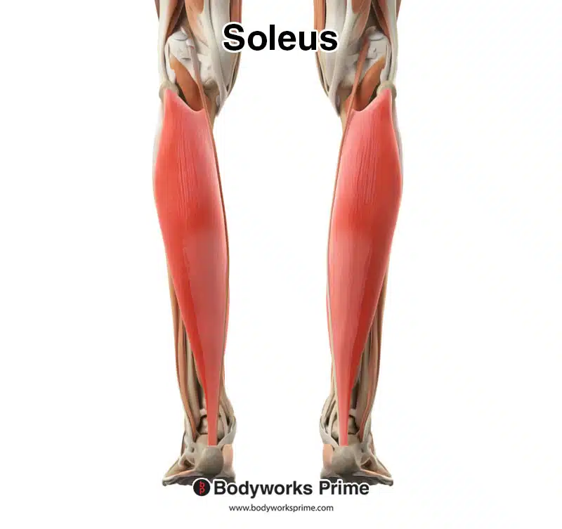
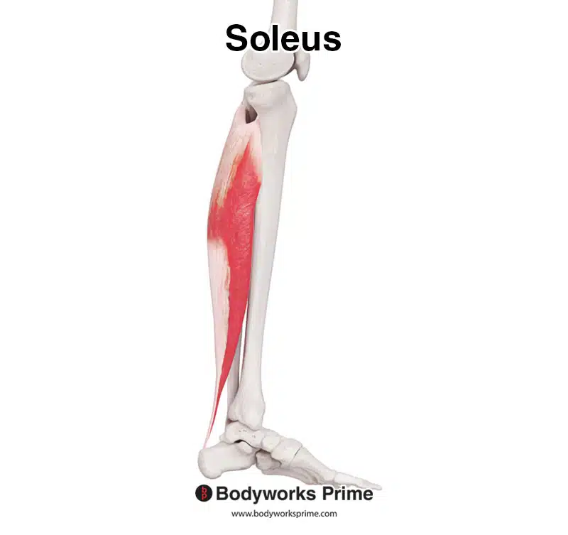
In this image, we have the soleus muscle highlighted amidst the other muscles of the leg. For clear visibility, the gastrocnemius has been removed, allowing us to see the soleus unobstructed.
Origin & Insertion
The soleus muscle originates from the head of the fibula, the posterior surface of the fibula, the soleal line, and the medial border of the tibia. As the soleus muscle extends distally down the lower leg, it merges with the gastrocnemius muscle, forming the calcaneal tendon, also known as the Achilles tendon. This tendon then inserts into the posterior surface of the calcaneus [5] [6] [7].
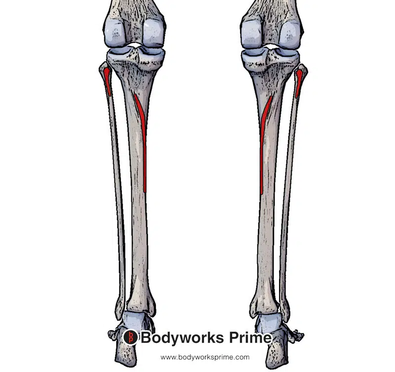
Here we can see the origin of the soleus muscle highlighted in red. The soleus originates from the head of fibula, posterior surface of fibula, soleal line of the tibia and the medial border of tibia.
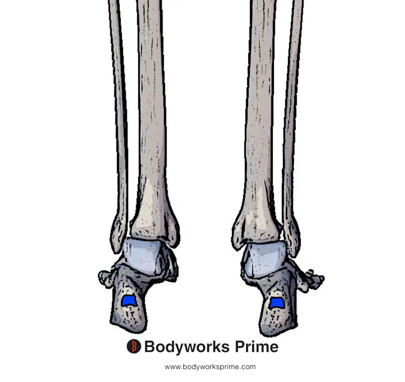
Here we can see the insertion of the soleus muscle highlighted in blue. The soleus inserts on the posterior surface of the calcaneus via the calcaneal tendon (also known as the Achilles tendon).
Actions
The soleus muscle is able to cause plantarflexion at the ankle joint. Plantarflexion involves extending the ankle joint (i.e extending foot away from the body or going onto your tip toes). The soleus works together with the other calf muscles in activities like running and jumping where powerful push-off forces are required. In addition, the soleus plays a role in maintaining postural stability and balance while standing, as it helps resist the force of gravity and stops the body from falling forward [8] [9] [10].
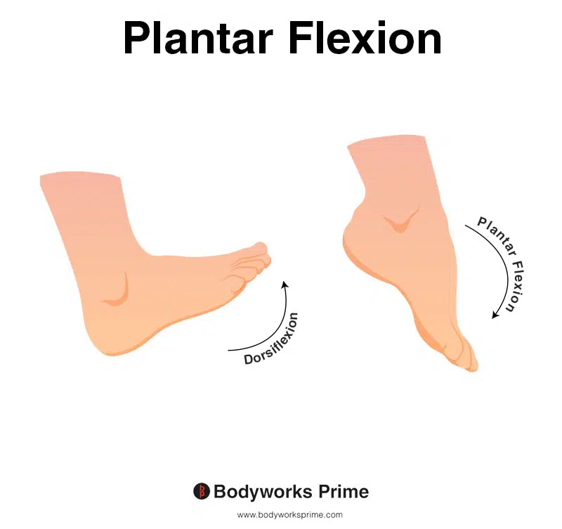
This image shows an example of foot plantar flexion, which involves extending the ankle. The opposite of plantar flexion is dorsiflexion. The soleus’ primary action is plantar flexion.
Innervation
The soleus muscle is innervated by the tibial nerve (S1- S2). This nerve is the larger branch of the sciatic nerve which divides into the tibial nerve. It also provides innervation for other muscles of the posterior lower leg compartment (e.g the gastrocnemius and the plantaris) [11].
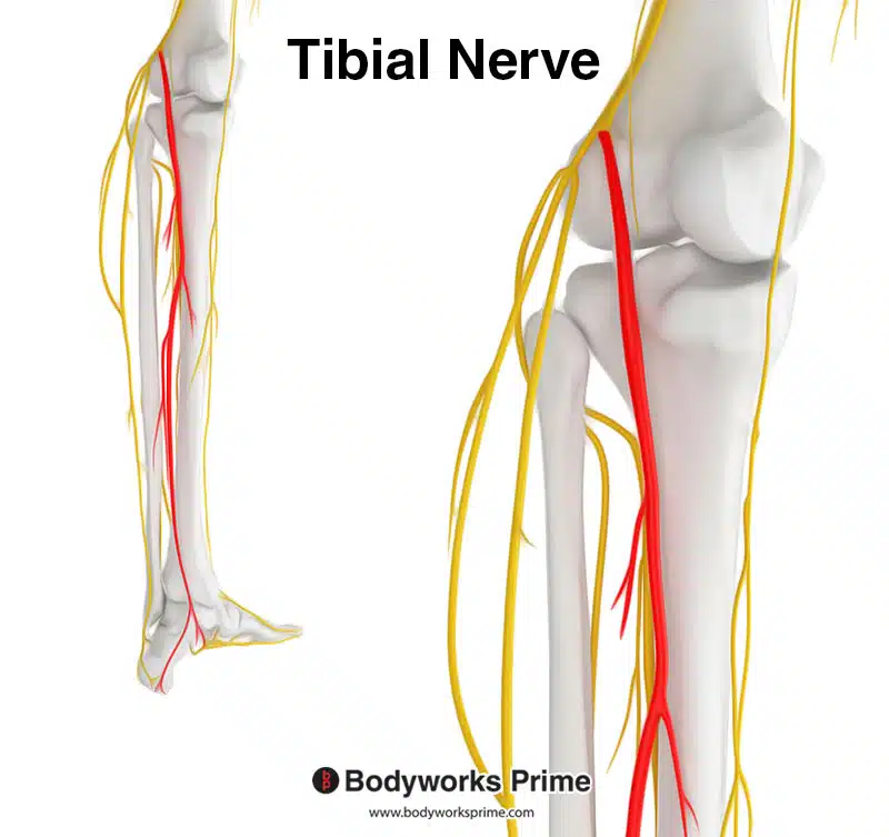
Highlighted in red we can see the tibial nerve, which is a branch of the sciatic nerve. The tibial nerve innervates the soleus muscle from the spinal nerve roots of S1 and S2. The tibial nerve also innervates the gastrocnemius and plantaris muscle too.
Blood Supply
Blood is supplied to the soleus muscle via the popliteal, fibular and posterior tibial arteries [12].
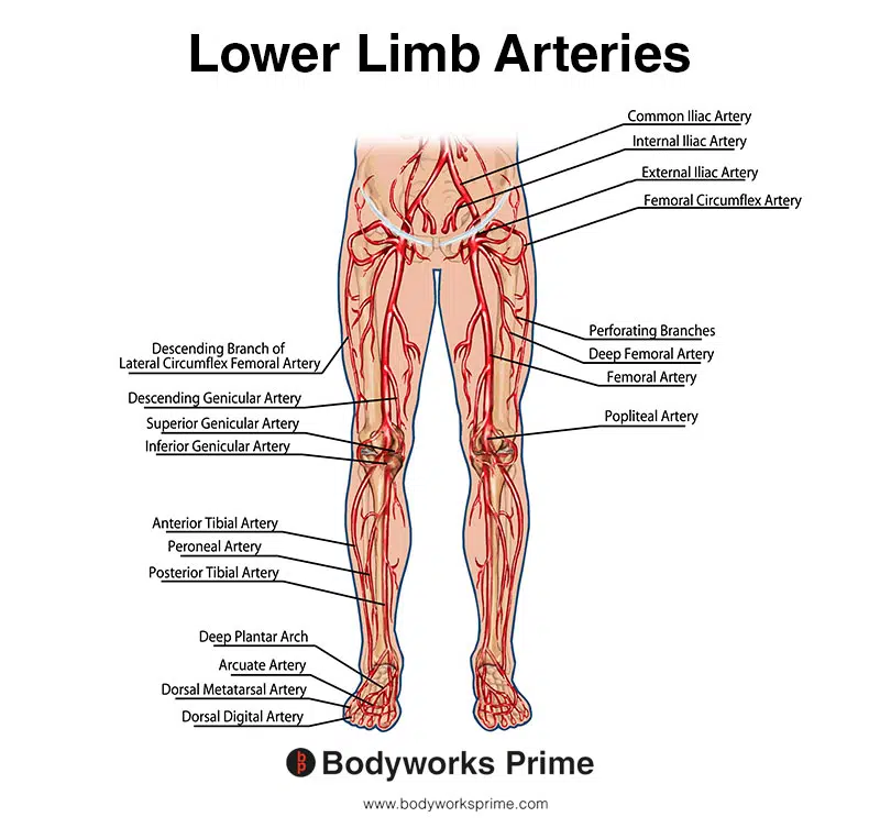
This image shows the arteries of the lower limb.
Want some flashcards to help you remember this information? Then click the link below:
Soleus Flashcards
Support Bodyworks Prime
Running a website and YouTube channel can be expensive. Your donation helps support the creation of more content for my website and YouTube channel. All donation proceeds go towards covering expenses only. Every contribution, big or small, makes a difference!
References
| ↑1, ↑7, ↑10, ↑11 | Binstead JT, Munjal A, Varacallo M. Anatomy, Bony Pelvis and Lower Limb, Calf. [Updated 2021 Jun 3]. In: StatPearls [Internet]. Treasure Island (FL): StatPearls Publishing; 2022 Jan-. Available from: https://www.ncbi.nlm.nih.gov/books/NBK459362/ |
|---|---|
| ↑2 | Kimura N, Kato K, Anetai H, Kawasaki Y, Miyaki T, Kudoh H, Sakai T, Ichimura K. Anatomical study of the soleus: Application to improved imaging diagnoses. Clin Anat. 2021 Oct;34(7):991-1001. doi: 10.1002/ca.23667. Epub 2020 Sep 2. PMID: 32783229. |
| ↑3, ↑6, ↑9 | Moore KL, Agur AMR, Dalley AF. Clinically Oriented Anatomy. 8th ed. Philadelphia: Lippincot Williams & Wilkins; 2017. |
| ↑4 | Simpson SL, Hertzog MS, Barja RH. The plantaris tendon graft: an ultrasound study. J Hand Surg [Am] 1991;16:708–711. |
| ↑5, ↑8 | Mostafa E, Graefe S, Varacallo M. Anatomy, Bony Pelvis and Lower Limb, Leg Posterior Compartment. [Updated 2021 Jun 3]. In: StatPearls [Internet]. Treasure Island (FL): StatPearls Publishing; 2022 Jan-. Available from: https://www.ncbi.nlm.nih.gov/books/NBK537340/ |
| ↑12 | Fathi M, Hassanzad Azar M, Arab Kheradmand A, Shahidi S. Anatomy of arterial supply of the soleus muscle. Acta Med Iran. 2011;49(4):237-40. PMID: 21713734. |


