| Origin | Lateral and medial condyles of femur Popliteal surface of femur |
| Insertion | Posterior surface of the calcaneus via the calcaneal tendon |
| Action | Plantar flexes the foot at the ankle joint Flexes the knee joint |
| Nerve | Tibial nerve (S1-S2) |
| Artery | Sural arteries |
Location & Overview
The gastrocnemius muscle is an essential muscle in the lower leg responsible for walking and maintaining posture. It is composed of two heads, namely the medial and lateral heads, with the former being thicker and wider than the latter. These two heads are connected to the epicondyles of the femur and, when combined with the soleus muscle, form what is commonly known as the ‘calf muscle’. The technical term for this group of muscles is the ‘triceps surae’, which also includes the plantaris muscle. Together, these three muscles make up the superficial flexor group of the lower leg, with the gastrocnemius being the most superficial, or closest to the surface of the skin [1] [2] [3].
The aponeurosis of the gastrocnemius muscle attaches to the calcaneal tendon, which is more commonly known as the ‘Achilles tendon’. This is the longest and widest tendon in the human body, and it derives its name from Achilles, a character in Greek mythology. According to the myth, Achilles’ entire body was invulnerable, except for his calcaneal tendon, which became his weakness. This is the origin of the phrase “Achilles heel,” which refers to a weakness in spite of overall strength [4] [5] [6].
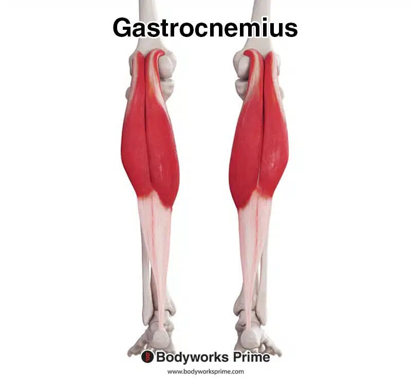
Here we can see the gastrocnemius from a posterior view.
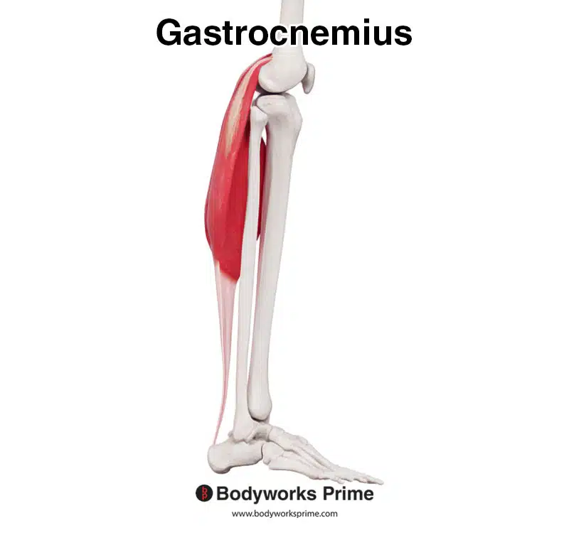
Here we can see the gastrocnemius from a lateral view.
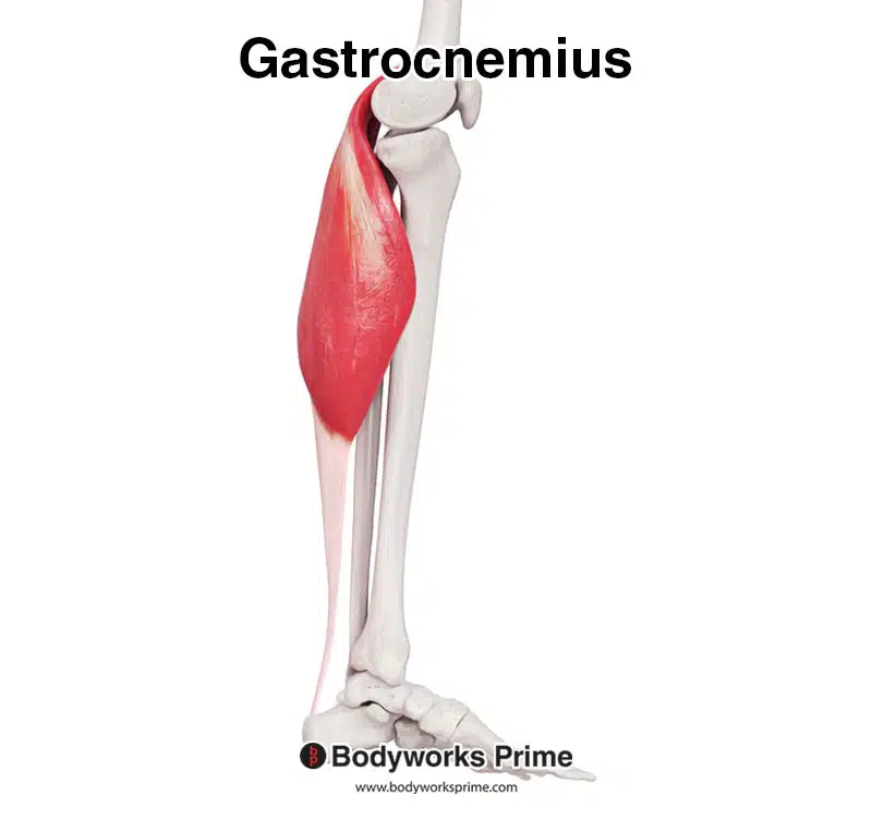
Here we can see the gastrocnemius from a medial view.
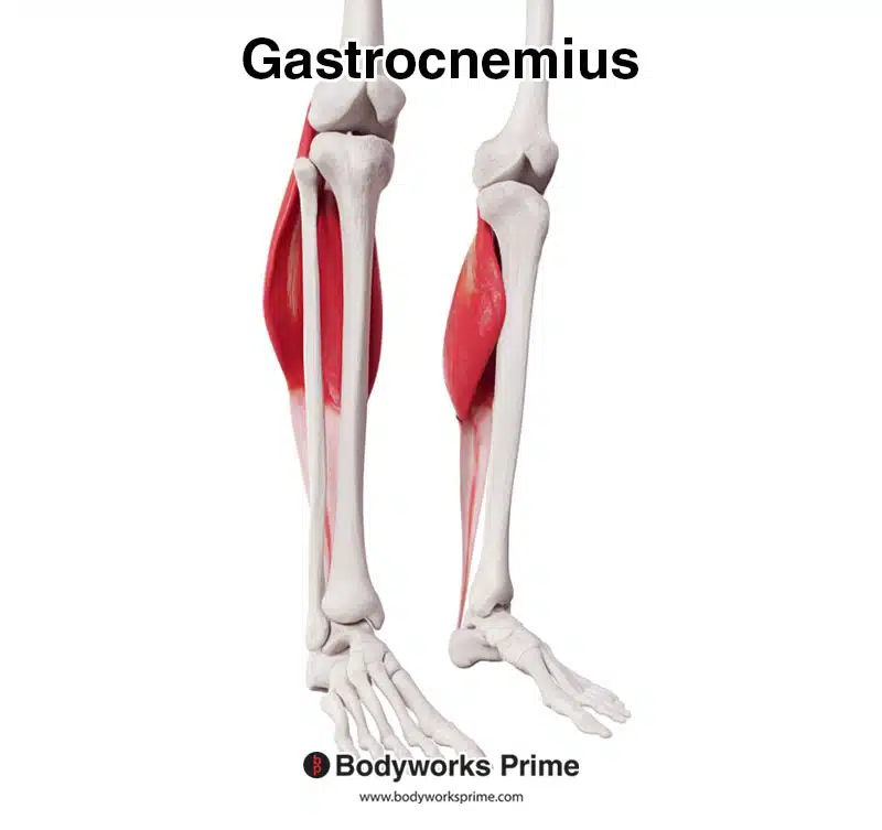
Here we can see the gastrocnemius from an anterior view.
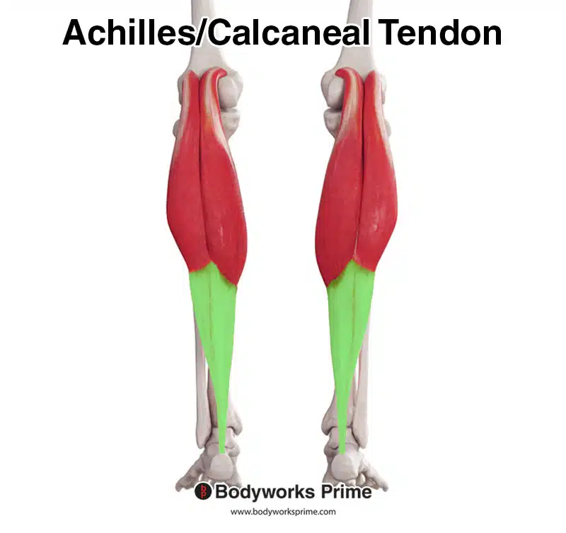
Here we can see pictured the gastrocnemius muscle, as well as the calcaneal tendon (also known as the Achilles tendon). The calcaneal tendon is highlighted in green.
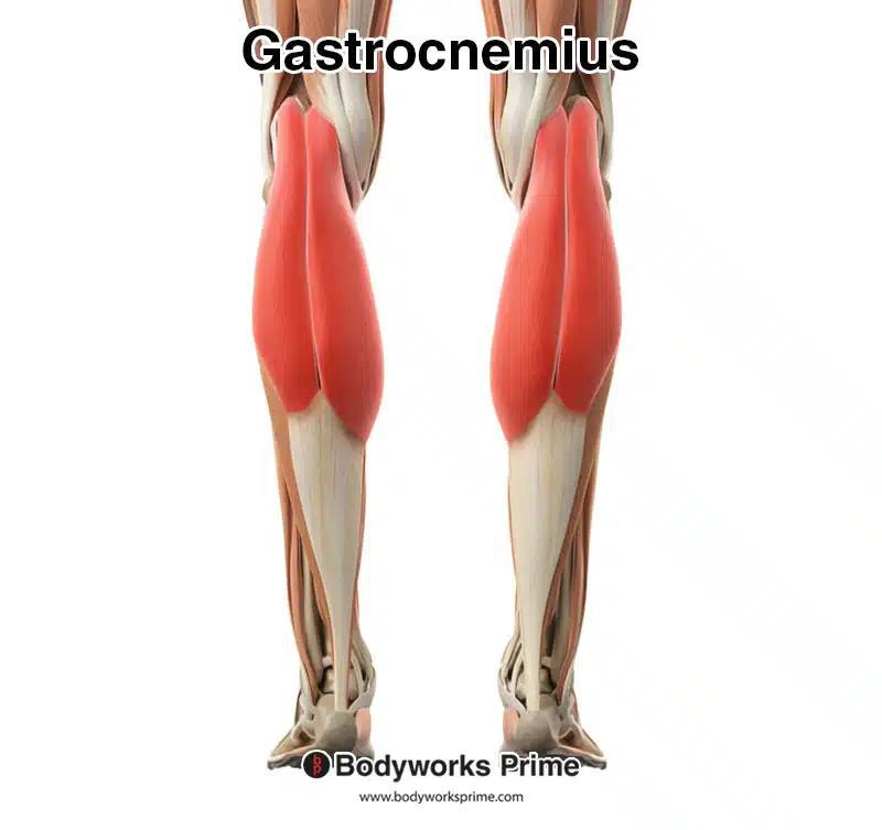
Here we can see the gastrocnemius muscle highlighted in red amongst the other muscles of the leg.
Origin & Insertion
The gastrocnemius muscle has two heads that originate from the lateral and medial condyles of the femur, with its origin also crossing onto the popliteal surface of the femur. The popliteal surface, also known as the popliteal fossa, is a shallow depression located on the posterior aspect of the knee joint. It is diamond-shaped and is bounded by the muscles and bones of the thigh and leg. As the gastrocnemius muscle extends downwards, the two heads converge and form a large aponeurosis that attaches to the calcaneal tendon. The calcaneal tendon, in turn, inserts onto the posterior surface of the calcaneus bone (heel bone) [7] [8].
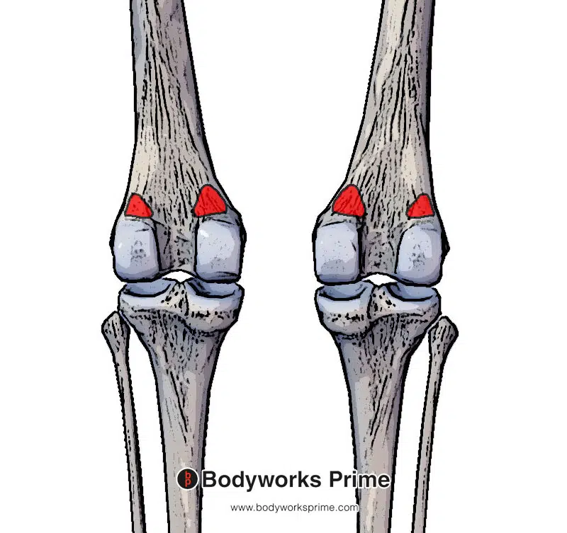
Here we can see the origin of the gastrocnemius muscle highlighted in red on the lateral and medial condyles of femur. You can also see how the origin spans slightly onto the popliteal surface of femur, as it approaches the middle of the femur.
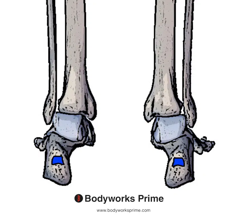
Here we can see the insertion of the gastrocnemius muscle highlighted in blue.
Actions
The primary action of the gastrocnemius muscle is to cause plantar flexion of the foot at the ankle joint, which is commonly used during activities such as walking, running, and jumping, especially during the “pushing away” forces are required by the ankle joint. It works in conjunction with the soleus muscle as part of the triceps surae group to achieve this action. The plantaris muscle can also weakly contribute to plantar flexion. Furthermore, the gastrocnemius contributes to maintaining postural stability and balance while standing, as it helps resist the force of gravity and stops the body from falling forward [9] [10] [11].
A secondary action of the gastrocnemius muscle is to cause flexion of the leg at the knee joint. However, the gastrocnemius muscle is not capable of exerting its full power when flexing the knee and plantarflexing the ankle simultaneously. Rather, it is most effective when performing its actions separately [12] [13] [14]. The hamstring muscles are primarily responsible for flexing the knee joint and are comprised of the semitendinosus, semimembranosus, and biceps femoris muscles [15].
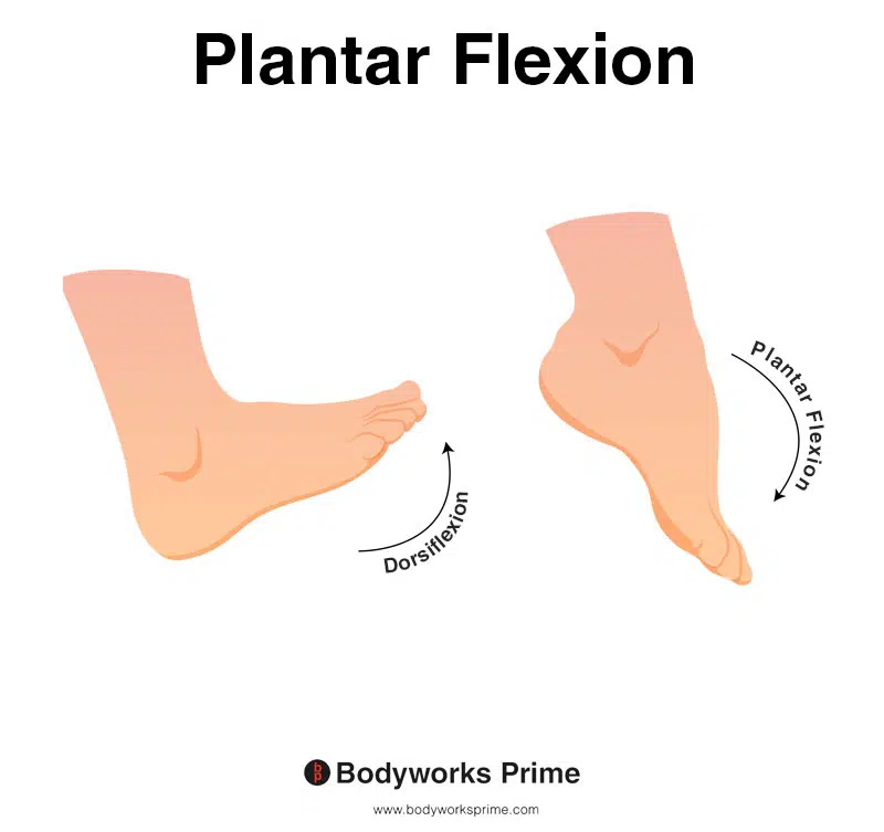
This image shows an example of foot plantar flexion, which involves extending the ankle. The opposite of plantar flexion is dorsiflexion. The gastrocnemius is a plantar flexor of the foot.
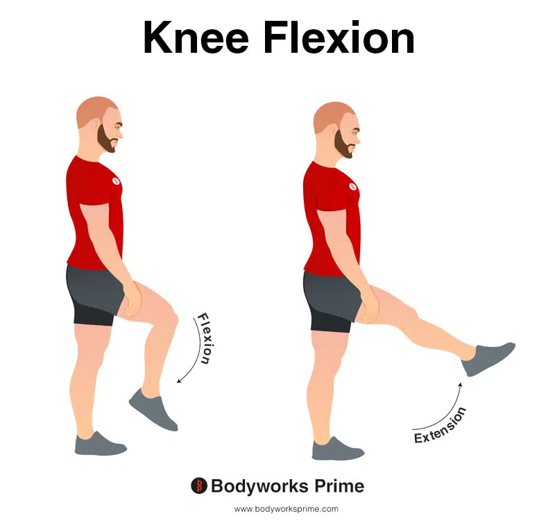
This image demonstrates knee flexion, which is the bending movement of the knee joint. The gastrocnemius muscle is involved in knee flexion, but its primary action is plantarflexion of the foot at the ankle joint. It’s important to note that the gastrocnemius muscle is not as effective when performing knee flexion and plantarflexion simultaneously, as it cannot exert its full power. Thus, the muscle is most powerful when these actions are performed separately.
Innervation
The gastrocnemius is innervated by the tibial nerve (S1-S2). The tibial nerve is a branch of the sciatic nerve. The tibial nerve innervates the calf muscles and the plantaris too [16] [17].
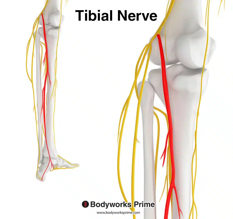
Highlighted in red we can see the tibial nerve, which is a branch of the sciatic nerve. The tibial nerve innervates the gastrocnemius muscle from the spinal nerve roots of S1 and S2. The tibial nerve also innervates the soleus and plantaris muscle too.
Blood Supply
Blood is supplied to the gastrocnemius via the sural arteries. The sural arteries are collateral arteries of the popliteal artery, which is derived from the femoral artery [18] [19].
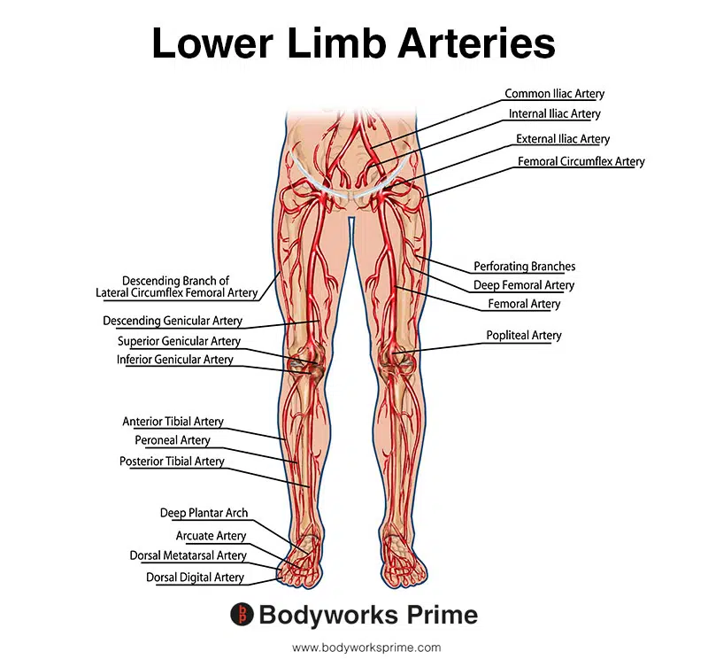
This image shows the arteries of the lower limb.
Want some flashcards to help you remember this information? Then click the link below:
Gastrocnemius Flashcards
Support Bodyworks Prime
Running a website and YouTube channel can be expensive. Your donation helps support the creation of more content for my website and YouTube channel. All donation proceeds go towards covering expenses only. Every contribution, big or small, makes a difference!
References
| ↑1, ↑4, ↑5, ↑9, ↑10, ↑12, ↑13, ↑17, ↑18 | Bordoni B, Varacallo M. Anatomy, Bony Pelvis and Lower Limb, Gastrocnemius Muscle. [Updated 2021 Jul 21]. In: StatPearls [Internet]. Treasure Island (FL): StatPearls Publishing; 2022 Jan-. Available from: https://www.ncbi.nlm.nih.gov/books/NBK532946/ |
|---|---|
| ↑2 | Andjelkov K, Atanasijevic TC, Popovic VM, Sforza M, Atkinson CJ, Soldatovic I. Anatomical aspects of the gastrocnemius muscles: A study in 47 fresh cadavers. J Plast Reconstr Aesthet Surg. 2016 Aug;69(8):1102-8. doi: 10.1016/j.bjps.2016.04.002. Epub 2016 May 4. PMID: 27292288. |
| ↑3, ↑6, ↑7, ↑11, ↑14, ↑16 | Binstead JT, Munjal A, Varacallo M. Anatomy, Bony Pelvis and Lower Limb, Calf. [Updated 2021 Jun 3]. In: StatPearls [Internet]. Treasure Island (FL): StatPearls Publishing; 2022 Jan-. Available from: https://www.ncbi.nlm.nih.gov/books/NBK459362/ |
| ↑8 | Moore KL, Agur AMR, Dalley AF. Clinically Oriented Anatomy. 8th ed. Philadelphia: Lippincot Williams & Wilkins; 2017. |
| ↑15 | Rodgers CD, Raja A. Anatomy, Bony Pelvis and Lower Limb, Hamstring Muscle. [Updated 2022 Jan 29]. In: StatPearls [Internet]. Treasure Island (FL): StatPearls Publishing; 2023 Jan-. Available from: https://www.ncbi.nlm.nih.gov/books/NBK546688/ |
| ↑19 | Aragão JA, Reis FP, Pitta GB, Miranda F Jr, Poli de Figueiredo LF. Anatomical study of the gastrocnemius venous network and proposal for a classification of the veins. Eur J Vasc Endovasc Surg. 2006 Apr;31(4):439-42. doi: 10.1016/j.ejvs.2005.10.022. Epub 2005 Dec 15. PMID: 16359881 |


