| Origin | Lateral condyle of the tibia Proximal two-thirds of the lateral surface of the tibia Anterior side of the interosseous membrane |
| Insertion | Medial cuneiform Base of the first metatarsal |
| Action | Dorsiflexion of ankle Inversion of foot Assists in maintaining the medial arch of the foot |
| Nerve | Peroneal nerve (L4, L5, S1) |
| Artery | Anterior tibial artery Medial tarsal arteries |
Location & Overview
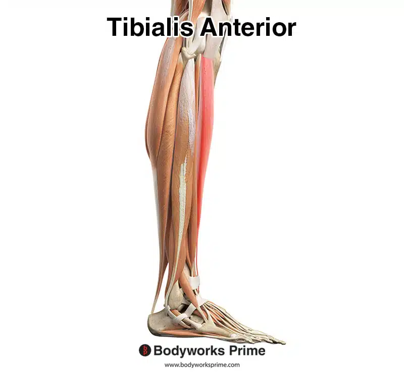
The tibialis anterior muscle, also referred to as the tibialis anticus, is a well-known superficial muscle located lateral to the anterior body of the tibia bone. Thanks to its prominent location, it is easily palpable and is the largest among the four muscles found in the anterior compartment of the lower leg. Along with the tibialis anterior, the other three anterior compartment muscles are the extensor hallucis longus, extensor digitorum longus, and the fibularis tertius [1] [2].
One of the key functions of the tibialis anterior muscle is dorsiflexion of the foot, which allows the foot to lift up towards the shin. This function is important during physical activities such as running and walking, as it helps the foot to pass over the ground and allows for a smooth, efficient gait. Without the tibialis anterior, activities like running and walking would be much more challenging and potentially even impossible [3].
In addition to its important role in dorsiflexion, the tibialis anterior muscle also provides medial support to the foot and ankle, aiding in the stabilization of the lower leg and the maintenance of proper body posture. This support is particularly important during weight-bearing activities, such as standing or walking, because it helps to distribute weight evenly across the foot and ankle, reducing the risk of injury or strain [4].
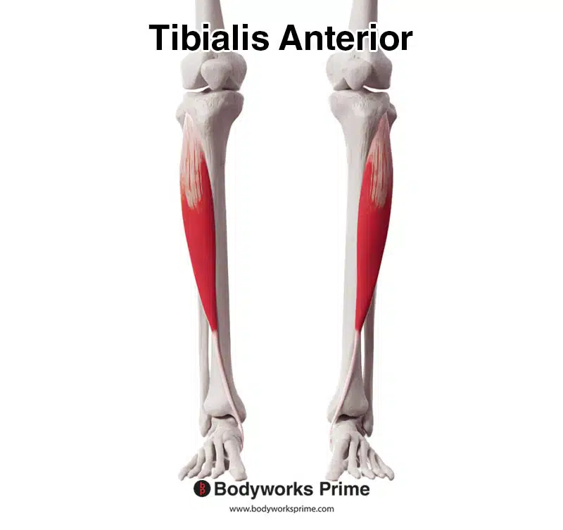
Here we can see the tibialis anterior muscle from an anterior view.
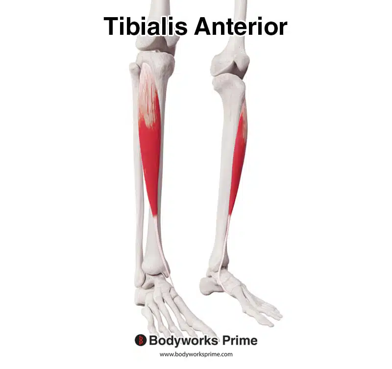
Here we can see the tibialis anterior muscle from a anterolateral view.

Here we can see the tibialis anterior muscle from a lateral view.
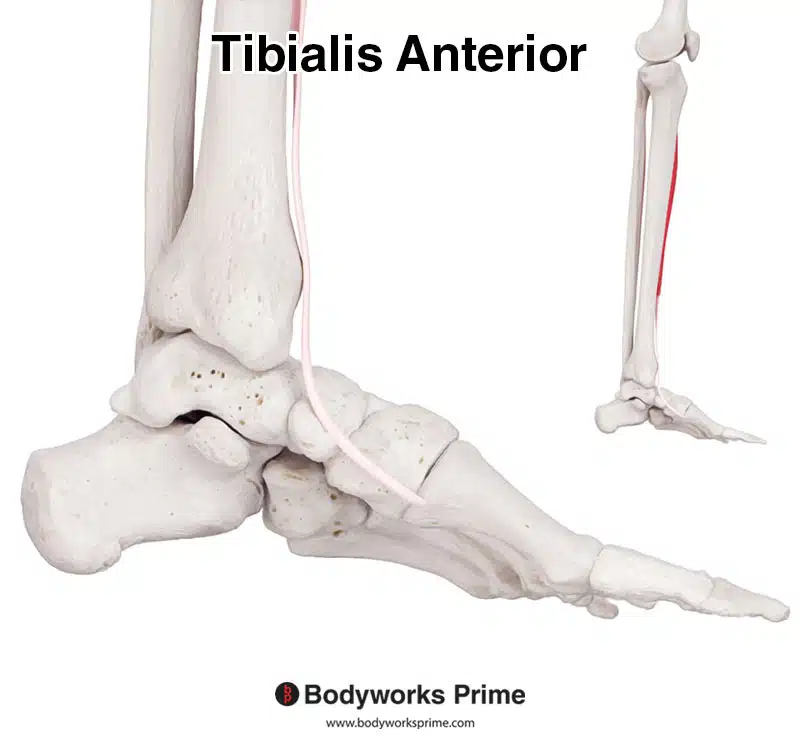
Here we can see a medial view of the foot, showing the tibialis anterior’s distal tendon.
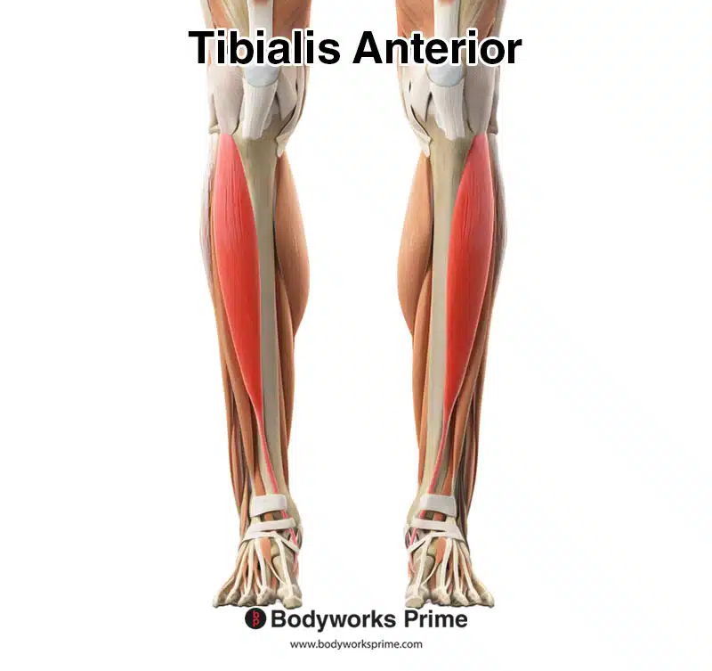
Here we can see the tibialis anterior muscle amongst the other muscles of the leg. The tibialis anterior is highlighted in red.
Here we can see the tibialis anterior muscle highlighted in red from a lateral view.
Origin & Insertion
The tibialis anterior originates from three places: the lateral condyle of the tibia, the proximal two-thirds of the lateral surface of the tibia, and the anterior side of the interosseous membrane (the interosseous membrane is a membrane in between the tibia and fibula). It works its way inferiorly from this origin where it then passes across the ankle joint and reaches the foot. On the foot, it then inserts on the medial cuneiform and the base of the first metatarsal. This tendinous insertion is the most medial tendon of the ankle and foot. This distal tendon is held against the front of the ankle and foot by the extensor retinaculum, which covers the anterior aspect of the ankle and foot [5] [6] [7].
The tendinous insertion of the tibialis anterior muscle exhibits individual variation, with the occurrence of one additional band being the most common variant, although two or more additional insertions have also been observed. Additionally, variations in the locations of attachment points for both the origin and insertion of this muscle have been documented. Nevertheless, the most commonly observed origin and insertion are as listed in the previous paragraph [8].
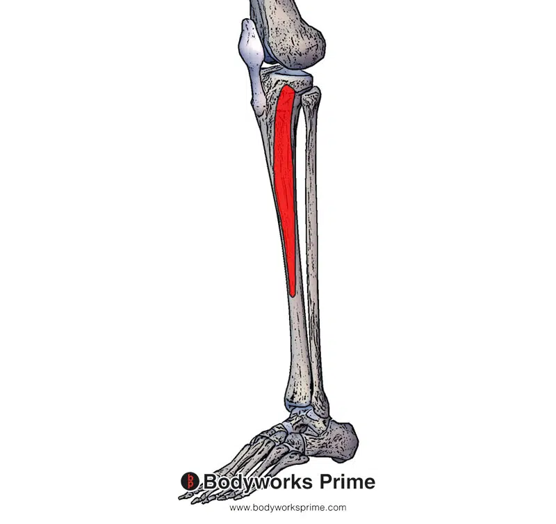
Pictured here we can see the first two origin points of the tibialis anterior muscle marked out in red on the lateral condyle of the tibia and proximal two-thirds of the lateral surface of the tibia.
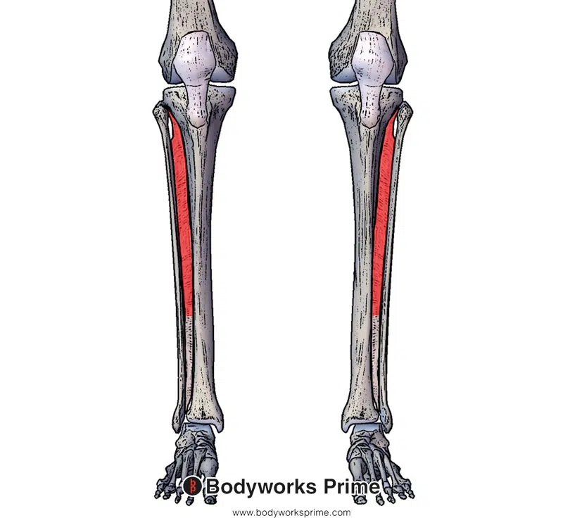
Pictured here we can see another origin point of the tibialis anterior marked out in red on the anterior side of the interosseous membrane.
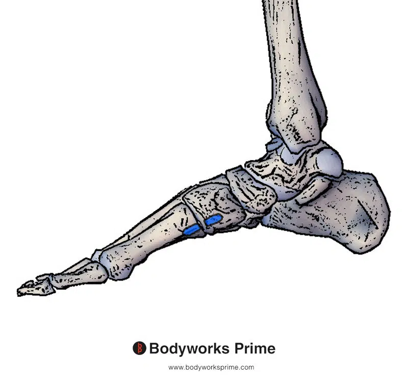
Pictured here we can see the insertion point of the tibialis anterior marked out in blue on the medial cuneiform and the base of the first metatarsal.
Actions
The primary role of the tibialis anterior muscle is to cause dorsiflexion of the foot, which involves lifting the front of the foot towards the shin and thereby causing flexion of the ankle joint. Among the muscles responsible for dorsiflexion, the tibialis anterior is the strongest. In addition, the tibialis anterior can act in conjunction with the tibialis posterior to produce inversion of the foot, which entails turning the sole of the foot medially (as opposed to eversion). Moreover, because the tibialis anterior inserts onto the medial border of the foot, it can assist in supporting the medial arch of the foot [9] [10] [11] [12].
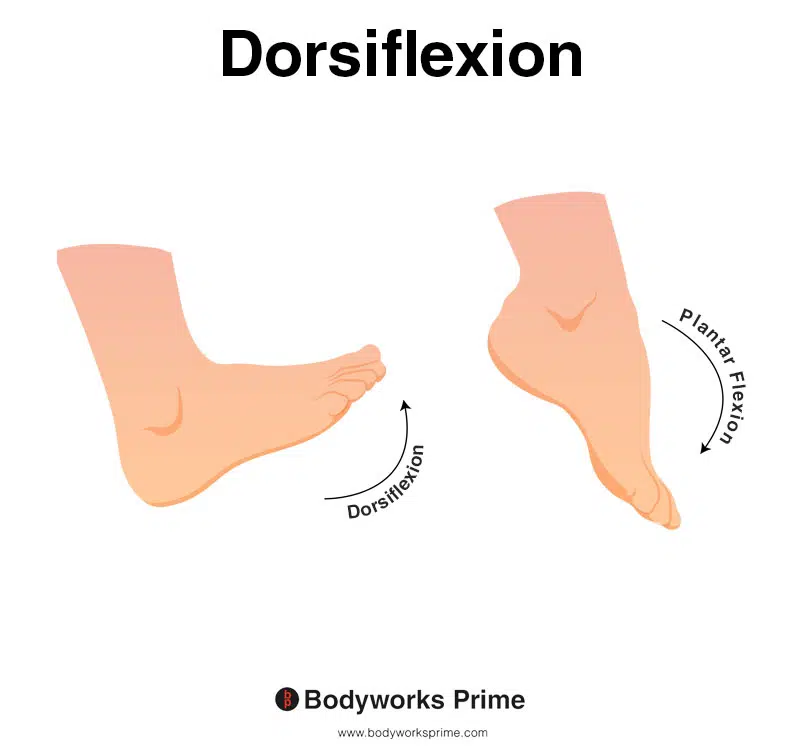
This image shows an example of dorsiflexion of the foot, which involves flexing the ankle joint, thereby lifting the foot upwards and towards the shins. The opposite of dorsiflexion is plantar flexion. The primary role of the tibialis anterior dorsiflexion of the foot.
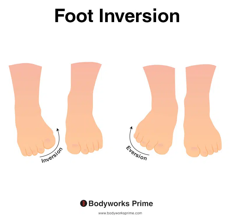
This picture demonstrates foot inversion, which means rotating the ankle inwards, causing the sole of the foot to turn towards the body. The opposite of inversion is eversion. The tibialis anterior muscle can aid in inversion of the foot and works alongside the tibialis posterior during this action. Both the tibialis anterior and posterior also assist in maintaining the medial arch of the foot.
Innervation
The tibialis anterior muscle is innervated by the deep peroneal nerve (L4, L5, S1). The deep peroneal nerve is also called the deep fibular nerve. The deep peroneal nerve arises from the splitting of the common peroneal nerve, which occurs between the head of the fibula and the superior portion of the peroneus longus muscle [13] [14] [15] [16] [17].
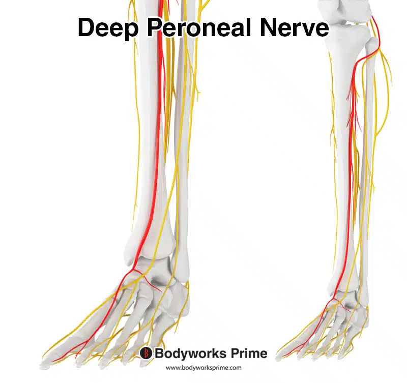
Pictured here we can see the deep peroneal nerve which innervates the tibialis anterior muscle.
Blood Supply
Blood is supplied to the proximal area of the tibialis anterior via the anterior tibial artery. Blood is supplied to the distal tendon via the medial tarsal arteries, which are branch of the dorsalis pedis artery [18] [19] [20] [21] .
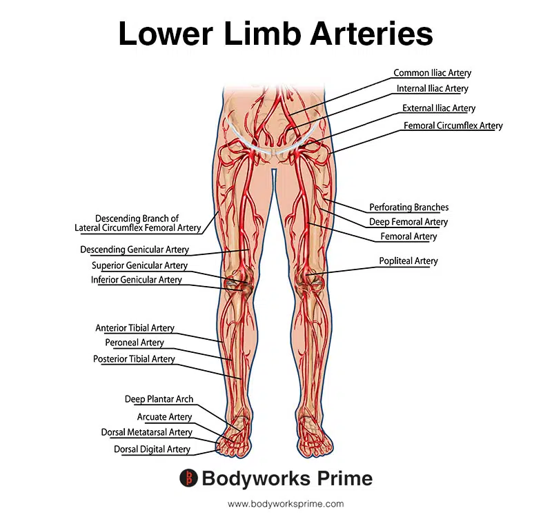
This image shows the arteries of the lower limb.
Want some flashcards to help you remember this information? Then click the link below:
Tibialis Anterior Flashcards
Support Bodyworks Prime
Running a website and YouTube channel can be expensive. Your donation helps support the creation of more content for my website and YouTube channel. All donation proceeds go towards covering expenses only. Every contribution, big or small, makes a difference!
References
| ↑1, ↑5, ↑9, ↑13, ↑18 | Juneja P, Hubbard JB. Anatomy, Bony Pelvis and Lower Limb, Tibialis Anterior Muscles. [Updated 2021 Aug 13]. In: StatPearls [Internet]. Treasure Island (FL): StatPearls Publishing; 2022 Jan-. Available from: https://www.ncbi.nlm.nih.gov/books/NBK513304/ |
|---|---|
| ↑2, ↑6, ↑8, ↑10, ↑15, ↑20 | Zielinska N, Tubbs RS, Paulsen F, Szewczyk B, Podgórski M, Borowski A, Olewnik Ł. Anatomical Variations of the Tibialis Anterior Tendon Insertion: An Updated and Comprehensive Review. J Clin Med. 2021 Aug 19;10(16):3684. doi: 10.3390/jcm10163684. PMID: 34441980; PMCID: PMC8396864. |
| ↑3, ↑4 | Moore KL, Agur AMR, Dalley AF. Clinically Oriented Anatomy. 8th ed. Philadelphia: Lippincot Williams & Wilkins; 2017. |
| ↑7, ↑12, ↑16, ↑21 | Varghese A, Bianchi S. Ultrasound of tibialis anterior muscle and tendon: anatomy, technique of examination, normal and pathologic appearance. J Ultrasound. 2013 Dec 19;17(2):113-23. doi: 10.1007/s40477-013-0060-7. PMID: 24883132; PMCID: PMC4033720. |
| ↑11 | Day JT, Lichtwark GA, Cresswell AG. Tibialis anterior muscle fascicle dynamics adequately represent postural sway during standing balance. J Appl Physiol (1985). 2013 Dec;115(12):1742-50. doi: 10.1152/japplphysiol.00517.2013. Epub 2013 Oct 17. PMID: 24136108. |
| ↑14 | Becciolini M, Pivec C, Riegler G. Ultrasound Imaging of the Deep Peroneal Nerve. J Ultrasound Med. 2021 Apr;40(4):821-838. doi: 10.1002/jum.15455. Epub 2020 Sep 3. PMID: 32881065. |
| ↑17 | Garrett A, Geiger Z. Anatomy, Bony Pelvis and Lower Limb: Calf Deep Peroneal Nerve (Deep Fibular Nerve) [Updated 2022 Aug 8]. In: StatPearls [Internet]. Treasure Island (FL): StatPearls Publishing; 2023 Jan-. Available from: https://www.ncbi.nlm.nih.gov/books/NBK526033/ |
| ↑19 | Azam M, Wehrle CJ, Shaw PM. Anatomy, Bony Pelvis and Lower Limb, Tibial Artery. [Updated 2021 Aug 11]. In: StatPearls [Internet]. Treasure Island (FL): StatPearls Publishing; 2022 Jan-. Available from: https://www.ncbi.nlm.nih.gov/books/NBK532871/ |


