| Origin | Lateral supracondylar line of the femur |
| Insertion | Posterior surface of the calcaneus |
| Action | Assists in plantar flexion of the foot Assists in flexion of the knee Potentially has an important proprioceptive role |
| Nerve | Tibial nerve (S1- S2) |
| Artery | Sural artery Popliteal artery Superior lateral genicular artery |
Location & Overview
The plantaris muscle is a long and thin muscle which runs down the posterior aspect of the lower leg. It is located superficial to the soleus muscle and deep to the gastrocnemius muscle [1]. Anatomical variation of this muscle can sometimes be present, such as: it being absent entirely (absent in only 7–20% of limbs [2]), terminating proximally into the calcaneal tendon, or even terminating into the gastrocnemius sometimes [3].
The plantaris muscle has become less significant during human evolution. The plantaris, a muscle once used for tree-climbing in primates, has become less important in human evolution due to the shift towards bipedal locomotion (using two legs for walking). In modern humans, the plantaris muscle mainly acts in conjunction with the gastrocnemius muscle, serving as an accessory to it. This understanding is based on the research conducted by Sichting et al. (2020) in their article “Evolutionary Anatomy of the Plantar Aponeurosis in Primates, Including Humans,” published in the Journal of Anatomy [4].
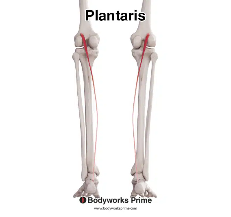
Pictured here we can see the plantaris muscle from a posterior view.
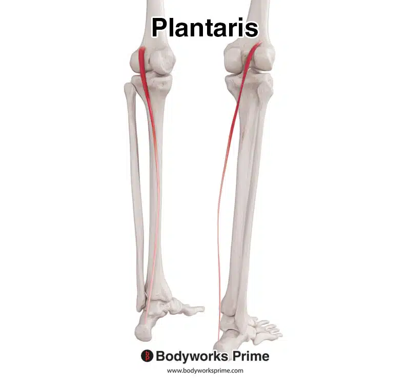
Here we can see the plantaris feom a posterolateral view.
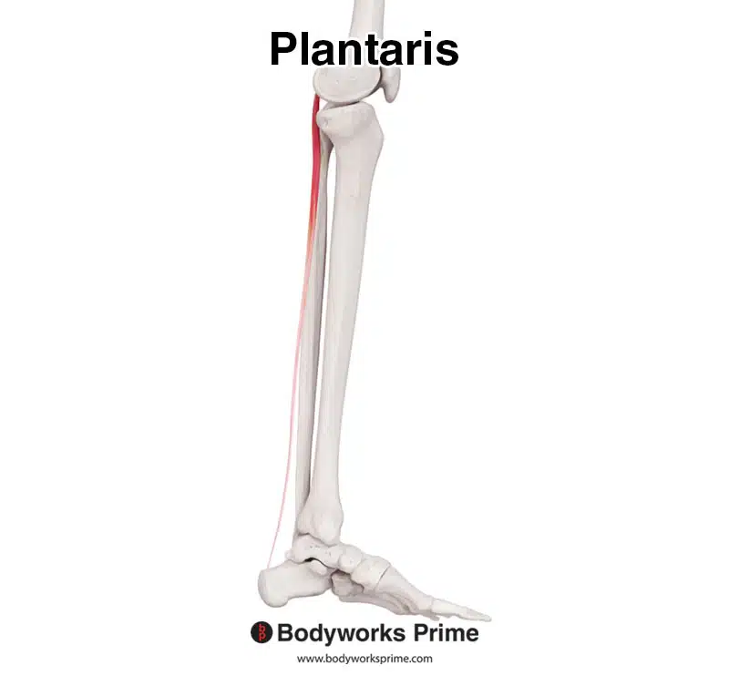
Here we can see the plantaris seen from a medial view.
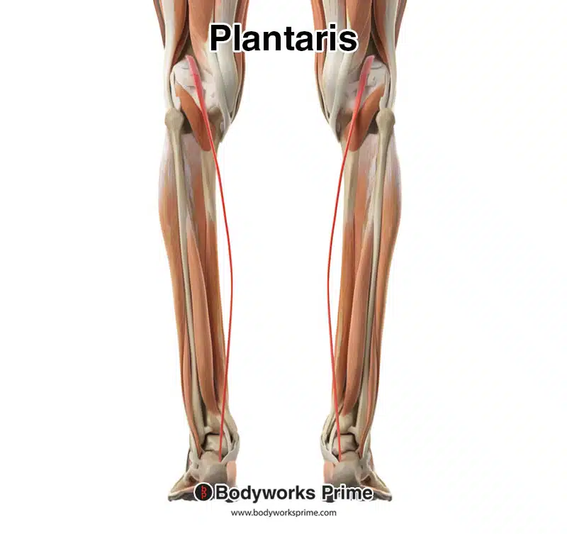
Here we can see the plantaris highlighted in red, amongst the other muscles of the leg, from a posterior view.
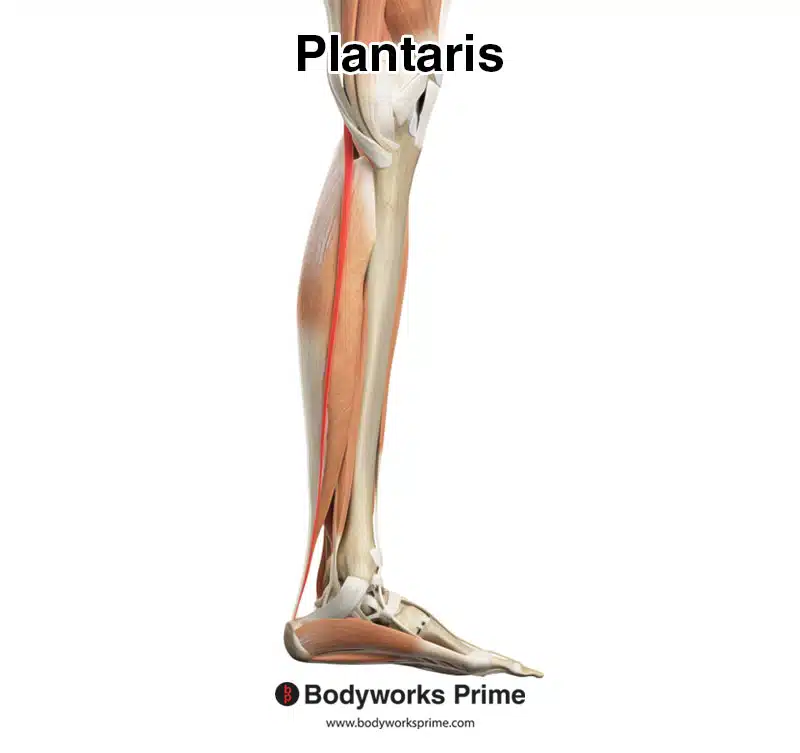
Here we can see the plantaris highlighted in red, amongst the other muscles of the leg, from a medial view.
Origin & Insertion
The plantaris muscle originates from the lateral supracondylar line of the femur. The origin is superior and medial to the gastrocnemius’ lateral head. The plantaris then continues down the leg located between the soleus and gastrocnemius and then along the medial aspect of the calcaneal tendon, which is inserts right next to, on the calcaneus [5] [6].
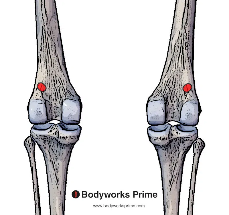
Here we can see the origin of the plantaris muscle on the lateral supracondylar line of the femur.
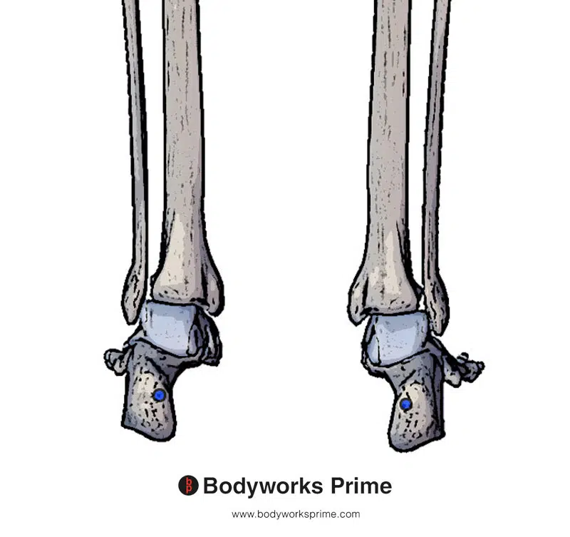
Here we can see the insertion of the plantaris muscle on the posterior surface of the calcaneus.
Actions
The action of the plantaris muscle is to provide weak plantar flexion at the ankle joint and weak flexion at the knee joint. The gastrocnemius is able to perform these actions much better and the soleus is a stronger plantar flexor too (though the soleus does not cross the knee joint so it can’t assist in knee flexion, unlike the gastrocnemius) [7] [8] Some studies have suggested the plantaris plays an important proprioceptive role too [9] [10] [11]. Proprioception is your body’s way to sense movement, action, and its location.
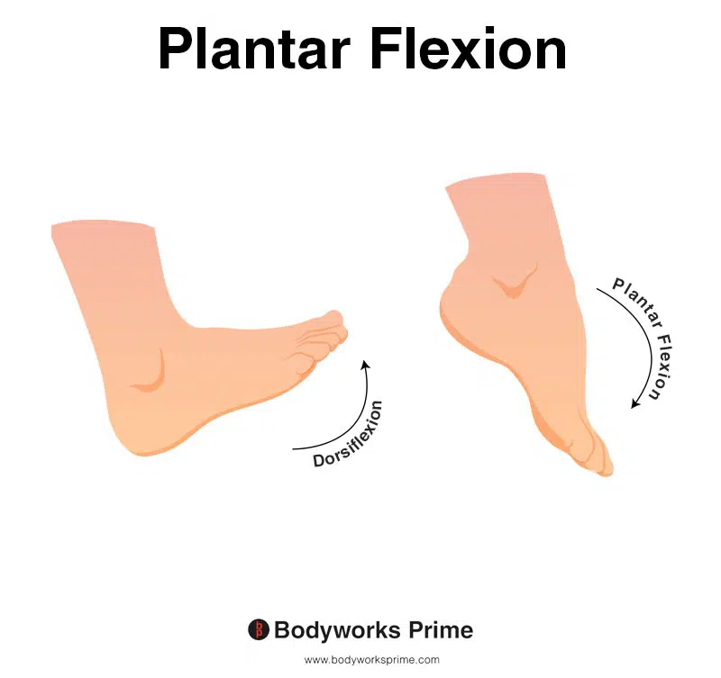
This image shows an example of foot plantar flexion, which involves extending the ankle. The opposite of plantar flexion is dorsiflexion. The plantaris muscle can weakly assist with plantar flexion.
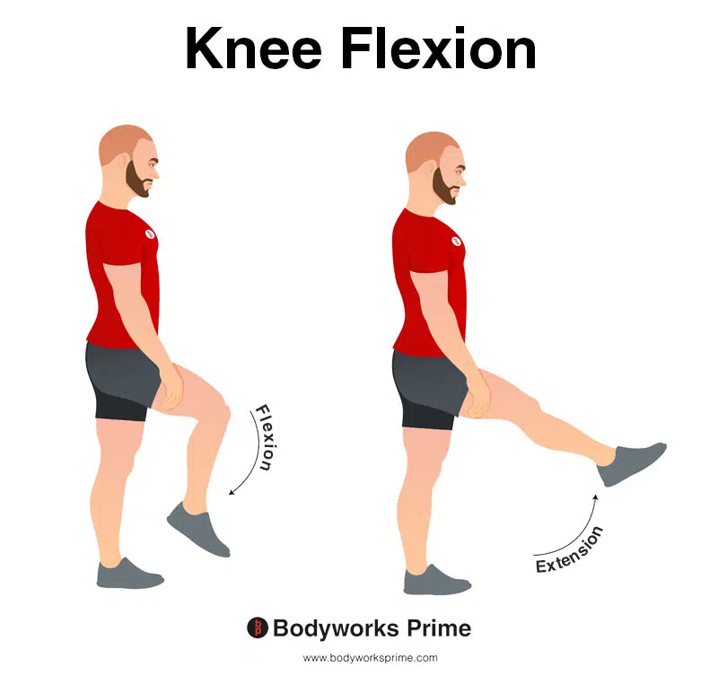
In this image, you can see an example of knee flexion, which is the action of bending your knee. The opposite movement of knee flexion is knee extension. The plantaris muscle can weakly assist with knee flexion.
Innervation
The plantaris muscle is innervated by the tibial nerve (S1- S2). This nerve is the larger branch of the sciatic nerve which divides into the tibial nerve. It also provides innervation for other muscles of the posterior lower leg compartment [12] [13].
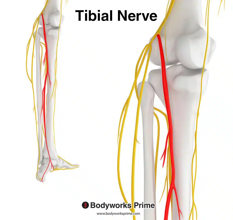
Highlighted in red we can see the tibial nerve, which is a branch of the sciatic nerve. The tibial nerve innervates the plantaris muscle from the spinal nerve roots of S1 and S2.
Blood Supply
Blood is supplied to the plantaris muscle via the sural, popliteal, and superior lateral genicular arteries [14].
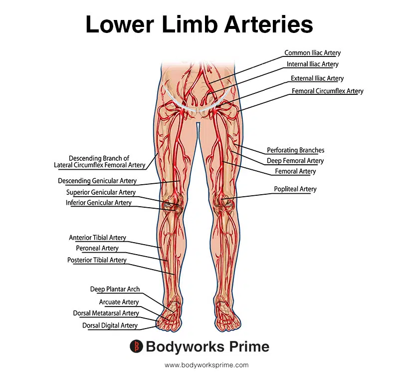
This image shows the arteries of the lower limb.
Want some flashcards to help you remember this information? Then click the link below:
Plantaris Flashcards
Support Bodyworks Prime
Running a website and YouTube channel can be expensive. Your donation helps support the creation of more content for my website and YouTube channel. All donation proceeds go towards covering expenses only. Every contribution, big or small, makes a difference!
References
| ↑1, ↑5, ↑7, ↑12 | Spina AA. The plantaris muscle: anatomy, injury, imaging, and treatment. J Can Chiropr Assoc. 2007;51(3):158-65. PMID: 17885678; PMCID: PMC1978447. |
|---|---|
| ↑2 | Simpson SL, Hertzog MS, Barja RH. The plantaris tendon graft: an ultrasound study. J Hand Surg [Am] 1991;16:708–711. |
| ↑3 | Egan KG, Emanuelli E, Przylecki W, Endress RD. Variant Plantaris Anatomy During Tendon Harvest. J Hand Surg Am. 2021 May;46(5):431.e1-431.e4. doi: 10.1016/j.jhsa.2021.01.004. Epub 2021 Mar 16. PMID: 33741215. |
| ↑4 | Sichting, Freddy; Holowka, Nicholas B.; Ebrecht, Florian; Lieberman, Daniel E. (2020-02-26). “Evolutionary anatomy of the plantar aponeurosis in primates, including humans”. Journal of Anatomy. Wiley. 237 (1): 85–104. doi:10.1111/joa.13173. ISSN 0021-8782. PMC 7309290. PMID 32103502 |
| ↑6, ↑14 | Moore KL, Agur AMR, Dalley AF. Clinically Oriented Anatomy. 8th ed. Philadelphia: Lippincot Williams & Wilkins; 2017. |
| ↑8, ↑10 | Moore KL, Dalley AF, editors. Clinically Oriented Anatomy. 5. Philadelphia: Lippincott Williams & Wilkins; 2006. pp. 648–649. |
| ↑9 | Vlaic J, Josipovic M, Bohacek I, Jelic M. The plantaris muscle: too important to be forgotten. A review of evolution, anatomy, clinical implications and biomechanical properties. J Sports Med Phys Fitness. 2019 May;59(5):839-845. doi: 10.23736/S0022-4707.18.08816-3. PMID: 30936418. |
| ↑11 | Greene BD, Smith SE, Smith JT. Snapping Plantaris Tendon: A Rare Case in a Competitive Dancer. J Am Acad Orthop Surg Glob Res Rev. 2021;5(5):e21.00008. Published 2021 May 4. doi:10.5435/JAAOSGlobal-D-21-00008 |
| ↑13 | Binstead JT, Munjal A, Varacallo M. Anatomy, Bony Pelvis and Lower Limb, Calf. [Updated 2021 Jun 3]. In: StatPearls [Internet]. Treasure Island (FL): StatPearls Publishing; 2022 Jan-. Available from: https://www.ncbi.nlm.nih.gov/books/NBK459362/ |


