| Origin | Iliac crest (lateral aspect) Gluteus maximus & tensor fasciae latae muscle’s aponeuroses |
| Insertion | Iliotibial tubercle (also known as Gerdy’s tubercle) |
| Action | Stabilisation of the hip and knee Supports the actions of the gluteus maximus and tensor fasciae latae |
| Nerve | Superior gluteal nerve L4-S1 Inferior gluteal nerve L5-S2 |
| Artery | Ascending branch of the lateral femoral circumflex artery Superior gluteal artery |
Location & Overview
The iliotibial tract is also referred to as the iliotibial band (ITB for short) and is also sometimes called Maissiat’s band. Maissiat’s band is named after the person who discovered it, Jacques Maissiat, who was a French anatomist [1].
The iliotibial tract is located in the lateral thigh region and spans from the ilium to the proximal tibia. The iliotibial tract is a thick band of deep fascia. The fascia lata, gluteus maximus and tensor fasciae latae muscles all contribute to the formation of the iliotibial tract. The gluteus maximus and tensor fasciae latae connect to the iliotibial tract via an aponeurosis. The fascia lata is a fascial sheath which envelops the thigh like a sleeve and its fibres contribute to the iliotibial tract. The iliotibial tract is a type of connective tissue that lacks contractile properties, meaning it cannot cause any direct muscle contractions. Instead, it serves to support and aid other muscles in carrying out their actions [2] [3].
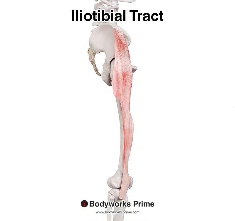
Here we can see the iliotibial tract in isolation from a lateral view.
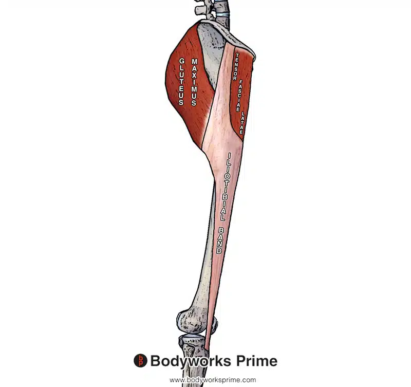
Pictured here you can see the iliotibial tract connecting via aponeuroses to the gluteus maximus and the tensor fasciae latae.
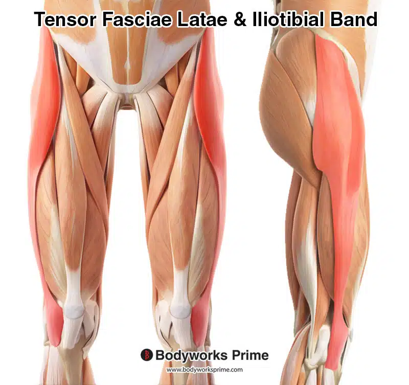
Here we can see both the tensor fasciae latae and iliotibial band highlighted in red amongst the other muscles of the thigh.
Origin & Insertion
The iliotibial tract originates on the ilium’s lateral aspect. Aponeuroses from the gluteus maximus and the tensor fasciae latae (TFL) also connect to the proximal iliotibial tract. These aponeuroses serve as further origin points for the iliotibial tract. The iliotibial tract then continues down the lateral thigh, crossing the knee joint. It then inserts onto the iliotibial tubercle, which is located on the tibia. The iliotibial tubercle is sometimes referred to as ‘Gerdy’s tubercle’ [4].
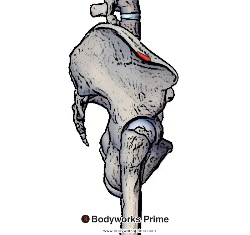
Highlighted in red you can see the origin of the iliotibial tract on the lateral aspect of the Iliac crest.
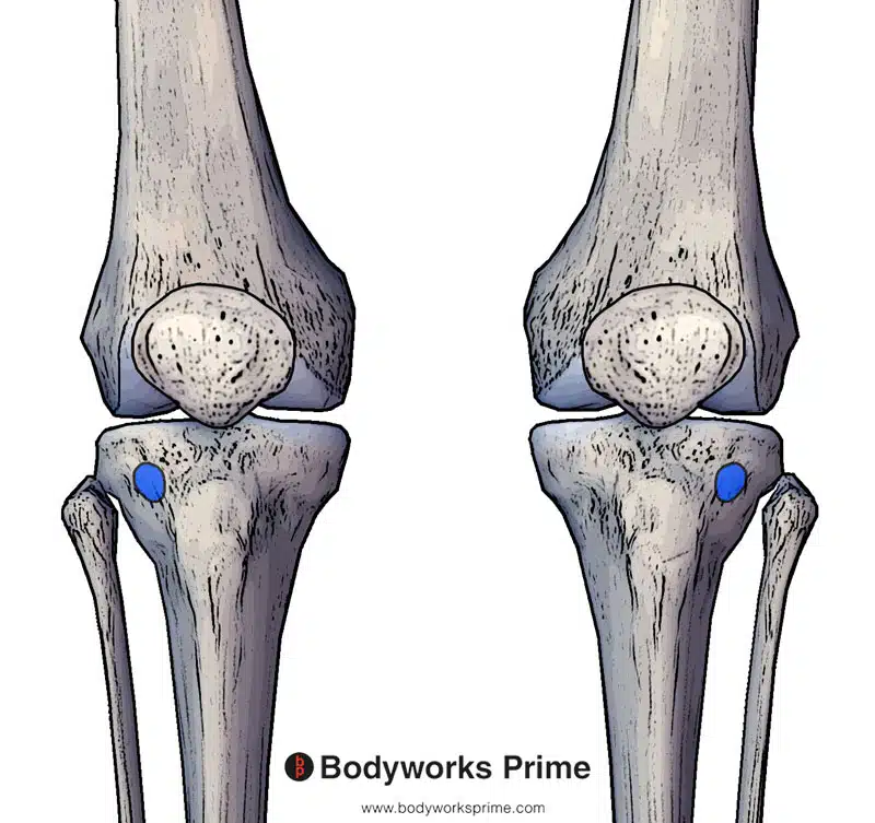
Highlighted in blue you can see the insertion of the iliotibial tract on the Iliotibial tubercle (also known as Gerdy tubercle).
Actions
The primary action of the iliotibial tract is stabilisation of the hip and knee. However, it also works together with gluteus maximus and tensor fasciae latae (TFL) due to the aponeuroses which connects these muscles to the iliotibial tract. The iliotibial tract can assist in the actions of both the gluteus maximus and the tensor fasciae latae because it connects to them [5].
The gluteus maximus primarily extends the hip and contributes to hip abduction. Contraction of the TFL can maintain tension on the iliotibial tract providing stability. The TFL’s contraction can also pull the hip inferiorly (hip abduction), causing the hip on the opposite side to rise. This hip movement assists in walking by allowing the leg to swing without hitting the ground [6] [7] [8].
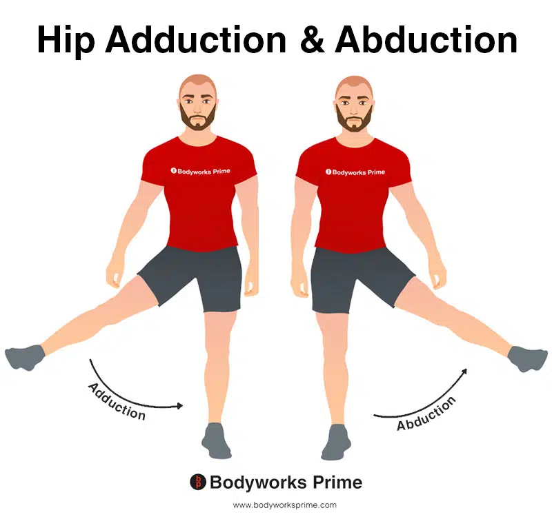
This image shows an example of hip adduction and abduction. Hip abduction involves moving the leg out to the side (laterally) whereas hip adduction involves moving the leg back to the body (medially). The iliotibial tract assists the TFL muscle and gluteus maximus muscle in their action of causing hip abduction.
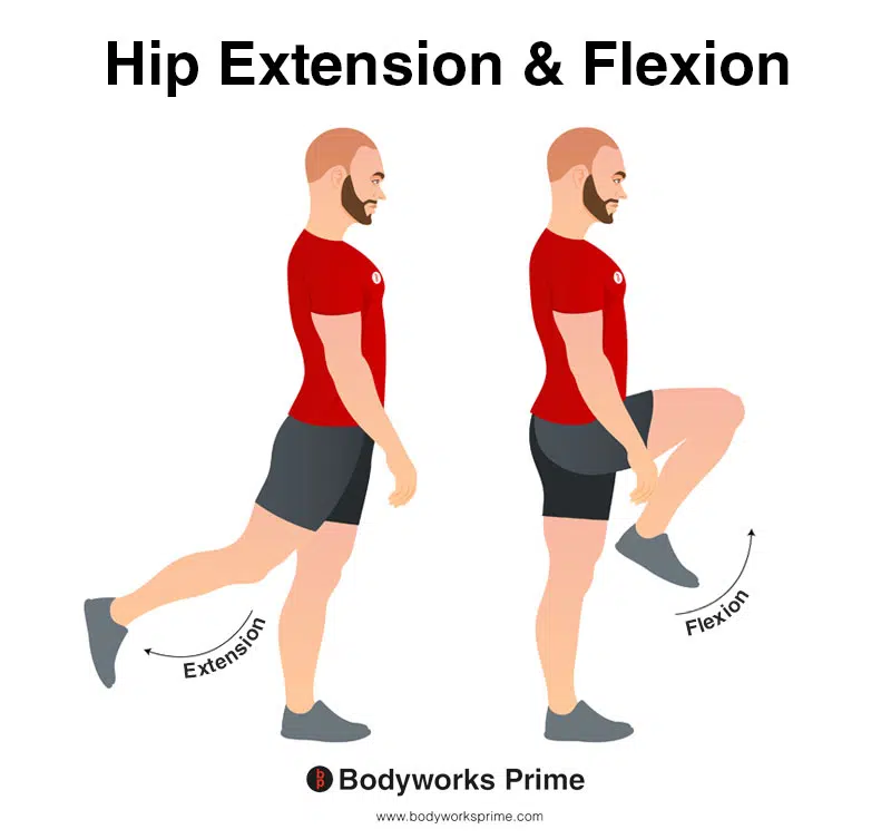
In this image, you can see an example of hip extension and flexion. Hip flexion is the action of raising your leg up in front of you and causing your hip to bend. Hip extension is the opposite of this and involves extending the hip from that flexed position. If both legs are fixed, the torso will lean forward towards the legs (hip flexion) or backwards away from the legs (hip extension). The iliotibial tract assists the TFL muscle and gluteus maximus muscle in their action of causing hip extension.
Innervation
The iliotibial band shares the same innervation as the gluteus maximus and the TFL. These innervations are the superior gluteal nerve (SGN, L4-S1) and inferior gluteal nerve (IGN, L5-S2) [9].
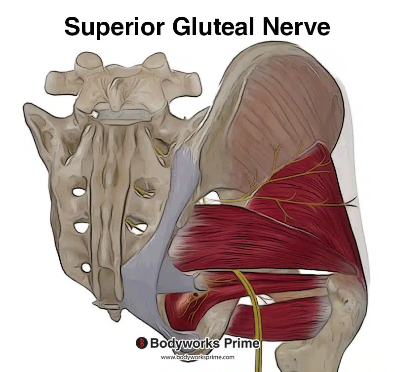
Pictured here you can see the superior gluteal nerve.
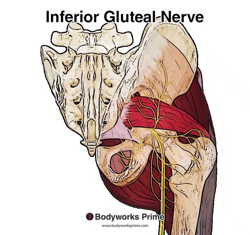
Pictured here you can see the inferior gluteal nerve.
Blood Supply
Blood is supplied to the iliotibial tract via the ascending branch of the lateral femoral circumflex artery and the superior gluteal artery [10].
Want some flashcards to help you remember this information? Then click the link below:
Iliotibial Tract Flashcards
Support Bodyworks Prime
Running a website and YouTube channel can be expensive. Your donation helps support the creation of more content for my website and YouTube channel. All donation proceeds go towards covering expenses only. Every contribution, big or small, makes a difference!
References
| ↑1, ↑2 | Flato R, Passanante GJ, Skalski MR, Patel DB, White EA, Matcuk GR Jr. The iliotibial tract: imaging, anatomy, injuries, and other pathology. Skeletal Radiol. 2017 May;46(5):605-622. doi: 10.1007/s00256-017-2604-y. Epub 2017 Feb 25. PMID: 28238018. |
|---|---|
| ↑3, ↑4, ↑5, ↑6, ↑9, ↑10 | Hyland S, Graefe SB, Varacallo M. Anatomy, Bony Pelvis and Lower Limb, Iliotibial Band (Tract) [Updated 2022 Aug 8]. In: StatPearls [Internet]. Treasure Island (FL): StatPearls Publishing; 2022 Jan-. Available from: https://www.ncbi.nlm.nih.gov/books/NBK537097/ |
| ↑7 | Trammell AP, Nahian A, Pilson H. Anatomy, Bony Pelvis and Lower Limb, Tensor Fasciae Latae Muscle. [Updated 2022 Aug 8]. In: StatPearls [Internet]. Treasure Island (FL): StatPearls Publishing; 2022 Jan-. Available from: https://www.ncbi.nlm.nih.gov/books/NBK499870/ |
| ↑8 | Elzanie A, Borger J. Anatomy, Bony Pelvis and Lower Limb, Gluteus Maximus Muscle. [Updated 2022 Mar 28]. In: StatPearls [Internet]. Treasure Island (FL): StatPearls Publishing; 2022 Jan-. Available from: https://www.ncbi.nlm.nih.gov/books/NBK538193/ |


