| Origin | Lateral epicondyle of the humerus & posterior border of the ulna |
| Insertion | Base of the fifth metacarpal (little finger) |
| Action | Extends and adducts the wrist |
| Nerve | Posterior interosseous nerve (C7, C8) |
| Artery | Ulnar artery & posterior interosseous artery |
Location & Overview
The extensor carpi ulnaris is located in the posterior compartment of the forearm and plays an important role in the movements of the forearm and wrist. This muscle extends from the elbow to the base of the little finger. It is situated in the most medial part of the extensor compartment of the forearm. At the wrist, the extensor carpi ulnaris runs within its tendinous sheath passing underneath the extensor retinaculum (the extensor retinaculum is a fibrous band on the back of the wrist) [1][2].
The extensor carpi ulnaris forms part of a group of extensor muscles located in the superficial layer of the posterior compartment of the forearm. This group includes the brachioradialis, extensor carpi radialis longus, extensor carpi radialis brevis, extensor digitorum, and extensor digiti minimi. They have a common origin on the lateral epicondyle via the common extensor tendon. As these muscles approach their distal insertion sites, they are firmly bound by the extensor retinaculum [3][4][5].
In relation to other muscles, the extensor carpi ulnaris’ superior fibers are located medially to the anconeus muscle, a small triangular shaped muscle at the back of the lower elbow. The flexor carpi ulnaris is also medial to the extensor carpi ulnaris. Located laterally to the extensor carpi ulnaris are the extensor digiti minimi and extensor digitorum muscles [6][7].
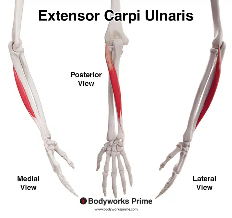
Here we can see the extensor carpi ulnaris muscle from a medial, posterior, and lateral view.
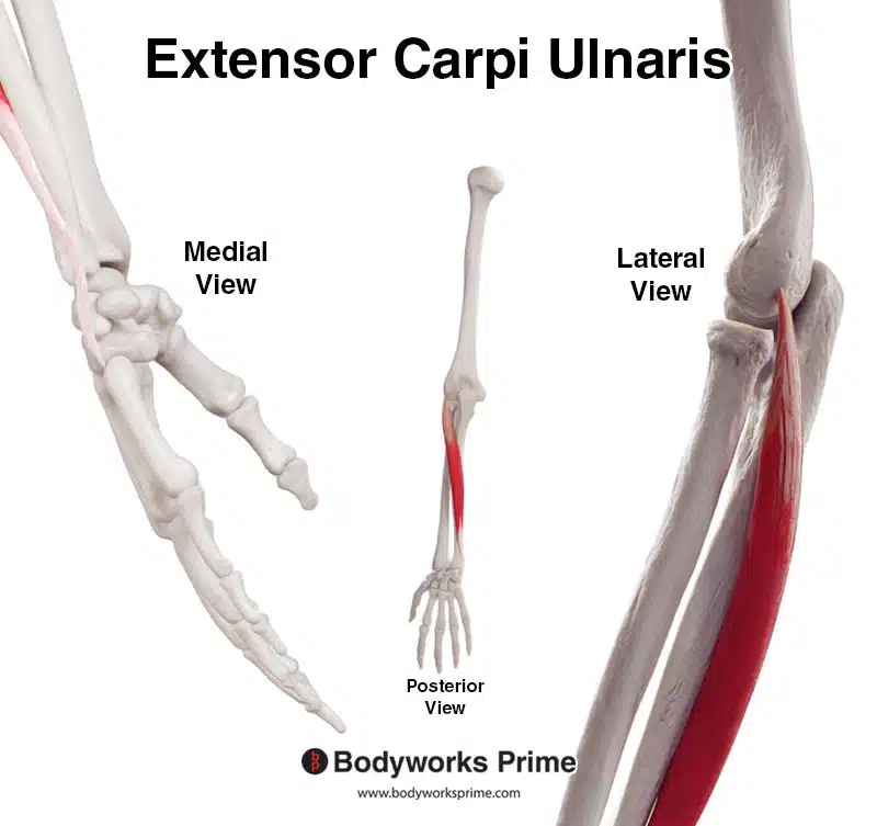
Here we can see a close up of the tendons of the extensor carpi ulnaris muscle.
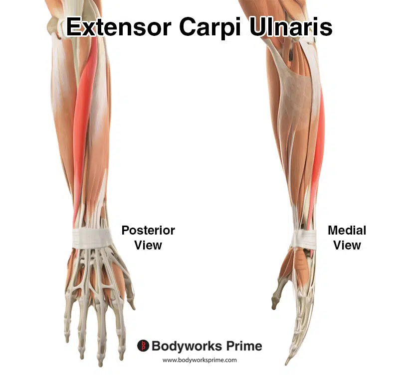
Here we can see the extensor carpi ulnaris muscle highlighted in red amongst the other muscles of the arm.
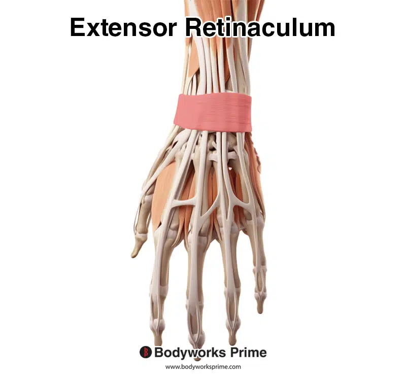
Highlighted in red we can see the extensor retinaculum, which is a fibrous band that holds the extensor tendons on the back of the wrist.
Origin & Insertion
The extensor carpi ulnaris has of two heads at its proximal end – the humeral and the ulnar head. The humeral head is attached to the lateral epicondyle of the humerus via the common extensor tendon, while the ulnar head originates from a common aponeurosis linked to the posterior border of the ulna. This shared common extensor tendon provides an anchor for the deep fascia of the forearm and also serves as a base for other muscles, including the extensor carpi radialis brevis, extensor digitorum, and the extensor digiti minimi [8][9][10].
From its origin points, the extensor carpi ulnaris travels down the arm towards the wrist. It passes under the extensor retinaculum, extending to its insertion point on the base of the fifth metacarpal (little finger) [11][12][13].
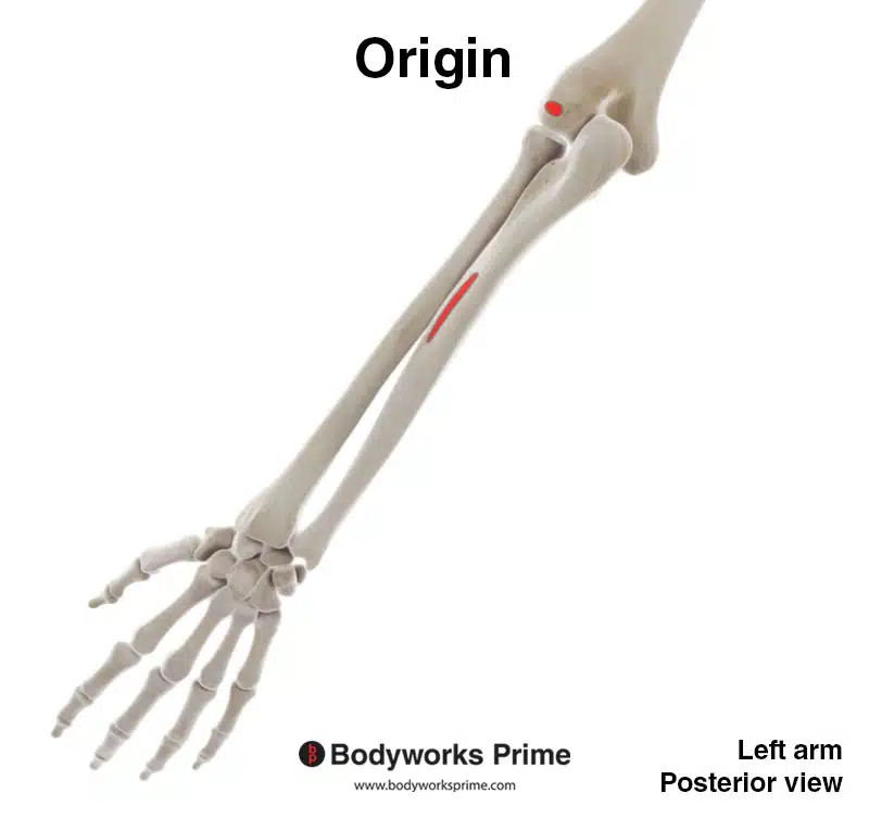
Here we can see the origins of the extensor carpi ulnaris highlighted in red.
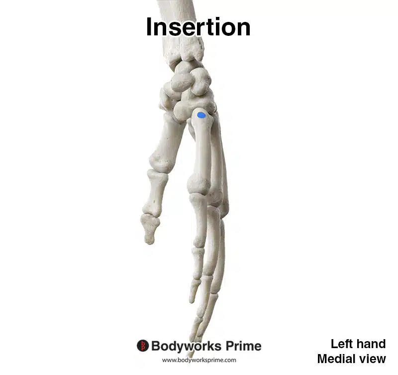
Here we can see the insertion of the extensor carpi ulnaris highlighted in blue.
Actions
When the extensor carpi ulnaris muscle contracts, it helps extend the wrist joint. This movement elevates the hand, allowing it to move away from the palm side of the forearm. The extensor carpi ulnaris muscle works in coordination with other extensor muscles of the wrist during this movement, such as the extensor carpi radialis longus and extensor carpi radialis brevis [14][15][16].
In addition to extension, the extensor carpi ulnaris contributes to the adduction of the wrist, a movement that draws the hand towards the midline of the body (essentially the wrist flexes sideways, towards the side the little finger is located on). In this action, it works collaboratively with the flexor carpi ulnaris muscle, which is a key flexor and adductor muscle of the wrist, located in the anterior compartment of the forearm [17][18][19].
Furthermore, when the extensor carpi ulnaris contracts simultaneously with the extensor carpi radialis longus and extensor carpi radialis brevis, it assists in providing medial stability to the wrist [20][21][22].
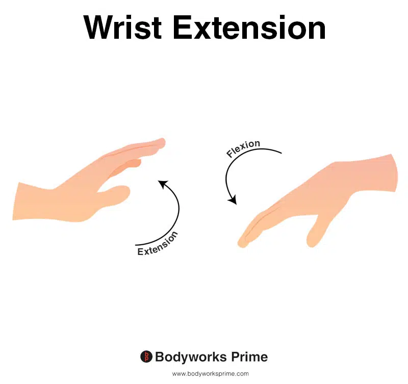
This image shows wrist extension, a motion that involves bending the wrist towards the posterior surface, or the top side of the forearm. Conversely, wrist flexion refers to the movement of bending the wrist towards the anterior surface, or the underside of the forearm. The extensor carpi ulnaris contributes to wrist extension.
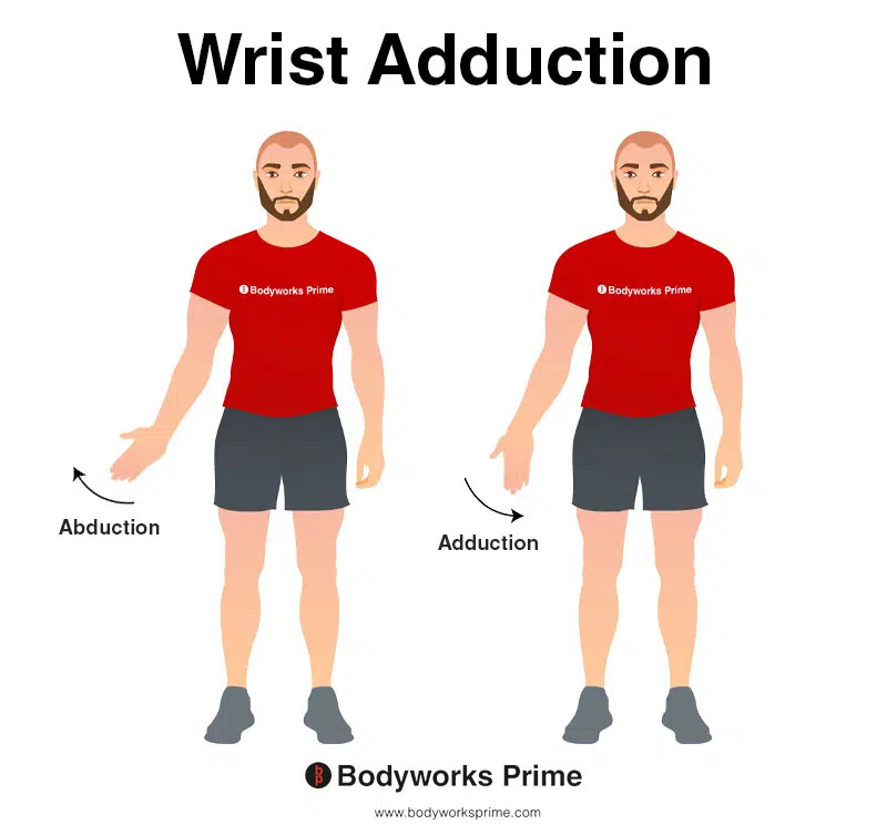
This image shows wrist adduction, which is the movement of bending the wrist to the side. During wrist adduction, the wrist bends away from the thumb, which would be towards the body when the wrist is in the anatomical position. The opposite movement of wrist adduction is wrist abduction. The extensor carpi ulnaris contributes to wrist adduction.
Innervation
The extensor carpi ulnaris muscle primarily receives its innervation from the posterior interosseous nerve, a branch of the radial nerve. The posterior interosseous nerve receives its innervation from spinal nerve roots C7 and C8 [23][24][25].
The radial nerve, the origin of the posterior interosseous nerve, originates from the brachial plexus, specifically the posterior cord. This complex network of nerves lies in the shoulder region, and the brachial plexus receives contributions from spinal nerve roots C5 to T1 [26][27][28].
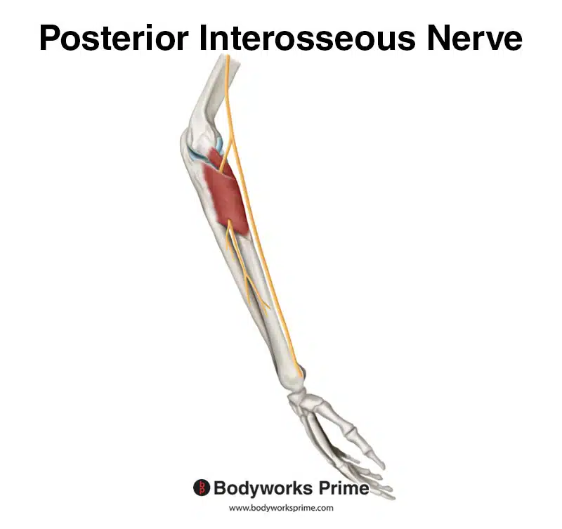
Pictured here, we can see the posterior interosseous nerve, which innervates the extensor carpi ulnaris from the nerve roots of C7 and C8. The muscle visible in the image is the supinator muscle. The deep branch of the radial nerve penetrates the supinator muscle. When it emerges from the supinator muscle, this part of the nerve is referred to as the posterior interosseous nerve.
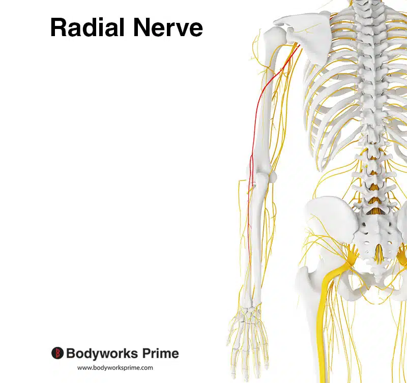
Here we can see the radial nerve highlighted in red. The posterior interosseous nerve is a branch of the radial nerve.
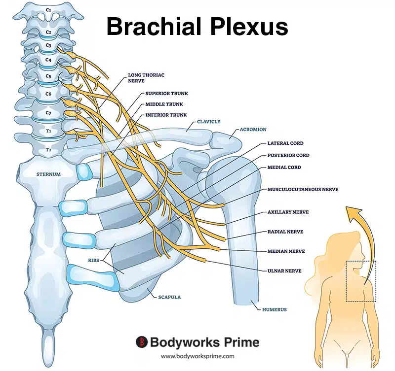
Here we can see the brachial plexus. the radial nerve is a part of the brachial plexus.
Blood Supply
Blood is mainly supplied to the extensor carpi ulnaris muscle via the ulnar artery. The ulnar artery is a significant branch of the brachial artery, which deviates near the antecubital fossa, the region in front of the elbow, and proceeds to supply the medial aspect of the extensor carpi ulnaris muscle. The ulnar artery and the ulnar nerve often follow a similar path [29][30][31].
In addition to the ulnar artery, the extensor carpi ulnaris muscle receives a supplemental blood supply from the posterior interosseous artery. This artery is a subsidiary branch of the radial artery and serves the muscles in the posterior compartment of the forearm, including the extensor carpi ulnaris, though its supply is lesser in comparison to the ulnar artery [32][33][34].
Want some flashcards to help you remember this information? Then click the link below:
Extensor Carpi Ulnaris Flashcards
Support Bodyworks Prime
Running a website and YouTube channel can be expensive. Your donation helps support the creation of more content for my website and YouTube channel. All donation proceeds go towards covering expenses only. Every contribution, big or small, makes a difference!
References
| ↑1, ↑3, ↑6, ↑9, ↑12, ↑14, ↑17, ↑20 | Moore KL, Agur AMR, Dalley AF. Clinically Oriented Anatomy. 8th ed. Philadelphia: Lippincot Williams & Wilkins; 2017. |
|---|---|
| ↑2, ↑4, ↑7, ↑8, ↑11, ↑23, ↑26, ↑29, ↑32 | Standring S. (2015). Gray’s Anatomy: The Anatomical Basis of Clinical Practice, 41st Edn. Amsterdam: Elsevier. |
| ↑5, ↑24, ↑27, ↑30, ↑33 | Mitchell B, Whited L. Anatomy, Shoulder and Upper Limb, Forearm Muscles. [Updated 2022 Jun 14]. In: StatPearls [Internet]. Treasure Island (FL): StatPearls Publishing; 2023 Jan-. Available from: https://www.ncbi.nlm.nih.gov/books/NBK536975/ |
| ↑10, ↑13, ↑16, ↑19, ↑22, ↑25, ↑28, ↑31, ↑34 | Sawyer E, Sajjad H, Tadi P. Anatomy, Shoulder and Upper Limb, Forearm Extensor Carpi Ulnaris Muscle. [Updated 2022 Sep 1]. In: StatPearls [Internet]. Treasure Island (FL): StatPearls Publishing; 2023 Jan-. Available from: https://www.ncbi.nlm.nih.gov/books/NBK539760/ |
| ↑15, ↑18, ↑21 | Campbell D, Campbell R, O’Connor P, Hawkes R. Sports-related extensor carpi ulnaris pathology: a review of functional anatomy, sports injury and management. Br J Sports Med. 2013 Nov;47(17):1105-11. doi: 10.1136/bjsports-2013-092835. Epub 2013 Oct 4. PMID: 24096897; PMCID: PMC3812850. |


