| Origin | Sternal Head: Manubrium of sternum (superior and anterior aspect) Clavicular Head: Medial quarter of clavicle’s superior surface |
| Insertion | Mastoid process of temporal bone & anterior portion of superior nuchal line |
| Action | Unilateral contraction: Ipsilateral flexion and contralateral rotation Bilateral contraction: Head/neck extension, head/neck flexion, & assists in inspiration by elevating clavicle and manubrium of sternum |
| Nerve | Accessory nerve (CN XI) Branches of cervical plexus (C2-C3) |
| Artery | Occipital artery Superior thyroid artery Suprascapular arteries |
Location & Overview
The sternocleidomastoid muscle (often abbreviated to SCM) is a neck muscle which is large, palpable, and has an easily recognisable shape. It is one of the largest and most superficial cervical muscles and plays an important role in movements of the neck, such as flexion, rotation, and additionally it can assist with inspiration [1] [2].
The sternocleidomastoid muscle has two heads, the clavicular head and the sternal head, which are distinguished by their origins. The sternal head arises from the sternum (breastbone), while the clavicular head arises from the clavicle (collarbone). The clavicular head is the largest of the two heads and the sternal head is the smallest of the two heads. The clavicular and sternal heads are often fused along the muscle’s length. Very occasionally, some sources break these sternocleidomastoid divisions down further into four portions: the sterno-mastoid, sterno-occipital, cleido-mastoid, and cleido-occipital [3] [4].
The sternocleidomastoid muscle is usually stronger and thicker in men than in women [5]. Approximately 65% of the sternocleidomastoid’s muscle fibers are comprised of fast twitch muscle fibers. The remaining fibers are slow twitch. This means the sternocleidomastoid is best at expressing a large amount of strength quite quickly and less adapt at endurance-based activities [6] [7].
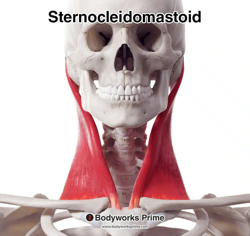
Here we can see the sternocleidomastoid muscle from an anterior view.
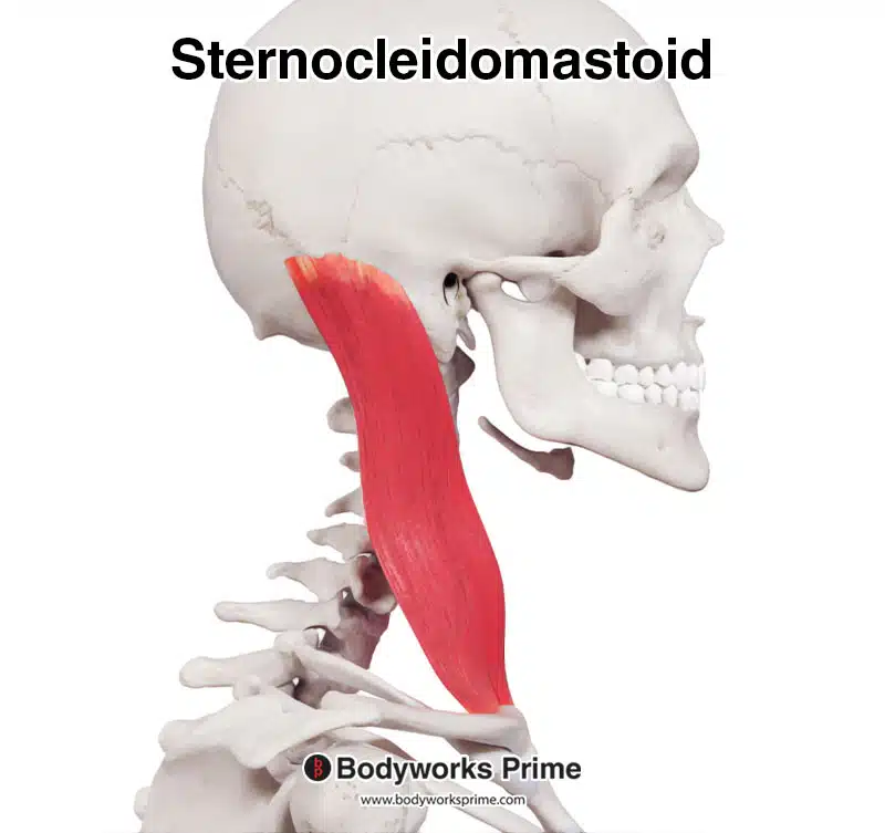
Here we can see the sternocleidomastoid muscle from a lateral view.
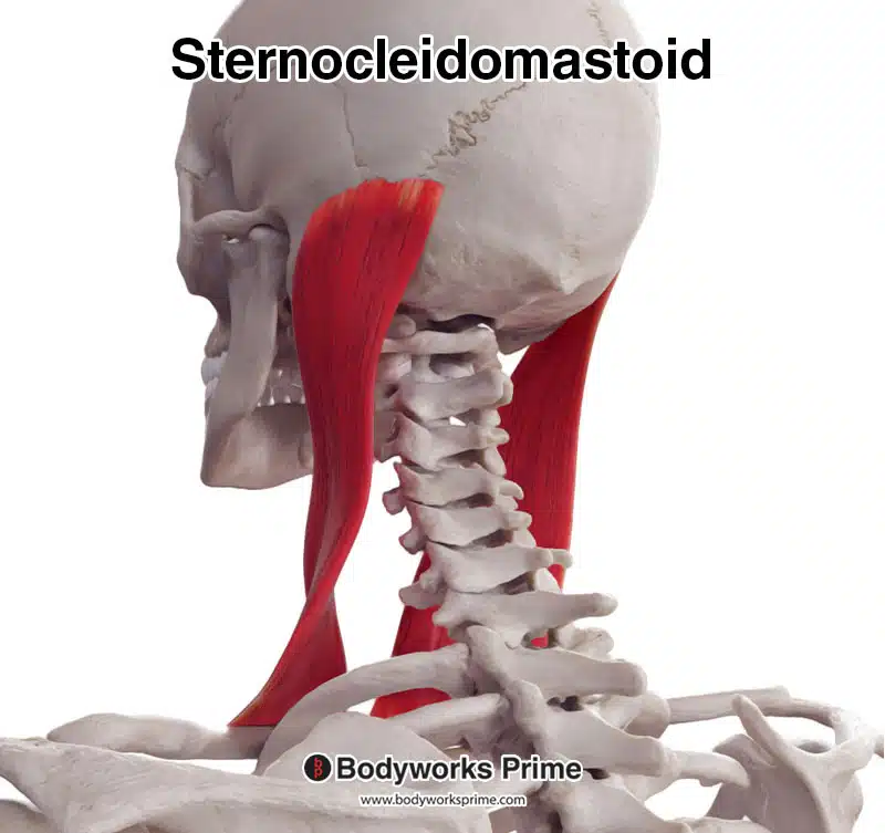
Here we can see the sternocleidomastoid muscle from a posterolateral view.
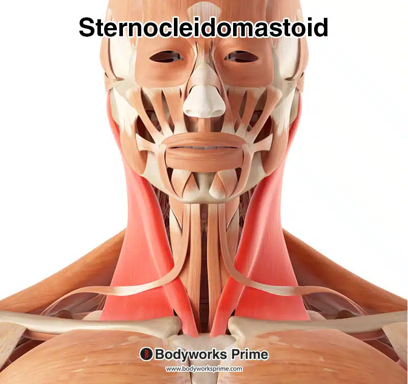
Here we can see the sternocleidomastoid muscle highlighted in red. Superficial muscles such as the platysma muscle have been removed to reveal the sternocleidomastoid.
Origin & Insertion
The sternocleidomastoid muscle originates from the superior edge of the manubrium and from the medial quarter of the superior surface of the clavicle. The two heads of this muscle (sternal and clavicular) merge to form a single muscle belly which continues up the neck in a superior and lateral direction. The sternocleidomastoid then inserts onto the mastoid process of the temporal bone and on the anterior portion of the superior nuchal line (located on the occipital bone of the skull) [8] [9] [10]. These origins and insertions are displayed in the images below.
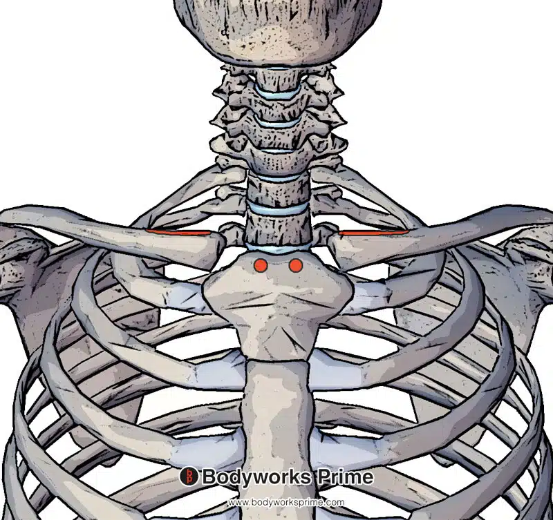
Here we can see the sternocleidomastoid muscle’s origin points on the superior and anterior aspect of the manubrium of the sternum (sternal head) and the medial quarter of clavicle’s superior surface (clavicular head).
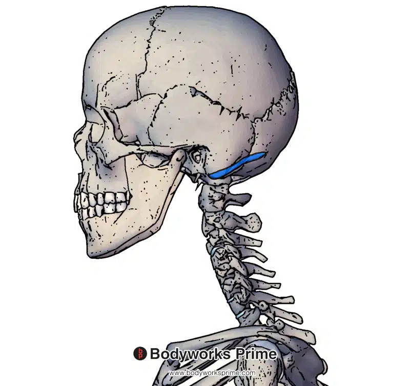
Here we can see the insertion of the sternocleidomastoid muscle on the mastoid process of the temporal bone and the anterior portion of the superior nuchal line. The mastoid process can be easily palpated, it is a bony bump behind the ear.
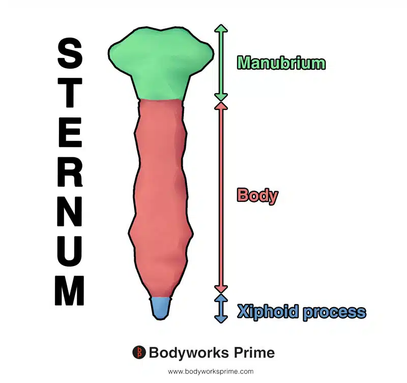
Here we can see the different sections of the sternum. The manubrium of the sternum is an origin point of the sternocleidomastoid’s sternal head.
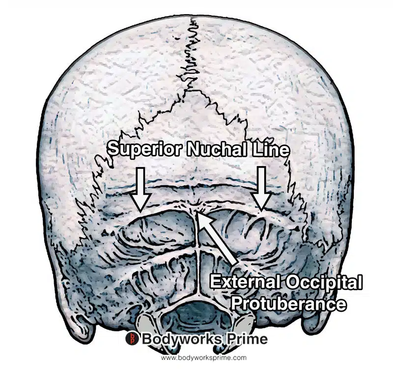
Here you can see the superior nuchal line which is part of the insertion point of the sternocleidomastoid muscle. The sternocleidomastoid muscle’s insertion is on the lateral and superior aspects of the superior nuchal line. It does not span the entirety of the superior nuchal line.
Actions
The sternocleidomastoid muscle can assist in head rotation when one side of the muscle contracts (unilateral contraction). This rotation causes the face side of the head to turn away from the contracting muscle (contralateral rotation). Similarly, a unilateral contraction can also cause the neck to bend towards the contracting muscle (ipsilateral flexion) [11] [12].
When both sides of the muscle contract (bilateral contraction), the cervical spine can either flex forward or extend backward depending on whether the spine is held in a fixed position or not. With a fixed spine, the muscle causes flexion of the cervical spine, leading to forward head lean. Without a fixed spine, it causes extension of the head and bending of the cervical spine [13] [14].
The sternocleidomastoid muscle can also assist with inspiration. Bilateral contraction of the muscle helps lift the sternum and clavicles, allowing for greater expansion of the chest during inhalation [15] [16].
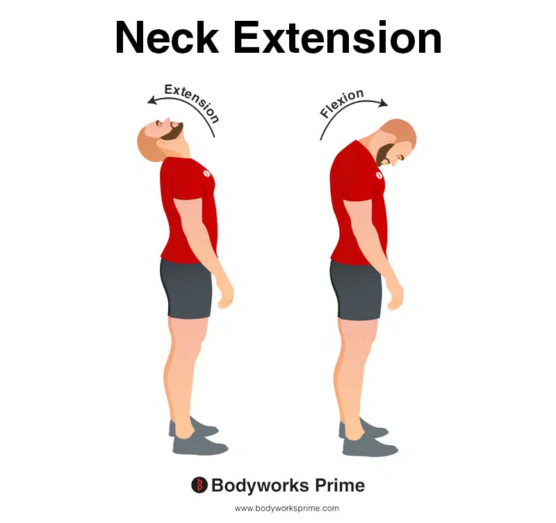
This image demonstrates neck extension, which involves bending the neck backwards. The opposite of neck extension is neck flexion.
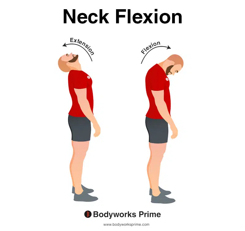
This image demonstrates neck flexion, which involves bending the neck forwards. The opposite of neck flexion is neck extension.
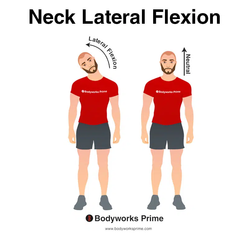
This image shows neck lateral flexion, which involves bending the neck to one side. This action is caused by the contraction of the sternocleidomastoid muscle. Specifically, it’s called ipsilateral flexion because the muscle on the same side as the neck bend contracts to bring the neck towards that side. The opposite of ipsilateral flexion is contralateral flexion, which occurs when the neck bends away from the side of the contracting muscle.
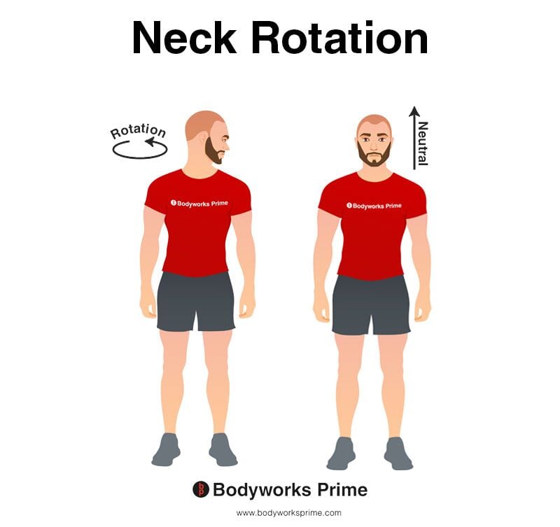
This image demonstrates neck rotation, which involves turning the neck/head to the side. This type of rotation can also be referred to as contralateral rotation, the sternocleidomastoid’s contraction head to turn away from the side of the muscle contracting. When the action happens at the opposite side to the muscle’s contraction, this is referred to as contralateral. The opposite of contralateral is ipsilateral.
Innervation
The posterior edge of the sternocleidomastoid muscle is where the cutaneous branches of the cervical plexus emerge. The nerve endings assist the muscle in its proprioceptive functions. Moreover, the cranial nerve or XI passes into the posterior triangle to innervate the trapezius and the sternocleidomastoid muscle[17].
The sternocleidomastoid is also innervated by branches of the cervical plexus. The cervical plexus lies deep to the sternocleidomastoid muscle on the anterolateral aspect of the neck[18].
Blood Supply
Blood is supplied to the sternocleidomastoid via the occipital artery and superior thyroid artery which are branches of the external carotid artery. Inferior to the sternocleidomastoid, is the jugular vein, which is where it drains venous blood [19] [20].
The middle third of the sternocleidomastoid muscle receives around 42% of its blood from the superior thyroid artery, 23% from the external carotid artery, and 27% from branches of both. In the majority of cases, the lower third of the sternocleidomastoid receives blood from a branch arising from the suprascapular artery (>80%)[21].
Want some flashcards to help you remember this information? Then click the link below:
Sternocleidomastoid Flashcards
Support Bodyworks Prime
Running a website and YouTube channel can be expensive. Your donation helps support the creation of more content for my website and YouTube channel. All donation proceeds go towards covering expenses only. Every contribution, big or small, makes a difference!
References
| ↑1, ↑3, ↑7, ↑8, ↑11, ↑13, ↑15, ↑17, ↑20 | Bordoni B, Varacallo M. Anatomy, Head and Neck, Sternocleidomastoid Muscle. [Updated 2022 Apr 5]. In: StatPearls [Internet]. Treasure Island (FL): StatPearls Publishing; 2022 Jan-. Available from: https://www.ncbi.nlm.nih.gov/books/NBK532881/ |
|---|---|
| ↑2, ↑4, ↑5, ↑9 | Kennedy E, Albert M, Nicholson H. The fascicular anatomy and peak force capabilities of the sternocleidomastoid muscle. Surg Radiol Anat. 2017 Jun;39(6):629-645. doi: 10.1007/s00276-016-1768-9. Epub 2016 Nov 2. PMID: 27807639. |
| ↑6 | Meznaric M, Eržen I, Karen P, Cvetko E. Effect of ageing on the myosin heavy chain composition of the human sternocleidomastoid muscle. Ann Anat. 2018 Mar;216:95-99. doi: 10.1016/j.aanat.2017.12.001. Epub 2017 Dec 28. PMID: 29289708. |
| ↑10, ↑12, ↑14, ↑18 | Moore KL, Agur AMR, Dalley AF. Clinically Oriented Anatomy. 8th ed. Philadelphia: Lippincot Williams & Wilkins; 2017. |
| ↑16, ↑19 | Kohan EJ, Wirth GA. Anatomy of the neck. Clin Plast Surg. 2014 Jan;41(1):1-6. doi: 10.1016/j.cps.2013.09.016. PMID: 24295343. |
| ↑21 | Kierner AC, Aigner M, Zelenka I, Riedl G, Burian M. The blood supply of the sternocleidomastoid muscle and its clinical implications. Arch Surg. 1999 Feb;134(2):144-7. doi: 10.1001/archsurg.134.2.144. PMID: 10025452. |


