| Origin | External surfaces of the fifth to twelfth ribs |
| Insertion | Iliac crest Pubic crest Pubic tubercle Linea alba |
| Action | Lateral flexion of the trunk Rotation of the trunk Flexion of the trunk Assists in forced expiration |
| Nerve | Intercostal nerves (T8-T11) Subcostal nerve (T12) |
| Artery | Lower posterior intercostal artery Subcostal artery Deep circumflex iliac artery The superior and inferior epigastric arteries |
Location & Overview
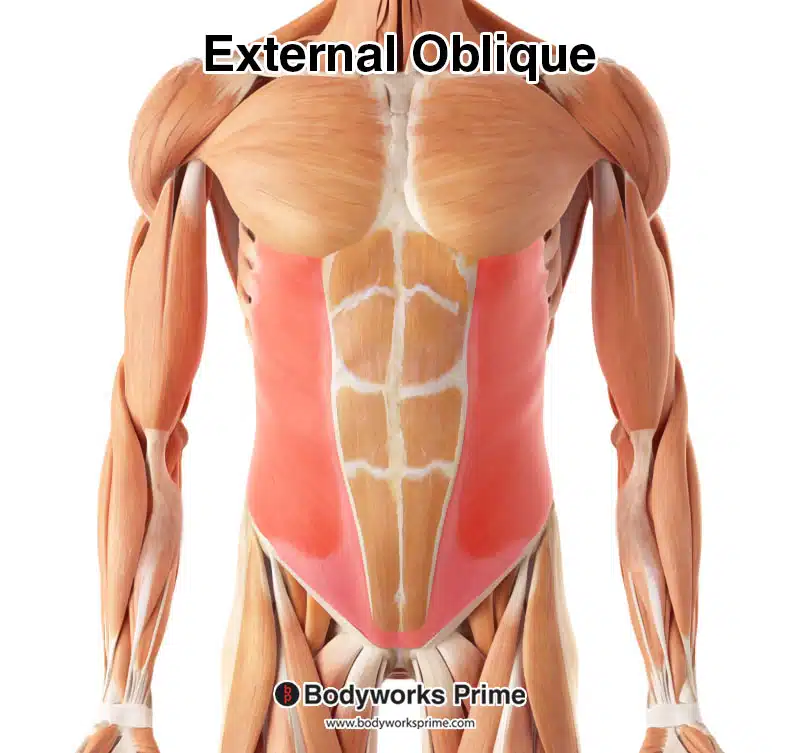
The external oblique muscle, located superficially to the internal oblique muscle (closer to the skin’s surface), is one of the five abdominal muscles. The other four include the internal oblique, rectus abdominis, transversus abdominis, and the pyramidalis. The external oblique is the most superficial and largest of the anterolateral abdominal wall muscles. Along with the internal oblique and transversus abdominis, it is one of the three flat muscles that compose the abdominal wall. These flat muscles also contribute to the formation of the rectus sheath [1] [2].
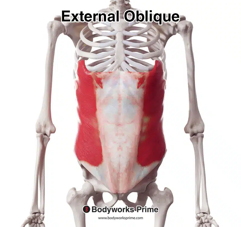
Here we can see the external oblique from an anterior view.
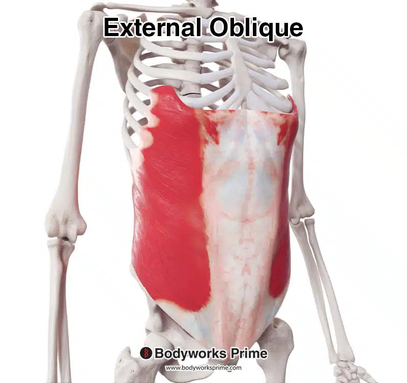
Here we can see the external oblique from an anterolateral view.
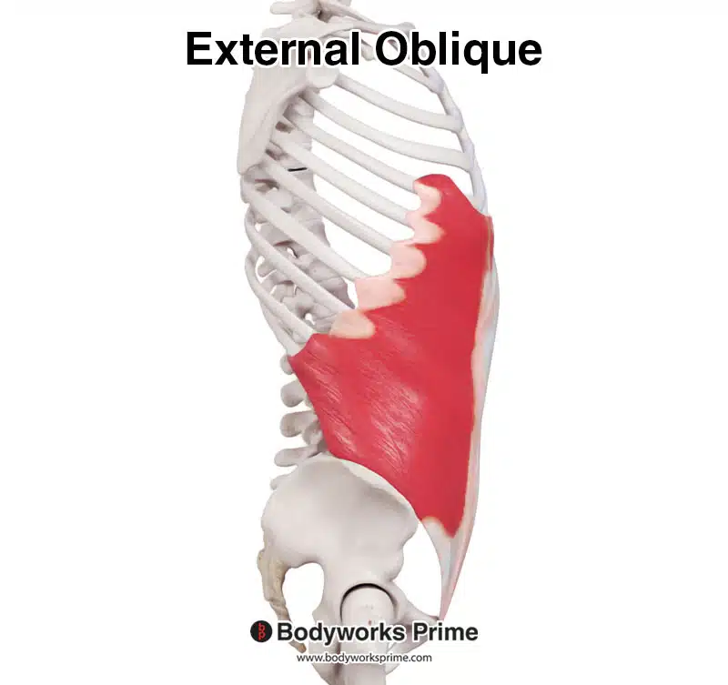
Here we can see the external oblique from a lateral view.
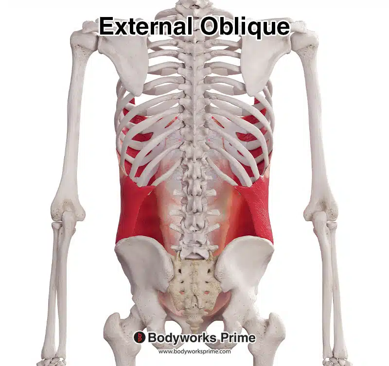
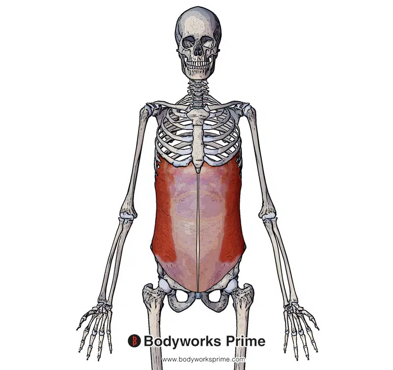
Here we can see the external oblique from a posterior view.
Here we can see the external oblique highlighted in red amongst the other muscles of the body. The muscle we can see in the middle is the rectus abdominis muscle.
Pictured here we can see the external oblique and its aponeurosis which is connecting into the linea alba.
Origin & Insertion
The external oblique originates from the fifth through twelfth ribs. Its fibres travel in an inferior, medial and anterior direction from its origin. When these fibres reach the midline, they form an aponeurosis which merges with the linea alba, which contributes to the anterior rectus sheath. This aponeurosis also inserts onto the iliac crest, pubic crest and pubic tubercle [3] [4] [5] [6].
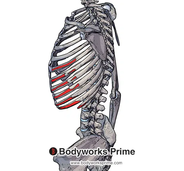
Pictured here we can see the origin of the external oblique highlighted in red on the external surfaces of the fifth to twelfth ribs.
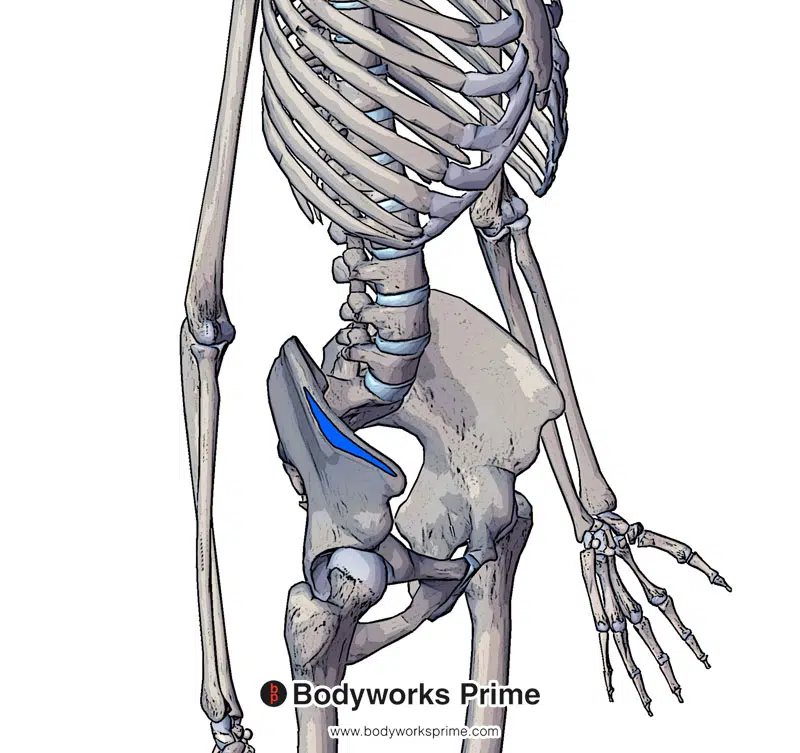
Highlighted in blue we can see one of the insertions of the external oblique on the iliac crest.
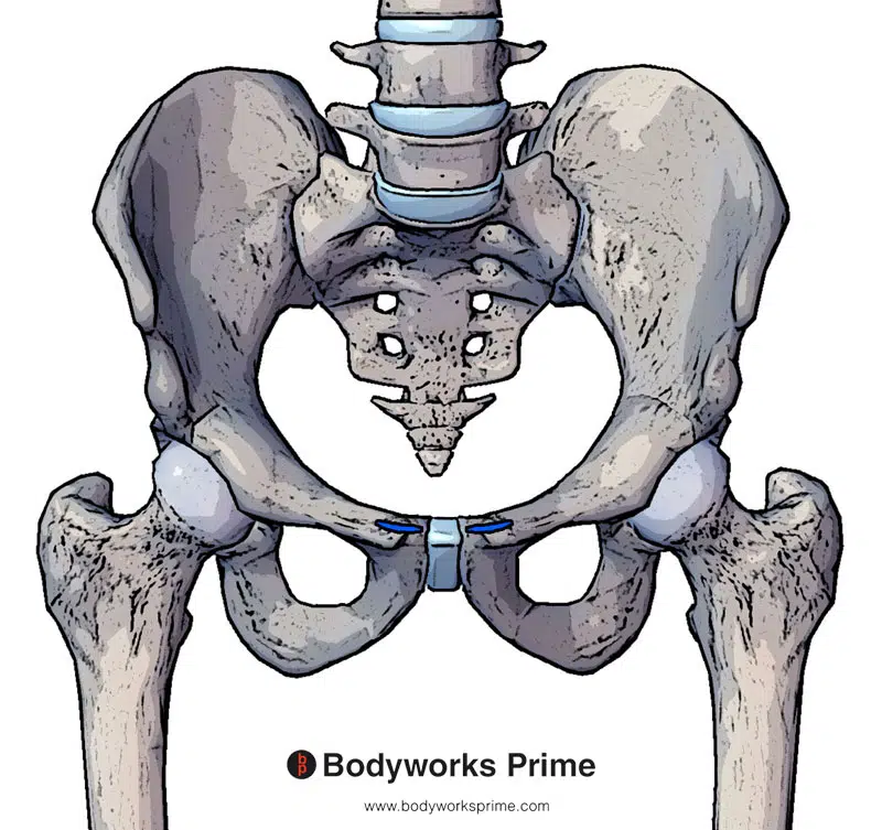
Highlighted in blue we can see one of the insertions of the external oblique on the pubic crest and pubic tubercle.
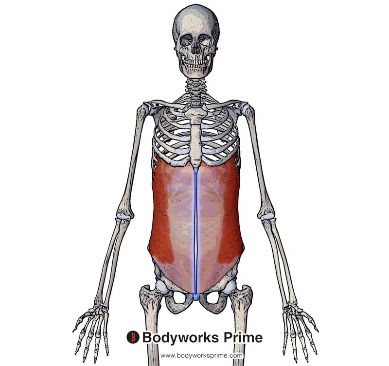
Highlighted in blue we can see the final insertion of the external oblique on the linea alba.
Actions
The external and internal oblique muscles work together to enable trunk rotation and lateral flexion. When only one side of the external oblique muscle contracts (unilateral contraction), it results in lateral flexion of the trunk. Unilateral contraction also causes trunk rotation in the opposite direction (e.g., if the right external oblique contracts, the trunk rotates to the left). When both sides of the external oblique contract simultaneously (bilateral contraction), it leads to trunk flexion. In addition to flexion and rotation, the external oblique muscle also provides support to nearby abdominal structures and assists in forced expiration by depressing the ribs, pulling on the fifth to twelfth rib origins [7] [8].
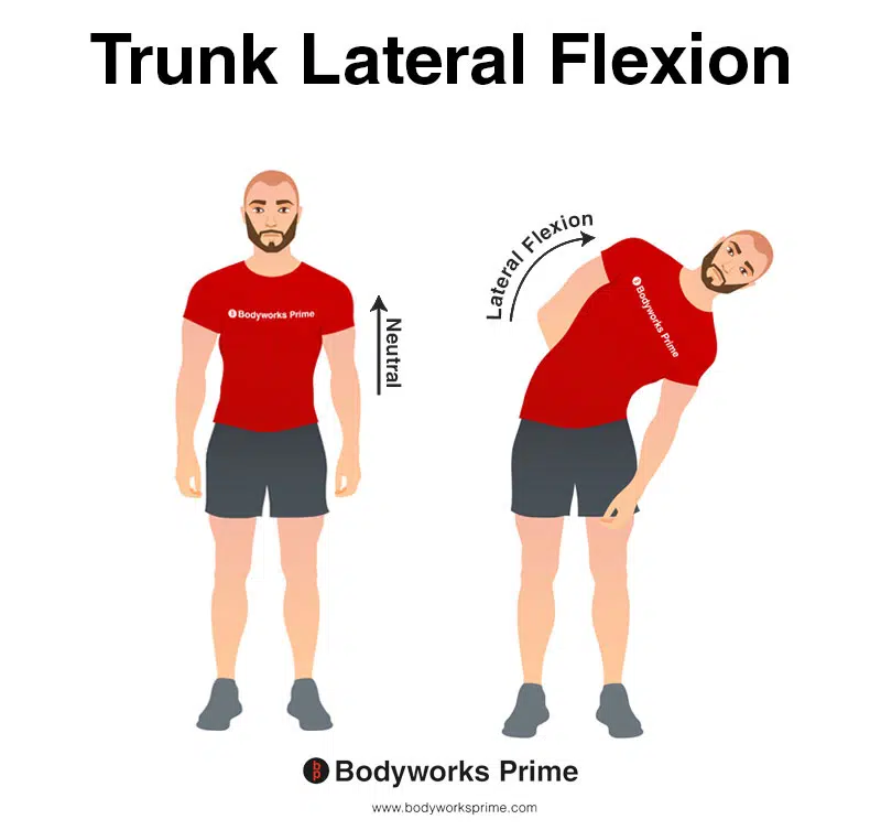
This image demonstrates trunk lateral flexion, which involves bending the torso to the side. Unilateral contraction of the external oblique can cause lateral flexion of the trunk.
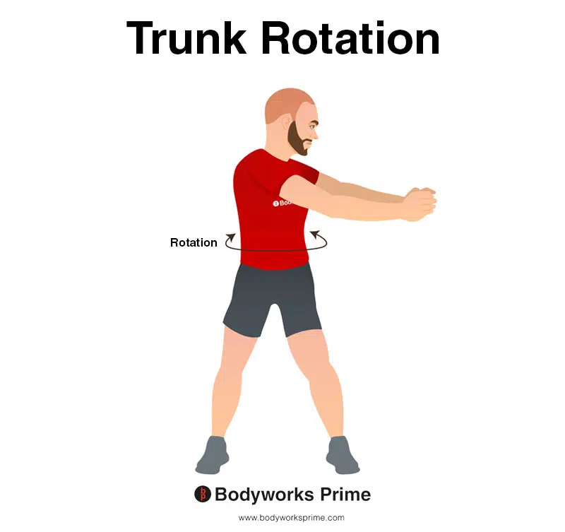
This image demonstrates trunk rotation, which involves twisting the waist/torso to the side. Unilateral contraction of the external oblique can cause rotation of the trunk.
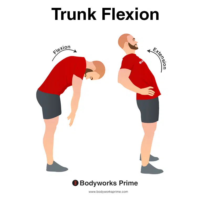
This image shows an example of trunk flexion, which involves bending the torso forwards. The opposite of trunk flexion is trunk extension. Bilateral contraction of the external oblique can cause flexion of the trunk.
The external oblique muscle also provides support to nearby abdominal structures and assists in forced expiration
Innervation
The external abdominal oblique is innervated by the intercostal nerves (T8-T11) and the subcostal nerve (T12) [9] [10].
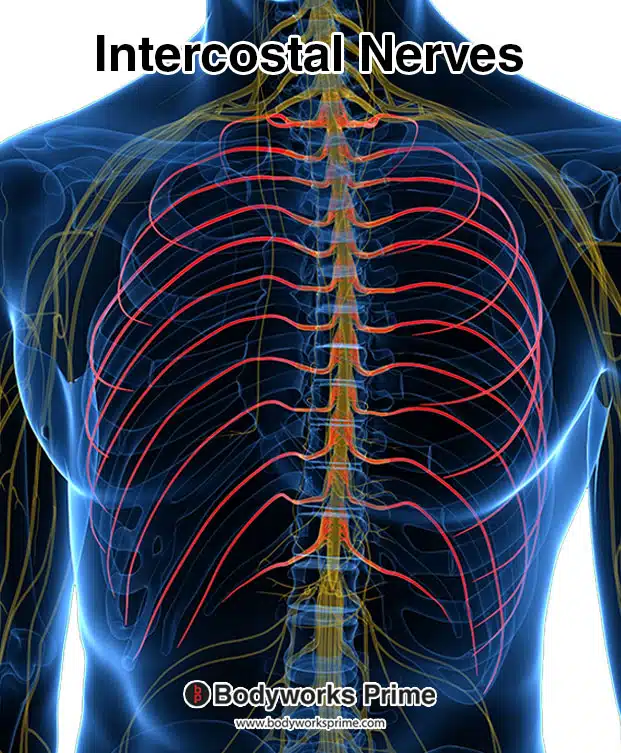
Here we can see the intercostal nerves highlighted in red. These nerves are an innervation of the external oblique muscle.
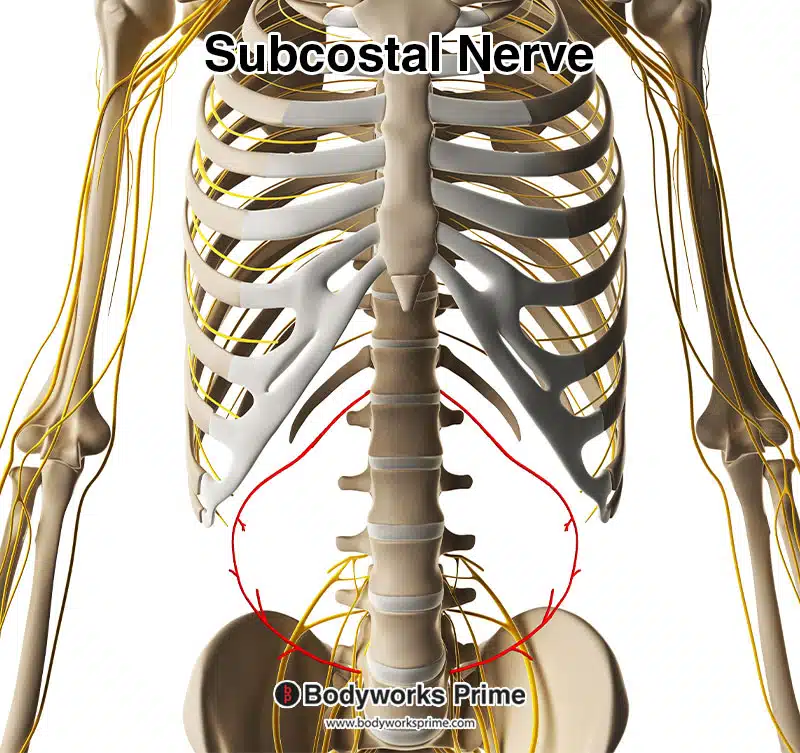
Here we can see the subcostal nerve highlighted in red. The subcostal nerve innervates the external oblique muscle.
Blood Supply
The external abdominal oblique gets its blood supply from: the lower posterior intercostal artery, subcostal artery, deep circumflex iliac artery, and the superior and inferior epigastric arteries [11] [12].
Want some flashcards to help you remember this information? Then click the link below:
External Oblique Flashcards
Support Bodyworks Prime
Running a website and YouTube channel can be expensive. Your donation helps support the creation of more content for my website and YouTube channel. All donation proceeds go towards covering expenses only. Every contribution, big or small, makes a difference!
References
| ↑1 | Flynn W, Vickerton P. Anatomy, Abdomen and Pelvis, Abdominal Wall. [Updated 2021 Jul 31]. In: StatPearls [Internet]. Treasure Island (FL): StatPearls Publishing; 2022 Jan-. Available from: https://www.ncbi.nlm.nih.gov/books/NBK551649/ |
|---|---|
| ↑2, ↑5, ↑9, ↑11 | Varacallo M, Scharbach S, Al-Dhahir MA. Anatomy, Anterolateral Abdominal Wall Muscles. [Updated 2021 Jul 31]. In: StatPearls [Internet]. Treasure Island (FL): StatPearls Publishing; 2022 Jan-. Available from: https://www.ncbi.nlm.nih.gov/books/NBK470334/ |
| ↑3, ↑7 | Flynn W, Vickerton P. Anatomy, Abdomen and Pelvis, Abdominal Wall. [Updated 2021 Jul 31]. In: StatPearls [Internet]. Treasure Island (FL): StatPearls Publishing; 2022 Jan-. Available from: https://www.ncbi.nlm.nih.gov/books/NBK551649/ |
| ↑4, ↑8 | Moore KL, Agur AMR, Dalley AF. Clinically Oriented Anatomy. 8th ed. Philadelphia: Lippincot Williams & Wilkins; 2017 |
| ↑6, ↑10, ↑12 | Seeras K, Qasawa RN, Ju R, et al. Anatomy, Abdomen and Pelvis, Anterolateral Abdominal Wall. [Updated 2021 Jul 26]. In: StatPearls [Internet]. Treasure Island (FL): StatPearls Publishing; 2022 Jan-. Available from: https://www.ncbi.nlm.nih.gov/books/NBK525975/ |


