| Origin | Pubic crest Pubic symphysis |
| Insertion | Xiphoid process Costal cartilages 5-7 |
| Action | Flexes the trunk Tenses the anterior abdominal wall |
| Nerve | Anterior rami of T6-T12 spinal nerves |
| Artery | Superior and inferior epigastric arteries |
Location & Overview
The rectus abdominis is one if the five abdominal muscles. The other four are: the external oblique, internal oblique, transversus abdominis and the pyramidalis [1]. The rectus abdominis muscle is long and somewhat narrow set of paired vertical muscles located on either side of the midline of the abdomen. It is located in the rectus sheath, running both parallel and vertically to the linea alba. The linea alba is a long white fibrous line running down the midline of the rectus sheath (linea alba translated from Latin to English means white line) [2].
The rectus sheath is a compartment which is fibrous, resilient and durable. It extends upwards from the pubic crest to the inferior costal margin and the costal cartilages of ribs 5 to 7. Contained within the rectus sheath is the rectus abdominus and the pyramidalis muscles. The rectus sheath is comprised of the aponeurosis of the transversus abdominis, internal oblique and external oblique muscles [3].
The belly of the rectus abdominis muscle has divisions which are created by three tendinous intersections. This creates four muscle segments sometimes referred to as a ‘six pack’ (or an ‘eight pack’ for those rare individuals who are genetically blessed enough to have clear visible surface separations for all eight segments!). These tendinous intersections are connected to the anterior rectus sheath which covers them [4].
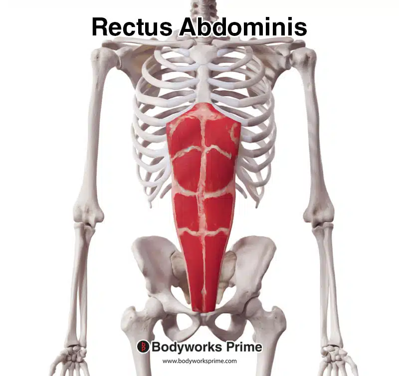
Pictured here we can see the rectus abdominis muscle from an anterior view.
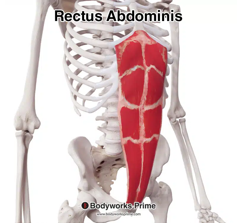
Here we can see the rectus abdominis muscle from an anterolateral view.
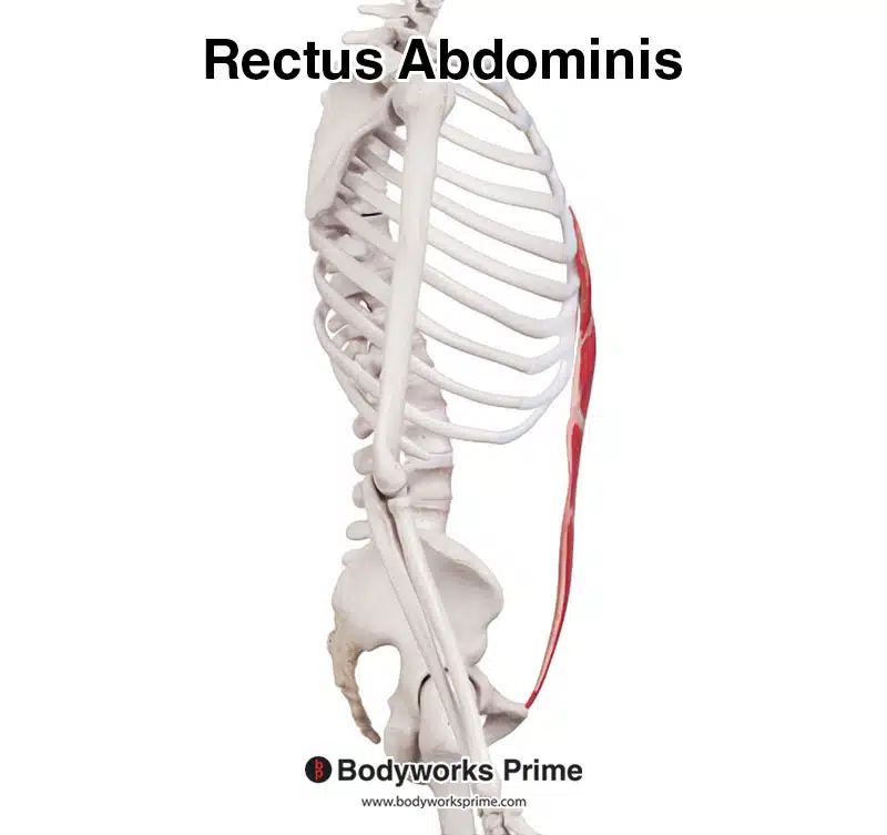
Here we can see the rectus abdominis muscle seen from a lateral view.
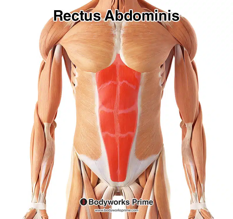
In this image we can see the rectus abdominis highlighted in red amongst the other muscles of the body.
Origin & Insertion
The rectus abdominis originates from the pubic symphysis and pubic crest located on the pelvis. It then inserts superiorly at the fifth to seventh costal cartilages of the ribs and the xiphoid process of the sternum [5].
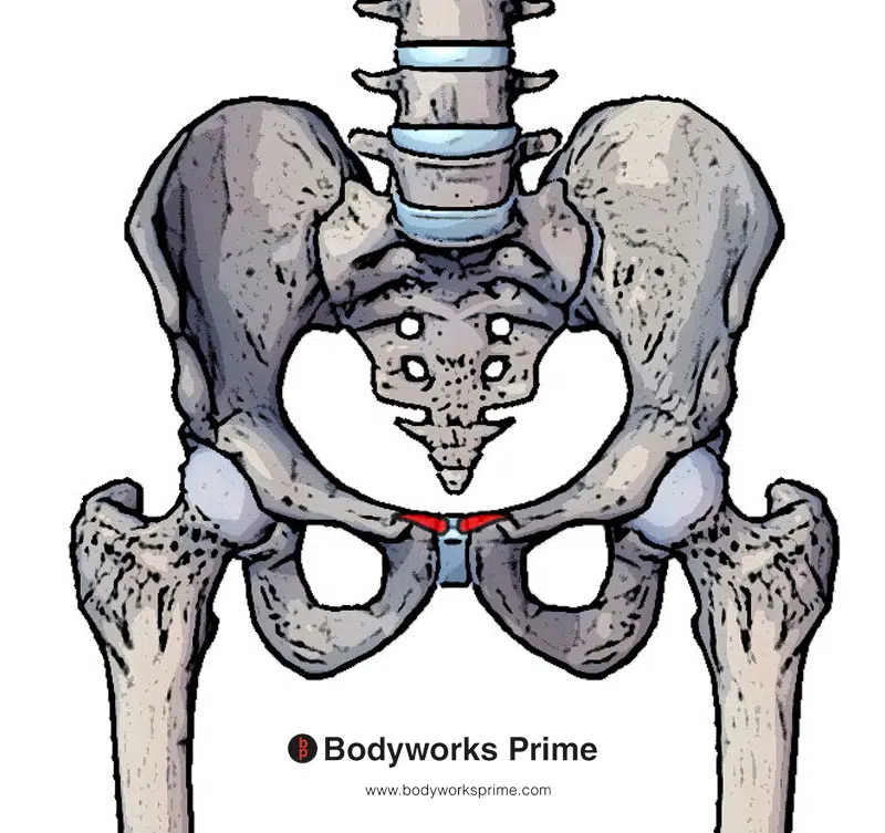
Here we can see the origin of the rectus abdominis highlighted in red. This origin is on the pubic crest and pubic symphysis.
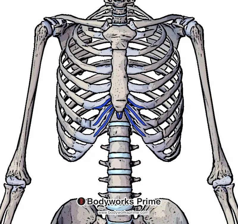
Here we can see the insertion of the rectus abdominis highlighted in blue. This insertion is on the xiphoid process of the sternum and the costal cartilages of ribs 5-7.
Actions
The primary action of the rectus abdominis muscle is to cause flexion of the trunk. It can also tense the abdominal wall which provides stability to the torso and also helps prevent herniation. Tension of the abdominal wall assists in increasing intrabdominal pressure which in turn creates a more rigid and stable torso under load. Intrabdominal pressure created via abdominal muscle contraction (or the Valsalva maneuverer) can also help expel faeces more rapidly [6].
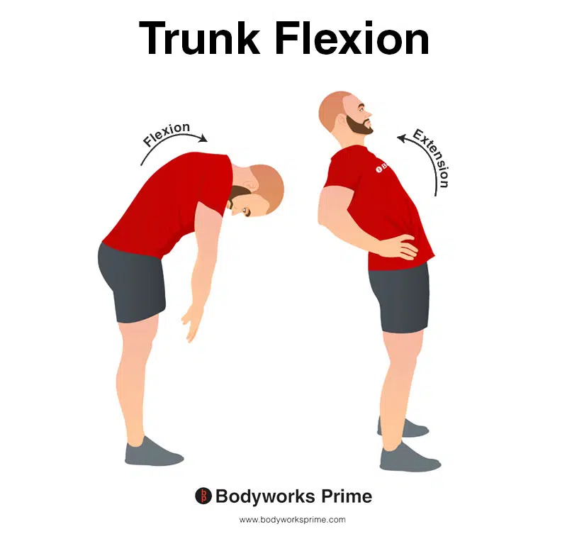
This image shows an example of trunk flexion, which involves bending the torso forwards. The opposite of trunk flexion is trunk extension. The rectus abdominis’ primary action is flexion of the trunk.
It can also tense the abdominal wall which provides stability to the torso and also helps prevent herniation.
Innervation
The rectus abdominis muscle is innervated by the thoraco-abdominal nerves. Specifically, the anterior rami of T6-T12 spinal nerves. The thoraco-abdominal nerves are branches of the intercostal nerves [7] [8].
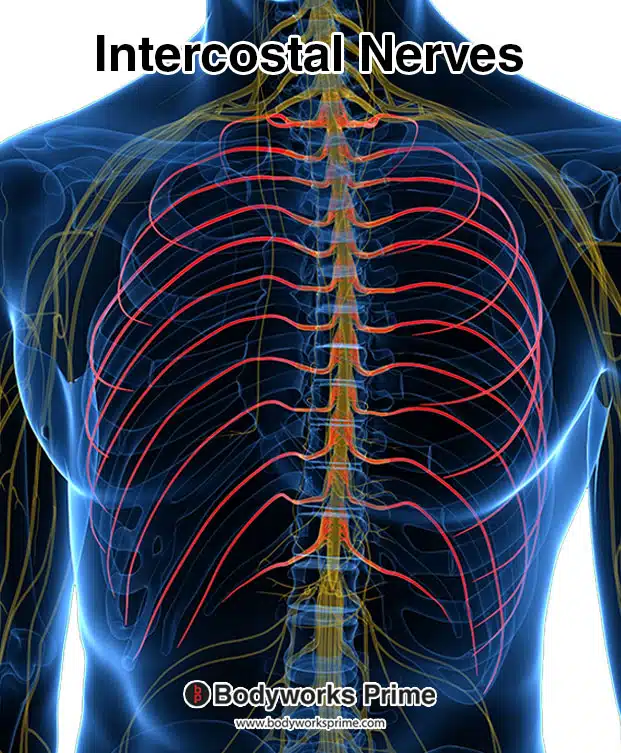
In this image, the intercostal nerves are highlighted in red. These nerves give rise to branches known as the thoraco-abdominal nerves, which directly innervate the rectus abdominis muscle.
Blood Supply
Blood is supplied to the rectus abdominus via the superior and inferior epigastric arteries. These two arteries also supply blood to the rectus sheath [9] [10].
Want some flashcards to help you remember this information? Then click the link below:
Rectus Abdominis Flashcards
Support Bodyworks Prime
Running a website and YouTube channel can be expensive. Your donation helps support the creation of more content for my website and YouTube channel. All donation proceeds go towards covering expenses only. Every contribution, big or small, makes a difference!
References
| ↑1, ↑6 | Varacallo M, Scharbach S, Al-Dhahir MA. Anatomy, Anterolateral Abdominal Wall Muscles. [Updated 2021 Jul 31]. In: StatPearls [Internet]. Treasure Island (FL): StatPearls Publishing; 2022 Jan-. Available from: https://www.ncbi.nlm.nih.gov/books/NBK470334/ |
|---|---|
| ↑2, ↑4, ↑5 | Flynn W, Vickerton P. Anatomy, Abdomen and Pelvis, Abdominal Wall. [Updated 2021 Jul 31]. In: StatPearls [Internet]. Treasure Island (FL): StatPearls Publishing; 2022 Jan-. Available from: https://www.ncbi.nlm.nih.gov/books/NBK551649/ |
| ↑3, ↑9 | Sevensma KE, Leavitt L, Pihl KD. Anatomy, Abdomen and Pelvis, Rectus Sheath. [Updated 2021 Jul 28]. In: StatPearls [Internet]. Treasure Island (FL): StatPearls Publishing; 2022 Jan-. Available from: https://www.ncbi.nlm.nih.gov/books/NBK537153/ |
| ↑7 | Seeras K, Qasawa RN, Ju R, et al. Anatomy, Abdomen and Pelvis, Anterolateral Abdominal Wall. [Updated 2021 Jul 26]. In: StatPearls [Internet]. Treasure Island (FL): StatPearls Publishing; 2022 Jan-. Available from: https://www.ncbi.nlm.nih.gov/books/NBK525975/ |
| ↑8 | Moore KL, Agur AMR, Dalley AF. Clinically Oriented Anatomy. 8th ed. Philadelphia: Lippincot Williams & Wilkins; 2017. |
| ↑10 | El-Mrakby HH, Milner RH. The vascular anatomy of the lower anterior abdominal wall: a microdissection study on the deep inferior epigastric vessels and the perforator branches. Plast Reconstr Surg. 2002 Feb;109(2):539-43; discussion 544-7. doi: 10.1097/00006534-200202000-00020. PMID: 11818833. |


