| Origin | Iliocostalis cervicis angle of ribs 3-6 Iliocostalis thoracis angle of ribs 7-12 Iliocostalis lumborum medial end of iliac crest, lateral crest of sacrum, & thoracolumbar fascia |
| Insertion | Iliocostalis cervicis posterior tubercle of the transverse process of C4-C6 Iliocostalis thoracis transverse process of C7 & angles of the ribs 1-6 Iliocostalis lumborum transverse processes of L1-L4 & angles of ribs 4-12 |
| Action | Unilateral contraction lateral flexion of the trunk/vertebral column & weak lateral flexion at the base of the neck (below C4) Bilateral contraction extends the trunk/vertebral column & weak extension of the base of the neck (below C4) |
| Nerve | Lateral branches of the posterior rami of the spinal nerves |
| Artery | Dorsal branches of the posterior intercostal, subcostal, lumbar & lateral sacral arteries |
Location & Overview
The iliocostalis muscle is a part of the deep musculature of the back and one of the three muscles that make up the erector spinae group, alongside the spinalis and longissimus. Out of these three, the iliocostalis is the most lateral. The iliocostalis is separated into three distinct regions based on their point of origin: iliocostalis cervicis, iliocostalis thoracis, and iliocostalis lumborum. The lumborum section is most inferior (lowest), and the capitis section is the most superior (highest). The cervicis section is in the middle. The different sections are displayed in the below images [1] [2].
The iliocostalis muscle plays an important role in various sporting activities, such as powerlifting, where it helps to maintain spinal extension during competition lifts like the deadlift. It also aids in stabilising and supporting the spine during gymnastics movements. In swimming, it helps to maintain proper body position, especially during freestyle and backstroke. The iliocostalis muscle is also important for day-to-day activities, such as lifting objects, by stabilizing the spine and maintaining proper posture. It also assists with less strenuous tasks, such as sitting upright and standing upright.
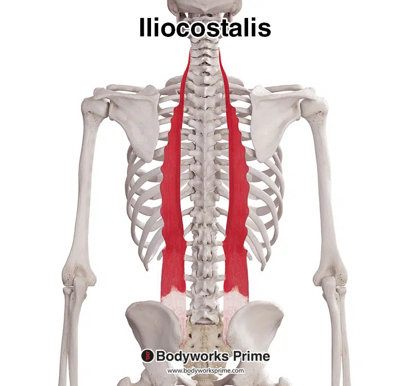
Pictured here, we can see the iliocostalis muscle by itself. The iliocostalis is the most lateral of the erector spinae muscles.
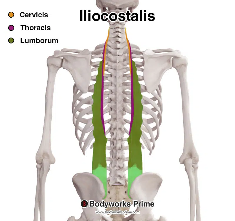
This image shows the different sections of the iliocostalis muscle. The cervicis is highlighted in yellow, the thoracis purple, and the lumborum in green.
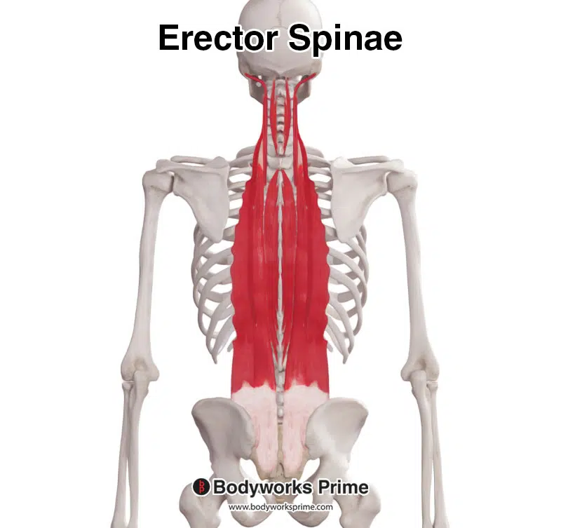
Displayed here are all of the erector spinae muscles together.
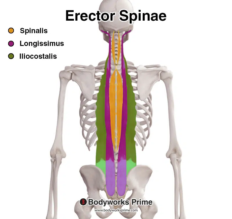
Pictured here, we can see the erector spinae muscle group. The spinalis is highlighted in yellow, the longissimus in purple, and the iliocostalis in green.
Origin & Insertion
The points of origin and insertion of the iliocostalis muscle differ among its various segments. The specifics are as follows:
The iliocostalis cervicis has its origin at the angles of ribs 3 to 6 and inserts at the posterior tubercle of the transverse process of vertebrae C4 to C6 [3] [4].
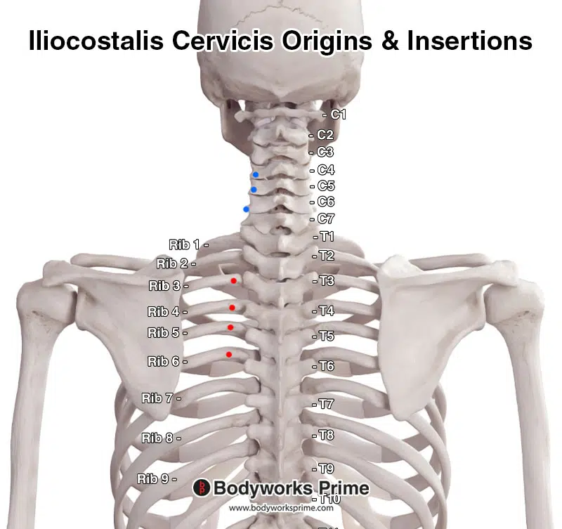
Displayed in this picture are the origins and insertions of the iliocostalis cervicis section, with origins highlighted in red and insertions highlighted in blue.
Note: For the sake of simplicity, only the left side of the muscle is displayed here. However, the origins and insertions are duplicated on the right-hand side as well.
The iliocostalis thoracis originates at the angles of the bottom six ribs and inserts at the transverse process of vertebra C7 and the angles of the top six ribs [5] [6].
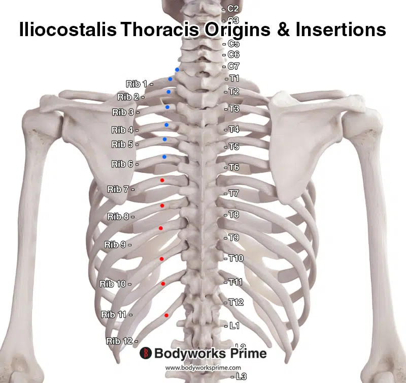
This image displays the origins and insertions of the iliocostalis thoracis section, with the origins highlighted in red and the insertions highlighted in blue.
Note: For the sake of simplicity, only the left side of the muscle is displayed here. However, the origins and insertions are duplicated on the right-hand side as well.
The iliocostalis lumborum originates at the medial end and dorsal segment of the iliac crest and the lateral crest of the sacrum. It inserts at the lumbar transverse processes of L1 to L4, the angles of ribs 4 to 12, and the thoracolumbar fascia [7] [8].
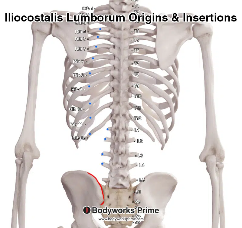
This image presents the origins and insertions of the iliocostalis lumborum section, with the origins highlighted in red and the insertions in blue.
Note: For the sake of simplicity, only the left side of the muscle is displayed here. However, the origins and insertions are duplicated on the right-hand side as well.
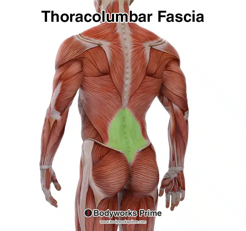
Pictured here, we can see the thoracolumbar fascia, another origin point of the iliocostalis lumborum. Due to the location of its attachments, some people also consider the iliocostalis lumborum to be an insertion as well. The classification of whether the thoracolumbar fascia is an origin or insertion point of the iliocostalis lumborum appears to vary among individuals and sources. However, it is most commonly considered an origin.
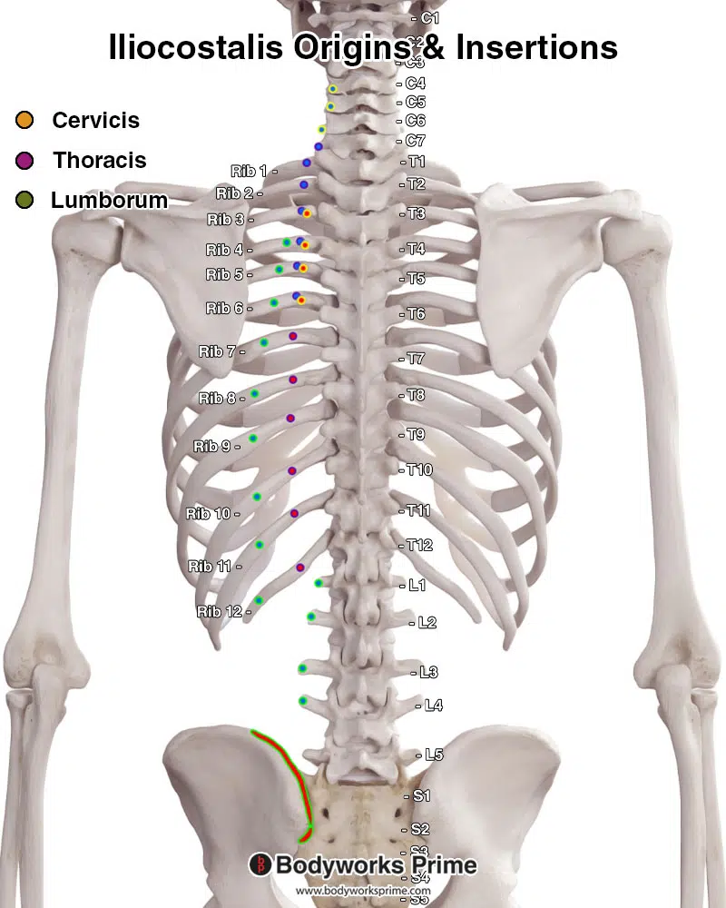
Pictured here are the origins and insertions of the iliocostalis lumborum, thoracis, and cervicis, with origins highlighted in red and insertions in blue. The cervicis has a yellow highlight around it, the thoracis has a purple highlight and the lumborum has a green highlight.
Note: For the sake of simplicity, only the left side of the muscle is displayed here. However, the origins and insertions are duplicated on the right-hand side as well.
Actions
The actions of the iliocostalis muscle vary based on whether it contracts unilaterally or bilaterally. When only one side contracts, the result is lateral flexion of the trunk and vertebral column. When both sides contract together, it causes extension of the trunk and vertebral column. The cervicis segment of the muscle extends from the cervical vertebrae to the fourth cervical vertebra (C4), offering weak support for lateral flexion at the neck’s base during unilateral contractions and weak extension at the neck’s base during bilateral contractions [9] [10].
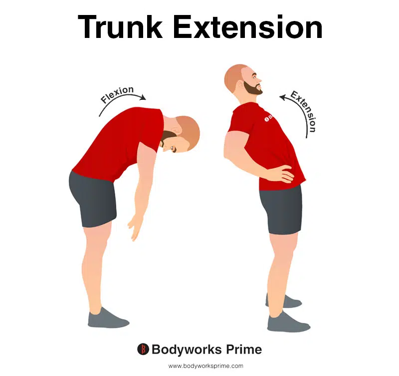
This image shows an example of trunk extension, which involves arching or bending the spine/torso/back backwards. The opposite of trunk extension is trunk flexion. The iliocostalis lumborum and thoracis sections are involved in trunk extension. The extension is the result of a bilateral contraction.
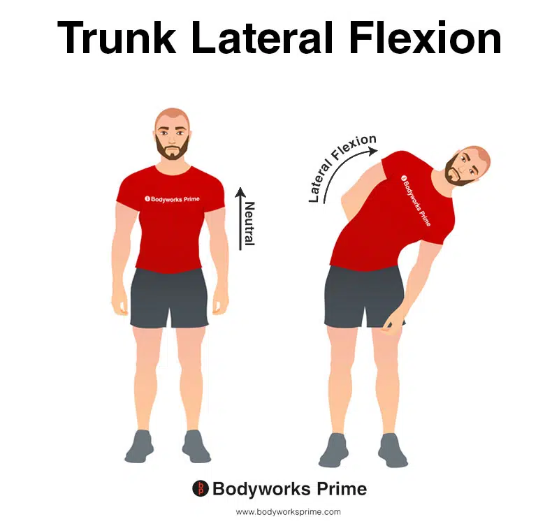
This image demonstrates trunk lateral flexion, which involves bending the torso to the side. The iliocostalis lumborum and thoracis sections are involved in trunk lateral flexion. The lateral flexion is the result of a unilateral contraction.
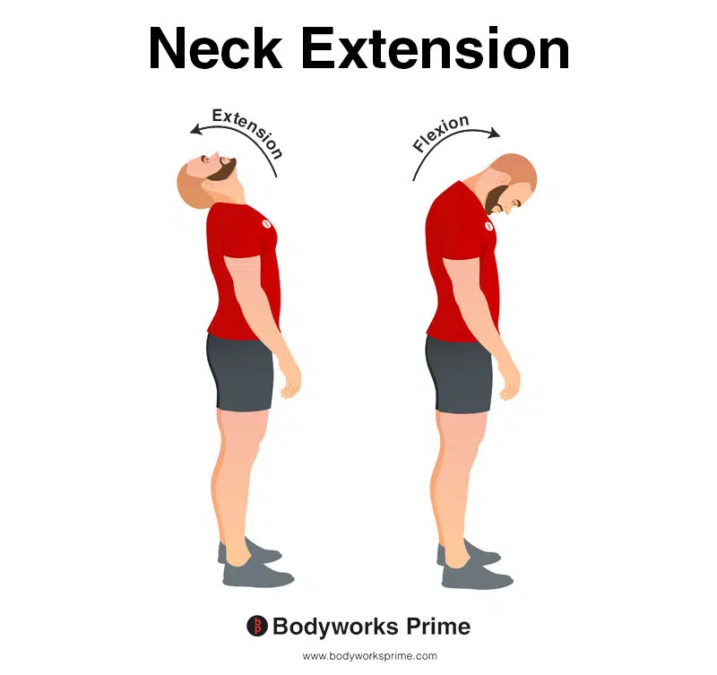
This image demonstrates neck extension, which involves bending the neck backwards. The iliocostalis cervicis section is involved in neck extension at the base of the neck. The extension is the result of a bilateral contraction. It provides other muscles with weak assistance during extension because it is not a strong extender of the neck.
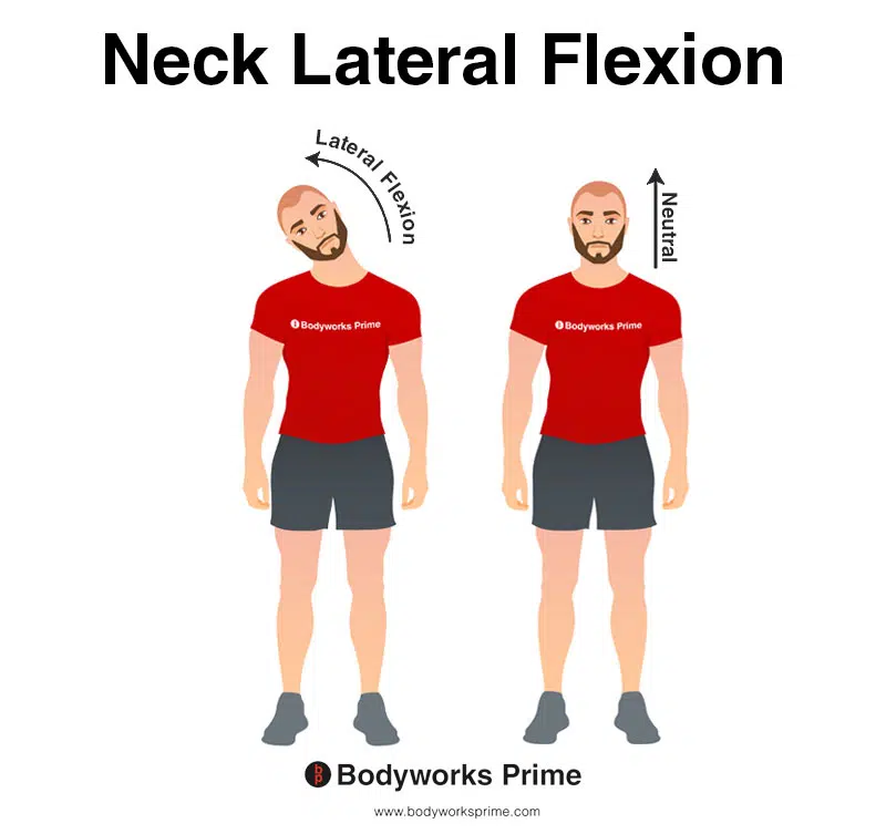
This image demonstrates neck lateral flexion, which involves bending the neck to the side. The iliocostalis cervicis section is involved in lateral flexion at the base of the neck. The lateral flexion is the result of a unilateral contraction. It provides other muscles with weak assistance during lateral flexion because it is not a strong lateral flexor of the neck.
Innervation
The iliocostalis muscle is supplied with nerve impulses by the lateral branches of the posterior rami of the spinal nerves, with each vertebral level corresponding to its own nerve supply [11] [12].
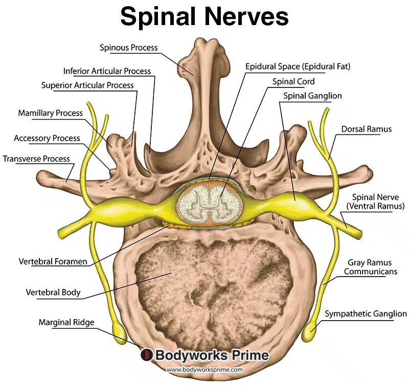
Pictured here is a cross-section of a lumbar vertebra. One of the labeled parts is the ‘dorsal ramus’, which is also known as the ‘posterior ramus’. Each segment of the erector spinae muscles receives innervation from the posterior ramus of the corresponding spinal nerve. In other words, the part of the muscle aligned with a specific spinal segment is innervated by that specific spinal nerve’s posterior ramus.
Blood Supply
Blood is supplies to the iliocostalis through dorsal branches of the posterior intercostal, subcostal, lumbar and lateral sacral arteries.
Want some flashcards to help you remember this information? Then click the link below:
Iliocostalis Flashcards
Support Bodyworks Prime
Running a website and YouTube channel can be expensive. Your donation helps support the creation of more content for my website and YouTube channel. All donation proceeds go towards covering expenses only. Every contribution, big or small, makes a difference!
References
| ↑1, ↑3, ↑5, ↑7 | Henson B, Kadiyala B, Edens MA. Anatomy, Back, Muscles. [Updated 2022 Aug 25]. In: StatPearls [Internet]. Treasure Island (FL): StatPearls Publishing; 2022 Jan-. Available from: https://www.ncbi.nlm.nih.gov/books/NBK537074/ |
|---|---|
| ↑2, ↑4, ↑6, ↑8, ↑10, ↑12 | Moore KL, Agur AMR, Dalley AF. Clinically Oriented Anatomy. 8th ed. Philadelphia: Lippincot Williams & Wilkins; 2017. |
| ↑9, ↑11 | Chaitow, Leon, and DeLany, Judith (2011), Clinical Application of Neuromuscular Techniques, Volume 2 (Second Edition), Oxford: Churchill Livingstone, ISBN 978-0-443-06815-7 |


