| Origin | Upper three-fourths of anterior & medial surfaces of ulna Interosseous membrane |
| Insertion | Palmar surface of distal phalanges of digits 2-5 |
| Action | Flexes the metacarpophalangeal and interphalangeal joints of digits 2-5 Weak assistance in wrist flexion |
| Nerve | Lateral part (digits 2-3): Anterior interosseous branch of the median nerve (C8-T1) Medial part (digits 4-5): Ulnar nerve (C8, T1) |
| Artery | Anterior interosseous artery |
Location & Overview
The flexor digitorum profundus is located deep to the flexor digitorum superficialis. The flexor digitorum profundus passes between a split/gap in the flexor digitorum superficialis’ distal tendon. This split/gap is located just before the proximal interphalangeal joint (The proximal interphalangeal joint is located between the middle and proximal phalanx sections of the finger, shown in the images below). The flexor digitorum profundus is the only muscle which can flex the distal interphalangeal joints of the fingers (the distal interphalangeal joints are the joints located at the tips of the fingers). The flexor digitorum profundus passes deep to the flexor retinaculum, within the common flexor sheath (i.e it passes through the carpal tunnel). The carpal tunnel is a narrow passageway formed by the bones and ligaments of the wrist joint. The flexor retinaculum forms the anterior ‘roof’ of the carpal tunnel [1] [2].
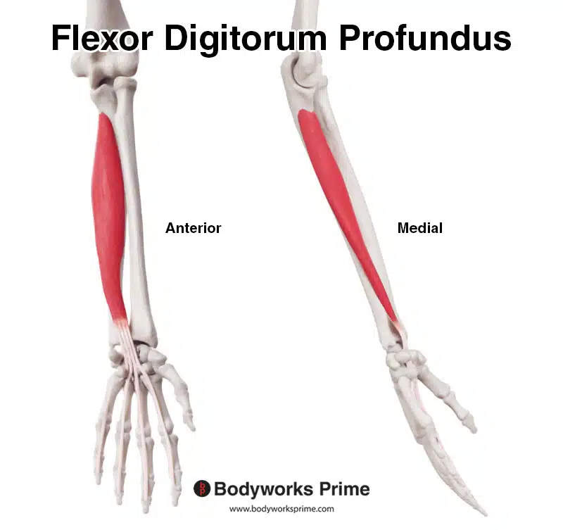
Here we can see the flexor digitorum profundus from a anterior and medial.
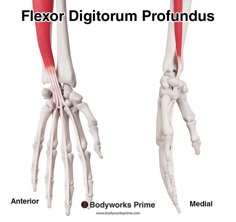
Here we can see a closer look at the flexor digitorum profundus’ distal tendon.
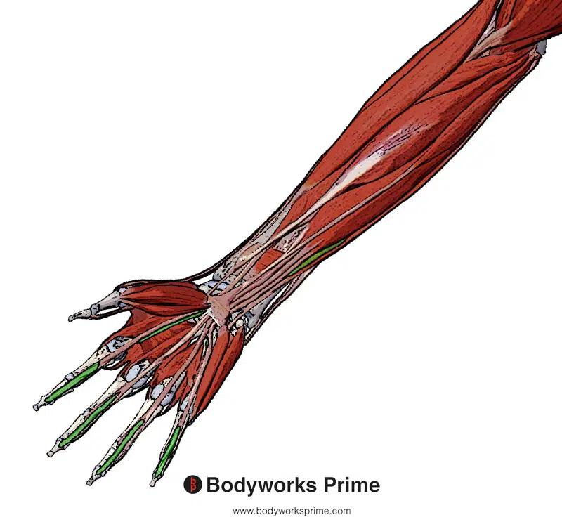
Highlighted in green, we can see the flexor digitorum profundus from a superficial view. We can see how it passes through a split in the flexor digitorum superficialis at its distal end.
Here we can see the flexor digitorum profundus muscle by itself.
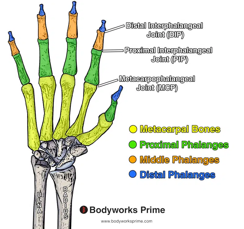
Here we can see a labelled diagram of the bones and the joints of the phalanges of the hand.
Origin & Insertion
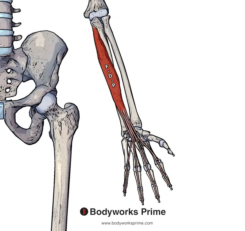
The flexor digitorum profundus originates from the upper three-fourths of the anterior and medial surfaces of the ulna. It also originates at the interosseous membrane and the deep fascia of the forearm. It then continues distally down the forearm, crossing the wrist joint via the carpal tunnel and finally connects to the tips of the fingers (distal phalanges). It inserts onto the bases of the distal phalanges of the 2nd, 3rd, 4th and 5th digits [3] [4].
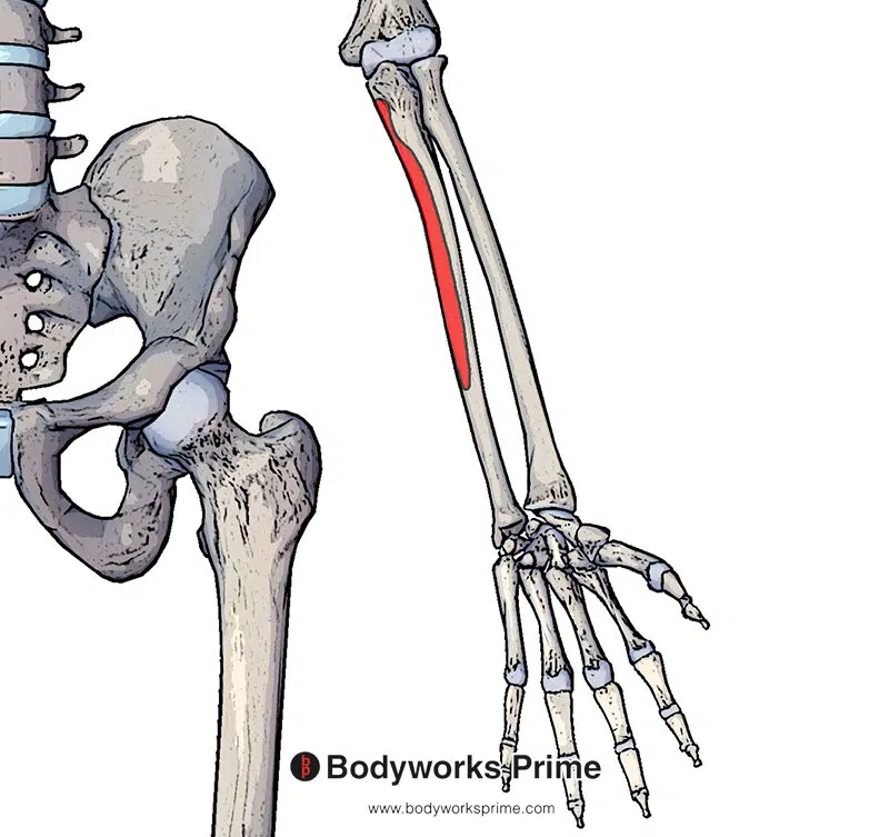
Here we can see the first origin point of the flexor digitorum profundus muscle highlighted in red. The origin is on the upper three-fourths of the anterior & medial surfaces of the ulna.
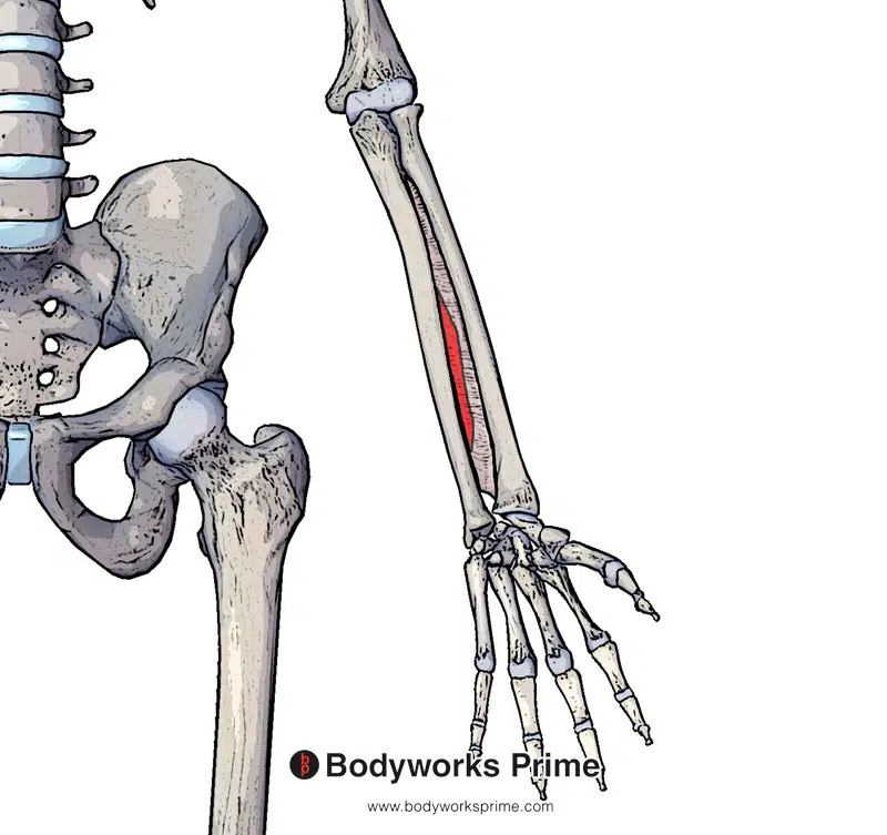
Here we can see the second origin point of the flexor digitorum profundus muscle highlighted in red on the interosseous membrane.
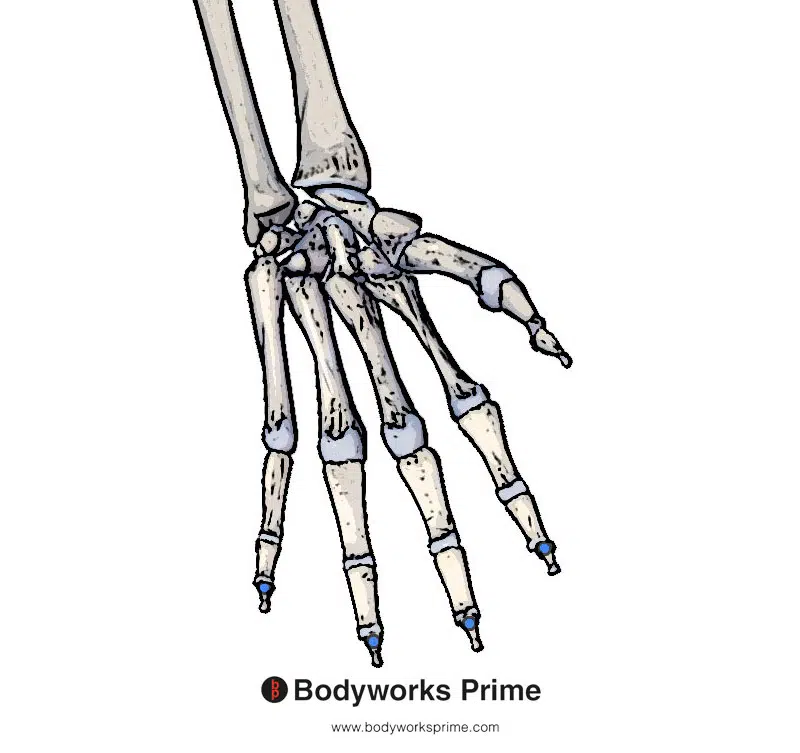
Here we can see the insertion points of the flexor digitorum profundus muscle highlighted in blue. The insertions are on the palmar surfaces of the distal phalanges of digits 2-5.
Actions
The flexor digitorum profundus is a muscle that enables the flexion of various joints in the fingers. It can flex the metacarpophalangeal and interphalangeal joints of the index, middle, ring, and little fingers. Additionally, it can aid in flexing the wrist at the midcarpal joint. The muscle’s medial part flexes the pinky/little and ring fingers (digits 4 & 5), while its lateral part flexes the index and middle fingers (digits 2 & 3) [5] [6]. To view a labelled diagram of the bones and joints of the phalanges of the hand, please check out the picture located at the end of the ‘Location & Overview’ section or press here to go straight to it.
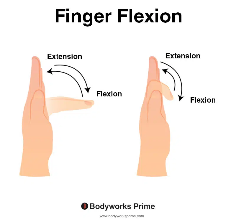
This image demonstrates finger flexion, which is the action of bending the fingers towards the palm of the hand. The left side of the image depicts flexion happening at the metacarpophalangeal joint, while the right side shows finger flexion occurring at the interphalangeal joints (specifically the proximal and distal interphalangeal joints). Flexion at all three of these joints is an action of the flexor digitorum profundus.
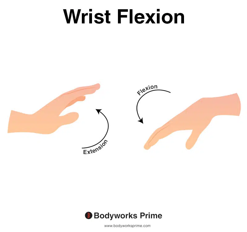
This image shows wrist flexion, which is the movement of bending the wrist in the direction of the underneath of the forearm (anterior surface of the forearm). The opposite of wrist flexion would be wrist extension which involves extending the wrist in the direction of the top of the forearm (posterior surface of the forearm). The flexor digitorum profundus muscle is able to provide weak assistance during wrist flexion.
Innervation
The flexor digitorum profundus’ lateral part is innervated by the anterior interosseous nerve which is a branch of the median nerve. The flexor digitorum profundus’ lateral part is located on the index and middle fingers. The flexor digitorum profundus’ medial part is innervated by the ulnar nerve. The flexor digitorum profundus’ medial part is located on the ring and little finger [7] [8].
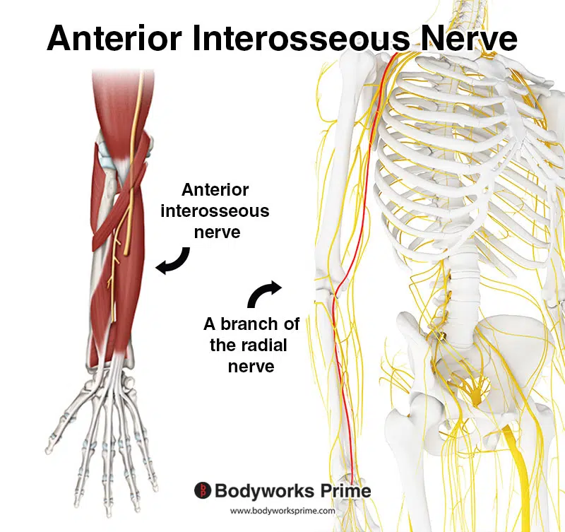
Pictrured here we can see the anterior interosseous nerve which is a branch of the radial nerve. The radial nerve is highlighted in red on the right hand side.
Blood Supply
Blood is supplied to the flexor digitorum profundus via the anterior interosseous artery. The anterior interosseous artery is a branch of the common interosseous artery [9].
Want some flashcards to help you remember this information? Then click the link below:
Flexor Digitorum Profundus Flashcards
Support Bodyworks Prime
Running a website and YouTube channel can be expensive. Your donation helps support the creation of more content for my website and YouTube channel. All donation proceeds go towards covering expenses only. Every contribution, big or small, makes a difference!
References
| ↑1, ↑3, ↑5, ↑7 | Moore KL, Agur AMR, Dalley AF. Clinically Oriented Anatomy. 8th ed. Philadelphia: Lippincot Williams & Wilkins; 2017 |
|---|---|
| ↑2, ↑4, ↑6, ↑8, ↑9 | Lung BE, Burns B. Anatomy, Shoulder and Upper Limb, Hand Flexor Digitorum Profundus Muscle. [Updated 2021 Oct 30]. In: StatPearls [Internet]. Treasure Island (FL): StatPearls Publishing; 2022 Jan-. Available from: https://www.ncbi.nlm.nih.gov/books/NBK526046/ |


