| Origin | Lateral femoral condyle Fibrous capsule of the knee joint Posterior horn of the knee’s lateral meniscus Head of the fibula via popliteofibular ligament |
| Insertion | Posterior surface of the tibia, proximal to the soleal line |
| Action | Lateral rotation of the femur on the tibia Medial rotation of the tibia on the femur Protection of the lateral meniscus Insignificant amount of knee flexion |
| Nerve | Tibial nerve (L5-S1) |
| Artery | Popliteal artery (medial inferior genicular branch) Posterior tibial artery (muscular branch) |
Location & Overview
The popliteus muscle is a small, somewhat triangular shaped muscle located at the posterior aspect of the knee joint. It plays an important role in the stability and functioning of the knee joint, as it aids in unlocking the knee during the initiation of knee flexion from a fully extended position [1]. The popliteus muscle is situated deep within the popliteal fossa, which is a diamond-shaped region found posterior to the knee [2]. The popliteus is unique because of its inverted origin and insertion, with the tendinous attachment originating from the proximal bone and the fleshy attachment representing the muscle’s insertion [3]. For most muscles, the origin is usually the more stable and fixed attachment point on a bone. While the insertion is the movable attachment point that pulls on a bone to create movement. However, in the case of the popliteus muscle, this relationship is inverted.
In relation to other muscles, the popliteus muscle is positioned deep to the gastrocnemius, plantaris, and the heads of the biceps femoris muscle in the popliteal fossa. It is also located medial to the lateral head of the gastrocnemius muscle and the tendon of the biceps femoris muscle [4].
The muscle is enveloped by the popliteal vessels and nerves, which include the popliteal artery, medial inferior genicular branch of the popliteal artery, muscular branch of the posterior tibial artery, tibial nerve, and the common peroneal nerve [5][6]. Furthermore, the popliteus muscle is in close association with the arcuate popliteal ligament, which originates from the posterior aspect of the fibular head and attaches to the lateral condyle of the femur [7]. This ligament lies superficial to the popliteus muscle and helps to stabilise the posterolateral corner of the knee joint [8].
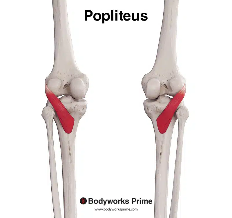
Pictured here we can see the popliteus muscle by itself from a posterior view.
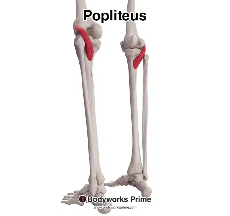
Pictured here we can see the popliteus muscle by itself from a more lateral view.
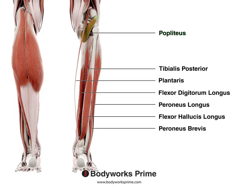
On the right, we can see the popliteus muscle highlighted in green amongst other deep muscles of the lower leg. The left leg shows a superficial view of all of the muscles, of which the popliteus is not visible, due to it being a deep muscle.
Origin & Insertion
The popliteus muscle has a unique morphology, with its origin and insertion points inverted compared to most muscles. The tendinous attachment serves as the origin, while the fleshy attachment acts as the insertion [9].
The origin of the popliteus muscle has several components. The primary origin is a small pit on the lateral surface of the lateral femoral condyle, just below the attachment of the fibular collateral ligament of the knee [10]. Additional origins include a fibrous attachment from the popliteus to the posterior aspect of the fibrous capsule of the knee joint, which is a tough, fibrous structure that encloses and protects the joint. Another component of the popliteus’ origin is a fibrous band that extends from the superior margin of the terminal portion of the tendon and attaches to the superior margin of the posterior horn of the lateral meniscus. The lateral meniscus is a crescent-shaped piece of cartilage in the knee joint that helps with shock absorption and stabilisation. The posterior horn of the meniscus refers to the rear part of the lateral meniscus [11]. Furthermore, there is also another origin which is fibrous band called the popliteofibular ligament, which extends between the head of the fibula and the popliteus tendon [12].
The insertion of the popliteus muscle is on the popliteal surface of the tibia, which is located on the posterior surface of the tibia, above the soleal line [13]. The muscle fibers of the popliteus run obliquely and inferiorly from this insertion to attach to the tibia, where they help produce the muscle’s actions and stabilise the knee joint during various movements.
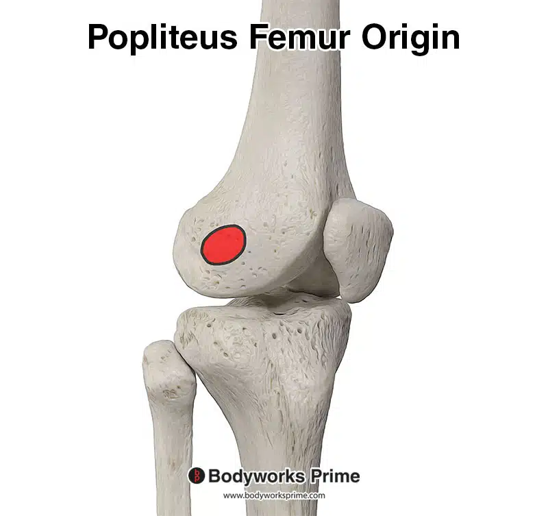
Marked in red we can see the primary origin of the popliteus muscle on the lateral femoral condyle.
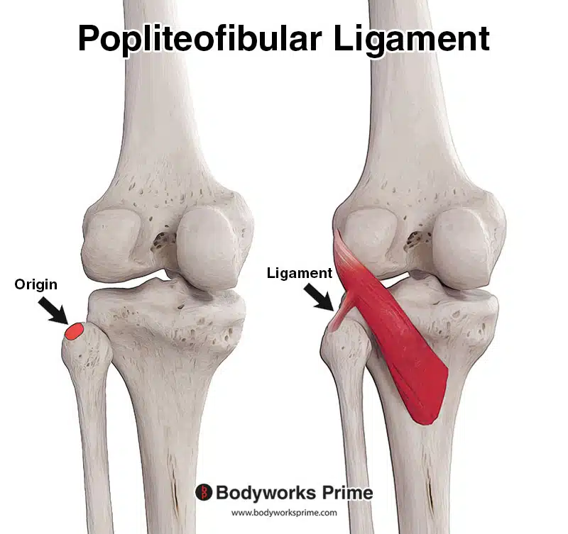
Pictured here we can see the popliteofibular ligament and how it originates from the head of the fibula. The head of the fibula is an origin of the popliteus muscle and it connects to the popliteus via the popliteofibular ligament.

Here, you can see the knee joint capsule. The knee joint capsule is a protective, fibrous structure that surrounds the knee joint. It serves to contain synovial fluid and maintain joint stability. The popliteus muscle passes through and attaches to the knee joint capsule before connecting to the lateral meniscus. This is the reason the lateral part of the knee joint capsule is considered an origin of the popliteus muscle.
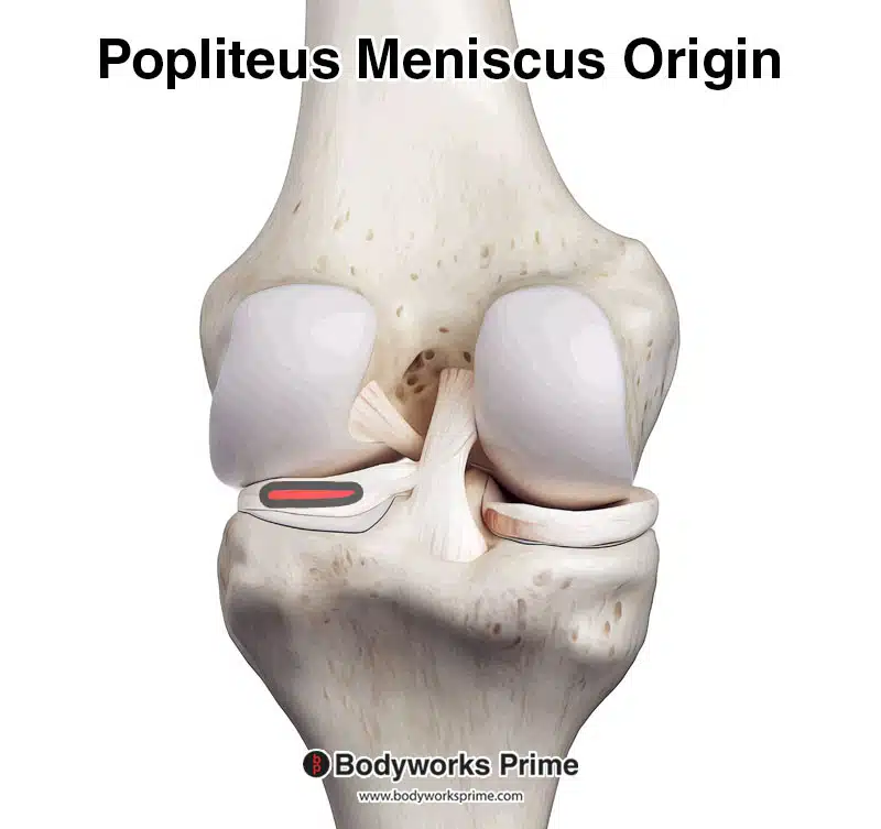
Highlighted in red you can see an origin of the popliteus muscle on the lateral meniscus. The lateral meniscus is a C-shaped, fibrocartilaginous structure located in the knee joint, on the lateral side. The next picture shows this in more detail.
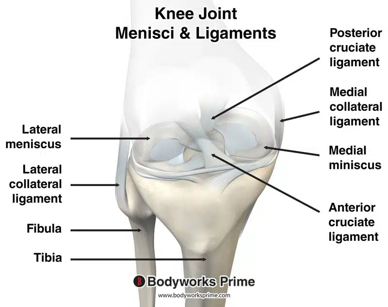
Pictured here you can see the knee joint along with the menisci and ligaments. This is useful for understanding the location of the popliteus origin on the lateral meniscus.
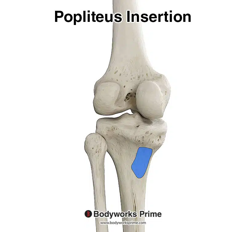
Pictured here we can see the popliteus muscle’s insertion on the posterior surface of the tibia, just above the soleal line.
Actions
The popliteus muscle plays an important role in maintaining knee stability and has several notable actions, which include:
Lateral rotation of the femur: At the beginning of knee flexion, the popliteus muscle laterally rotates the femur on the tibia, helping to “unlock” the knee joint. This action releases the “screw-home mechanism” that occurs during full knee extension, facilitating smoother knee flexion. This generally occurs when the foot is placed on the floor (e.g when the knee is locked out while standing) the popliteus then helps unlock the knee by rotating the femur laterally on the tibia [14] [15].
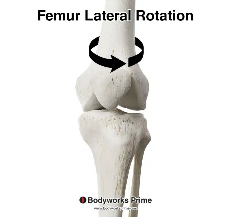
This picture shows the direction which the femur moves when rotating laterally, as depicted by the direction of the black arrow.
Medial rotation of the tibia: When the knee is flexed, the popliteus muscle contributes to the medial rotation of the tibia on the femur. This medial rotation generally occurs when the foot is not in contact with the ground and the knee is flexed. The popliteus can then help with rotating the tibia medially beneath the femoral condyles [16] [17].
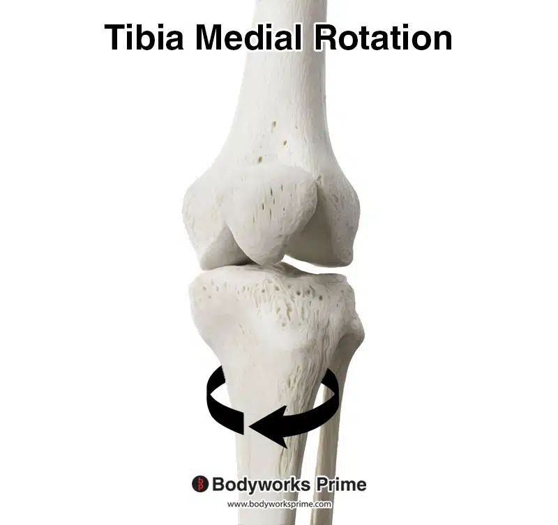
Here we can see the direction which the tibia moves when rotating medially, as depicted by the direction of the black arrow.
Protection of the lateral meniscus: The popliteus muscle helps to protect the lateral meniscus of the knee by pulling the lateral meniscus posteriorly during knee flexion [18] [19]. The lateral meniscus is a crescent-shaped piece of fibrocartilage which is located within the knee joint, specifically on the outer edge of the top surface of the tibia. The meniscus of the knee acts as a shock absorber between the femur and tibia, and it also helps distribute the body’s weight across the knee. This distribution of weight leads to increased stability and helps prevent damage to the joint. The lateral meniscus is more mobile and less prone to injury than its medial counterpart, this is likely due to the role the popliteus muscle plays in protecting the lateral meniscus. The medial meniscus does not have an equivalent muscle to pierce the joint capsule and connect to it like the lateral meniscus does. Therefore, the lateral meniscus benefits greatly from the role of the popliteus muscle. Conversely, the medial meniscus must depend solely on adjustments in tension, compression, and torsional forces to alter its shape and position, lacking the benefit of a dedicated supporting muscle.
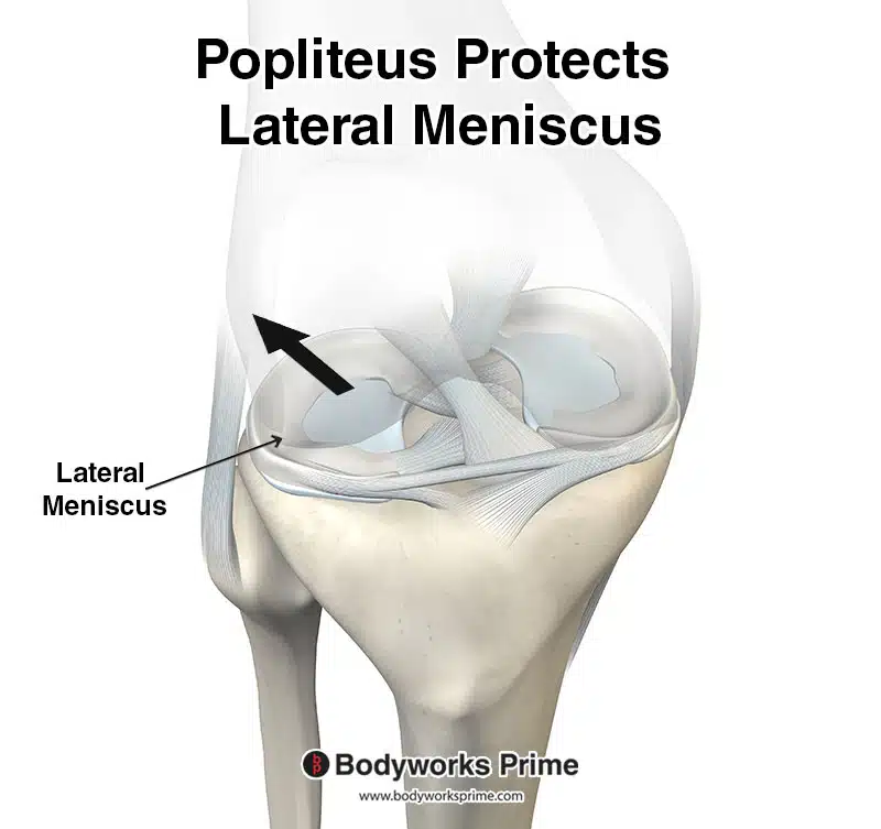
In this image depicts the lateral meniscus moving in a posterior direction. An action of the popliteus muscle is to protect the lateral meniscus of the knee by pulling the lateral meniscus posteriorly during knee flexion.
Insignificant knee flexion: The popliteus muscle provides an insignificant amount of knee flexion, working alongside other more primary knee flexors such as the hamstrings and gastrocnemius [20]. Knee flexion is in no way a primary action of this muscle, but it is worth mentioning as some sources overstate the contribution of the popliteus muscle in flexion of the knee joint. Its knee flexion contribution is very minimal, to the point of being considered insignificant.
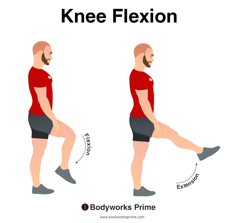
In this image, you can see an example of knee flexion, which is the action of bending your knee. The opposite movement of knee flexion is knee extension. The popliteus muscle contributes very little to knee flexion. Nonetheless, it is still considered an action, although an insignificant one.
Innervation
The popliteus muscle is primarily innervated by the tibial nerve, which is composed of one of the two terminal branches of the sciatic nerve. The sciatic nerve is the largest and longest nerve in the human body. The sciatic nerve, itself, is comprised of nerve fibers from the L4 to S3 segments of the spinal cord, and it separates into the tibial and common fibular (peroneal) nerves at the level of the knee. The tibial nerve is specifically responsible for the innervation of the muscles in the posterior compartment of the leg, including the popliteus [21] [22] [23].
The nerve roots that contribute to the innervation of the popliteus muscle are L5, S1. These segments form part of the lower region of the spinal cord, also known as the lumbosacral region. The neurons originating from these roots ultimately form the tibial nerve, which branches off the sciatic nerve in the lower part of the thigh, close to the knee. This nerve, after branching off, travels inferiorly along the posterior aspect of the leg, providing motor innervation to the muscles located there, such as the popliteus [24] [25] [26].
Upon reaching the popliteus muscle, the tibial nerve penetrates at the lateral distal margin of the muscle, located inferior to the head of the fibula. Following its entry into the muscle, the nerve divides into anterior, medial, and lateral branches, ensuring widespread distribution throughout the muscle. This comprehensive distribution of nerve fibers allows for the efficient control required for the popliteus muscle’s actions [27] [28] [29].
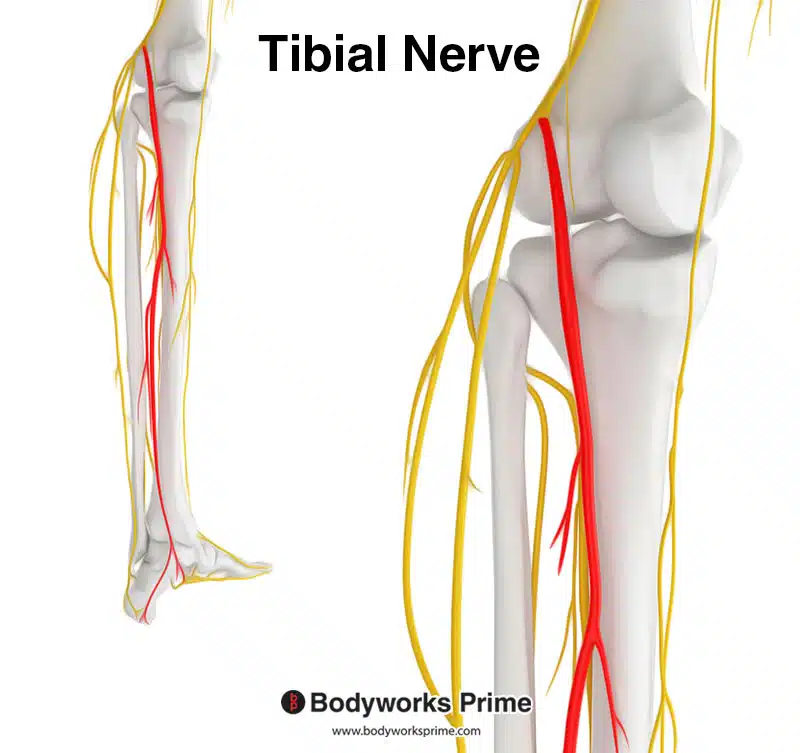
Highlighted in red is the tibial nerve which is a branch of the sciatic nerve. The tibial nerve innervates the popliteus muscle. The tibial nerve spinal roots are L5-S1.
Blood Supply
The blood supply to the popliteus muscle is principally provided by two arteries: the medial inferior genicular branch of the popliteal artery and the muscular branch of the posterior tibial artery [30].
The popliteal artery, which is a direct continuation of the femoral artery, travels through the popliteal fossa at the back of the knee. This artery gives rise to several branches, including the medial inferior genicular artery, which contributes to the vascular supply of the popliteus muscle [31].
Meanwhile, the posterior tibial artery, one of the terminal branches of the popliteal artery, also plays a role in supplying blood to this muscle. Specifically, the muscular branches of the posterior tibial artery, which diverge to supply various muscles in the posterior compartment of the leg, also extend their reach to the popliteus muscle [32].
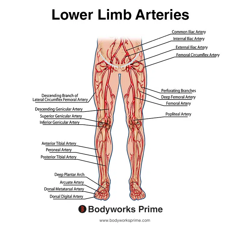
This image shows the arteries of the lower limb. You can see both the posterior tibial artery and the popliteal artery labelled here.
Want some flashcards to help you remember this information? Then click the link below:
Popliteus Flashcards
Support Bodyworks Prime
Running a website and YouTube channel can be expensive. Your donation helps support the creation of more content for my website and YouTube channel. All donation proceeds go towards covering expenses only. Every contribution, big or small, makes a difference!
References
| ↑1, ↑3, ↑7, ↑9, ↑10, ↑11, ↑12, ↑13 | Paraskevas G, Papaziogas B, Kitsoulis P, Spanidou S. A study on the morphology of the popliteus muscle and arcuate popliteal ligament. Folia Morphol (Warsz). 2006 Nov;65(4):381-4. PMID: 17171619. |
|---|---|
| ↑2, ↑6 | Hyland S, Sinkler MA, Varacallo M. Anatomy, Bony Pelvis and Lower Limb: Popliteal Region. [Updated 2022 Jul 25]. In: StatPearls [Internet]. Treasure Island (FL): StatPearls Publishing; 2023 Jan-. Available from: https://www.ncbi.nlm.nih.gov/books/NBK532891/ |
| ↑4, ↑5, ↑15, ↑30 | Hyland S, Varacallo M. Anatomy, Bony Pelvis and Lower Limb: Popliteus Muscle. [Updated 2022 Jun 6]. In: StatPearls [Internet]. Treasure Island (FL): StatPearls Publishing; 2023 Jan-. Available from: https://www.ncbi.nlm.nih.gov/books/NBK526084/ |
| ↑8 | LaPrade RF, Engebretsen AH, Ly TV, Johansen S, Wentorf FA, Engebretsen L. The anatomy of the medial part of the knee. J Bone Joint Surg Am. 2007 Sep;89(9):2000-10. doi: 10.2106/JBJS.F.01176. PMID: 17768198. |
| ↑14, ↑16, ↑18, ↑20, ↑31, ↑32 | Moore KL, Agur AMR, Dalley AF. Clinically Oriented Anatomy. 8th ed. Philadelphia: Lippincot Williams & Wilkins; 2017. |
| ↑17, ↑19 | Nyland J, Lachman N, Kocabey Y, Brosky J, Altun R, Caborn D. Anatomy, function, and rehabilitation of the popliteus musculotendinous complex. J Orthop Sports Phys Ther. 2005 Mar;35(3):165-79. doi: 10.2519/jospt.2005.35.3.165. PMID: 15839310. |
| ↑21, ↑24, ↑27 | Standring S. (2015). Gray’s Anatomy: The Anatomical Basis of Clinical Practice, 41st Edn. Amsterdam: Elsevier. |
| ↑22, ↑25, ↑28 | Yu D, Yin H, Han T, Jiang H, Cao X. Intramuscular innervations of lower leg skeletal muscles: applications in their clinical use in functional muscular transfer. Surg Radiol Anat. 2016 Aug;38(6):675-85. |
| ↑23 | Desai SS, Cohen-Levy WB. Anatomy, Bony Pelvis and Lower Limb: Tibial Nerve. [Updated 2022 Aug 8]. In: StatPearls [Internet]. Treasure Island (FL): StatPearls Publishing; 2023 Jan-. Available from: https://www.ncbi.nlm.nih.gov/books/NBK537028/ |
| ↑26, ↑29 | Day CP, Orme R. Popliteal artery branching patterns — an angiographic study. Clin Radiol. 2006 Aug;61(8):696-9. |


